. 2013 Sep 9;23(17):1707-12.
doi: 10.1016/j.cub.2013.07.017. Epub 2013 Jul 25.

Discs large links spindle orientation to apical-basal polarity in Drosophila epithelia

Affiliations

Discs large links spindle orientation to apical-basal polarity in Drosophila epithelia

Dan T Bergstralh et al. Curr Biol. .

Abstract

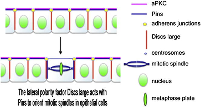
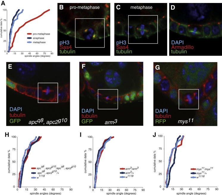
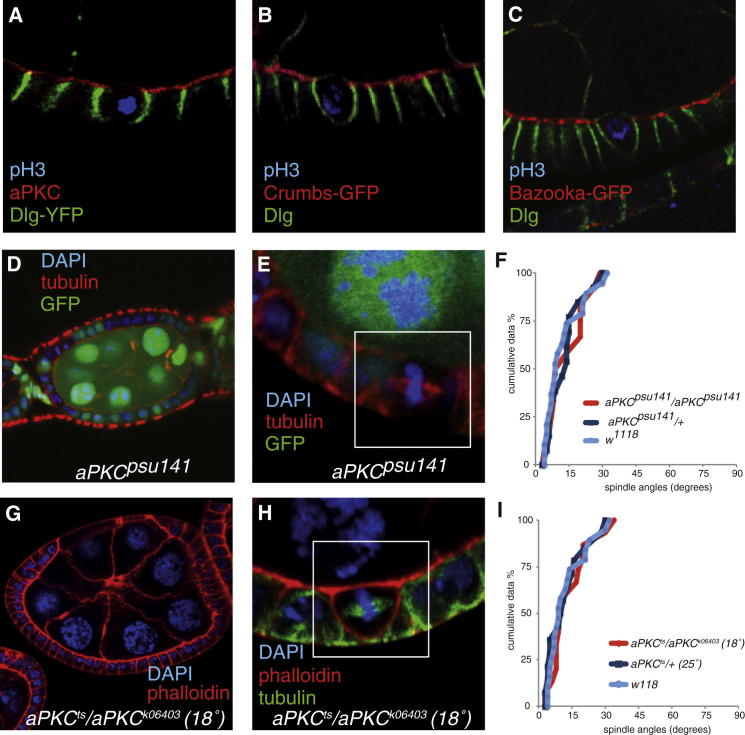
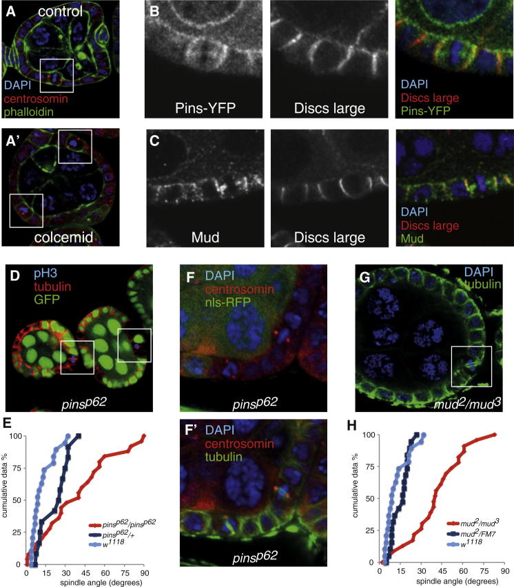
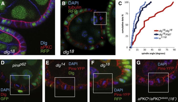
Mitotic spindles in epithelial cells are oriented in the plane of the epithelium so that both daughter cells remain within the monolayer, and defects in spindle orientation have been proposed to promote tumorigenesis by causing epithelial disorganization and hyperplasia. Previous work has implicated the apical polarity factor aPKC, the junctional protein APC2, and basal integrins in epithelial spindle orientation, but the underlying mechanisms remain unclear. We show that these factors are not required for spindle orientation in the Drosophila follicular epithelium. Furthermore, aPKC and other apical polarity factors disappear from the apical membrane in mitosis. Instead, spindle orientation requires the lateral factor Discs large (Dlg), a function that is separable from its role in epithelial polarity. In neuroblasts, Pins recruits Dlg and Mud to form an apical complex that orients spindles along the apical-basal axis. We show that Pins and Mud are also necessary for spindle orientation in follicle cells, as is the interaction between Dlg and Pins. Dlg localizes independently of Pins, however, suggesting that its lateral localization determines the planar orientation of the spindle in epithelial cells. Thus, different mechanisms recruit the conserved Dlg/Pins/Mud complex to orient the spindle in opposite directions in distinct cell types.


Figures

None
Graphical abstract
Figure 1
Figure 1
At Metaphase, Spindles Are Angled Roughly in Parallel to the Plane of the Follicle Cell Epithelium (A) Cumulative plot of spindle angles in the FCE at prometaphase (red line), metaphase (blue), and anaphase (dark blue). The spindle angles were measured relative to a line through the adherens junctions, which represents the plane of the epithelium, and were plotted in rank order from lowest to highest against the spindle angles along the x axis. (B and C) Mitotic spindles at prometaphase (B) and metaphase (C). Sas4-GFP (red) marks the spindle poles, and α-phospho-Histone 3 (pH3; blue) marks mitotic cells. (D) Mitotic spindles (green) in follicle cells orient below the level of the adherens junctions, which are marked by Armadillo (Drosophila β-catenin) in red. (E and F) Metaphase spindles are not misoriented in apc1q8, apc2g10 (E), or arm3 (F) homozygous mutant clones, marked by the absence of GFP (green). Metaphase cells are outlined by white boxes. (G) Metaphase spindles are normally oriented in single-layer mys11 clones (marked by the absence of RFP in green). (H–J) Cumulative plots of spindle angles in apc1q8, apc2g10 (H), arm3 (I), and mys11 (J) mutant follicle cells. See also Figure S1.
Figure 2
Figure 2
Spindle Orientation in Follicle Cells Does Not Depend on aPKC (A–C) The apical polarity determinants aPKC (A, red), Crumbs-GFP (B, red), and Bazooka (C, red), disappear from the apical cortex of mitotic follicle cells, although Dlg (green) remains along the lateral cortex. Dlg-YFP is shown in (A), and anti-Dlg staining is shown in (B) and (C). pH3, shown in blue, marks mitotic cells. (D) aPKCpsu14 mutant follicle cell clones have normal epithelial architecture. The aPKCpsu14 mutant cells are marked by the loss of GFP (green). (E) Metaphase spindles are not misoriented in aPKCpsu14 mutant follicle cell clones (marked by the absence of GFP). (F) Cumulative plot of metaphase spindle angles in aPKCpsu141 mutant follicle cells. (G) An apkcts/apkck06403 egg chamber at 18° showing a cyst encapsulation defect, as well as normal tissue. (H) Metaphase spindles are correctly oriented in an apkcts/apkck06403 egg chamber at 18°. (I) Cumulative plot of metaphase spindle angles in apkcts/apkck06403 follicle cells at 18°.
Figure 3
Figure 3
Pins and Mud Are Required for Spindle Orientation in the Follicle Cells (A and A′) The centrosomes (marked by centrosomin, red) align with the lateral cortex in untreated cells, and the metaphase plate (marked with DAPI, blue) is positioned vertically. The centrosomes and the metaphase plate are misoriented after treatment with the microtubule depolymerizing drug colcemid. (B and C) Pins-YFP (B, red) and Mud (C, red) colocalize with Dlg (green) along the lateral cortex in mitotic follicle cells (DAPI, blue). (D) Metaphase spindles are misoriented in the absence of pins function. Cells homozygous for pinsp62 are marked by the absence of GFP (green). Boxes highlight two metaphase cells with misaligned spindles. (E) Cumulative plot of metaphase spindle angles in pinsp62 mutant cells showing that the spindles are randomly oriented. Spindle angles in pinsp62 mutant cells (n = 32) differ significantly from the wild-type, with a p value of <0.005 as determined by the Kolmogorov-Smirnov test. (F and F′) pinsp62 mutant cells marked by the absence of RFP (green in F) form normal bipolar spindles (green in F′) with centrosomes at each pole (marked by Centrosomin, in red). (G) Metaphase spindles are misoriented in mud2/mud3 follicle cells. (H) Cumulative plot of metaphase spindle angles in mud2/mud3 mutant follicle cells showing spindle misorientation with clustering at approximately 45°. Spindle angles in mud2/mud3 mutant cells (n = 24) differ significantly from those in the wild-type (p value of <0.005 as determined by the Kolmogorov-Smirnov test). See also Figure S2.
Figure 4
Figure 4
Dlg Is Required for Mitotic Spindle Orientation (A) dlg18 mutant follicle cell clones (marked by the absence of RFP in green) maintain normal epithelial polarity until stage 6 of oogenesis, as shown by the normal localization of aPKC (red) apically and Dlg (blue) laterally. (B) Metaphase spindles are misoriented in dlg18 mutant follicle cells. (C) A cumulative plot of metaphase spindle angles in dlg18 mutant follicle cells. Spindle angles in dlg18 mutant cells (n = 27) differ significantly from those in the wild-type (p value of <0.005). (D) Dlg localization is normal in mitotic pinsp62 cells. Mutant cells are marked by the absence of GFP (in green). Dlg (red) localizes to the lateral cortex in a mitotic cell, which is outlined by a white box. DAPI is in blue. (E and F) Pins localizes to the apical and lateral cortex in dlg14 (E) and dlg18 (F) mutant cells. Mutant cells are marked by the absence of Dlg immunoreactivity (green in E) or RFP (green in F); Pins-YFP is shown in red. (G) Pins-YFP (in red) localizes normally during mitosis in apkcts/apkck06403 at 18°. See also Figure S3.

References

    1. McCaffrey L.M., Macara I.G. Epithelial organization, cell polarity and tumorigenesis. Trends Cell Biol. 2011;21:727–735. - PubMed
    1. Fernández-Miñán A., Martín-Bermudo M.D., González-Reyes A. Integrin signaling regulates spindle orientation in Drosophila to preserve the follicular-epithelium monolayer. Curr. Biol. 2007;17:683–688. - PubMed
    1. Lu B., Roegiers F., Jan L.Y., Jan Y.N. Adherens junctions inhibit asymmetric division in the Drosophila epithelium. Nature. 2001;409:522–525. - PubMed
    1. McCartney B.M., Price M.H., Webb R.L., Hayden M.A., Holot L.M., Zhou M., Bejsovec A., Peifer M. Testing hypotheses for the functions of APC family proteins using null and truncation alleles in Drosophila. Development. 2006;133:2407–2418. - PubMed
    1. Tanentzapf G., Smith C., McGlade J., Tepass U. Apical, lateral, and basal polarization cues contribute to the development of the follicular epithelium during Drosophila oogenesis. J. Cell Biol. 2000;151:891–904. - PMC - PubMed

Publication types

LinkOut - more resources