. 2013 Aug 22;20(8):991-1001.
doi: 10.1016/j.chembiol.2013.06.011. Epub 2013 Jul 25.

Peroxiredoxin-1 from the human hookworm Ancylostoma ceylanicum forms a stable oxidized decamer and is covalently inhibited by conoidin A

Affiliations

Peroxiredoxin-1 from the human hookworm Ancylostoma ceylanicum forms a stable oxidized decamer and is covalently inhibited by conoidin A

Jennifer B Nguyen et al. Chem Biol. .

Abstract

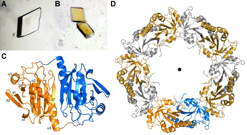
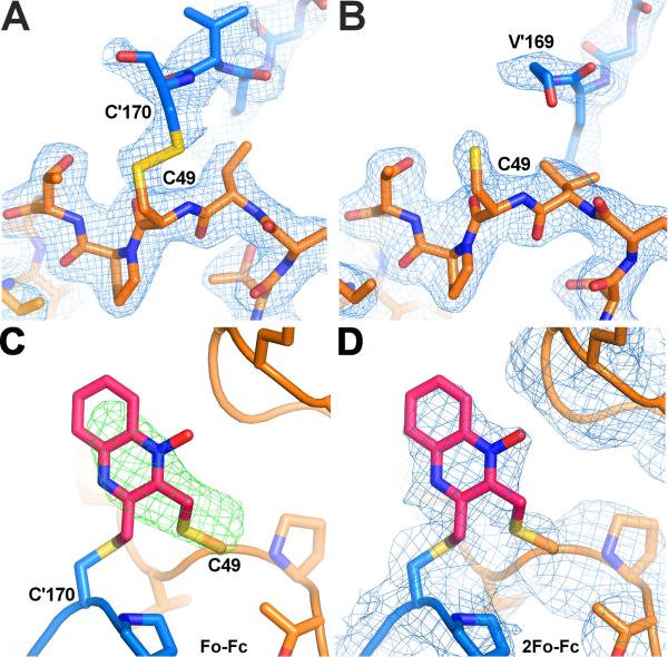
Hookworms are parasitic nematodes that have a devastating impact on global health, particularly in developing countries. We report a biochemical and structural analysis of a peroxiredoxin from the hookworm Ancylostoma ceylanicum, AcePrx-1. Peroxiredoxins provide antioxidant protection and act as signaling molecules and chaperones. AcePrx-1 is expressed in adult hookworms and can be inactivated by 2,3-bis(bromomethyl)quinoxaline-1,4-dioxide (conoidin A). Conoidin A inactivates AcePrx-1 by alkylating or crosslinking the catalytic cysteines, while maintaining the enzyme in the "locally unfolded" conformation. Irreversible oxidation of the resolving cysteine may contribute additional inhibitory activity. A crystal structure of oxidized AcePrx-1 reveals a disulfide-linked decamer. A helix macrodipole near the active site increases the reactivity of the catalytic cysteines to conoidin A. This work demonstrates the promise of conoidin compounds as probes to evaluate peroxiredoxins as drug targets in human parasites.


Figures

Figure 1
Figure 1. Sequence alignment of peroxiredoxin sequences
Genbank IDs are JX124321 (Ancylostoma ceylanicum Prx-1), 47499100 (Haemonchus contortus Prx), 193204376 (Caenorhabditis elegans Prx-2), 32189392 (human PrxII) and 5453549 (human PrxIV). Secondary structure elements in the AcePrx-1 crystal structure are overlaid on the alignment.
Figure 2
Figure 2. AcePrx-1 is expressed in adult hookworms and is inhibited by conoidin A
A. Analysis of AcePrx-1 mRNA levels and protein expression throughout the life cycle of A. ceylanicum shows that AcePrx-1 is highly expressed in adult hookworms compared to egg (E), early larval stage (L1) or infectious larvae (L3). B. Specific activity of AcePrx-1 as determined by monitoring the consumption of H2O2 in an iron-based colorimetric assay. Activity of human peroxiredoxins-II and -IV are provided for comparison, with the C49A/C73A/C170A AcePrx-1 mutant used as a negative control. C-D. Inhibition of AcePrx-1, hPrxII, and hPrxIV activity by conoidin A (C) and conoidin B (D). The lack of inhibitory activity of conoidin B in the concentration range assayed may be due in part to the low solubility of conoidin B.
Figure 3
Figure 3. SDS-PAGE analysis shows that both conoidin A and conoidin B react with AcePrx-1
AcePrx-1 was purified under reducing conditions and desalted immediately prior to sample preparation. A. Wild type and mutant AcePrx-1 under non-reducing conditions. Most of the wild type protein is a disulfide-linked dimer. Neither a C73A mutation nor a deletion of C-terminal residues 172-196 affect dimerization. Mutation of Cys49 or Cys170 renders the protein predominantly monomeric. B. Time course assay of wild type AcePrx-1 reactivity with conoidin A and conoidin B. The time points are 0 h, 0.5 h, 1 h, 3 h, 6 h, 24 h and 96 h. The samples were boiled in Laemmli buffer to quench reactivity, and then run under reducing conditions. Dimeric species appear within only 0.5 h and remain stable. After 96 h, appreciable degradation was observed in these samples in the presence of conoidin A but not of conoidin B over the same duration. Faint bands corresponding to higher-order oligomers were observed after 96 h. C. Reducing gel from dissolved crystals of the AcePrx-1(Δ171)/conoidin A complex. Conidin A covalently crosslinks AcePrx-1 into dimers. Traces of monomer and higher order oligomers are visible. D. Reducing gel showing that conoidin A and conoidin B cause covalent (or non-covalent SDS-resistant) oligomerization of wild type and mutant AcePrx-1 after incubation for 24 h. The appearance of higher-order oligomer bands in the C49A/C73A/C170A triple mutant suggest that side chains other than cysteine can react with conoidin A nonspecifically and lead to crosslinking of AcePrx-1. See also Table S1.
Figure 4
Figure 4. Conoidin A and conoidin B covalently modify AcePrx-1 by alkylation or crosslinking
Liquid chromatography and electrospray ionization mass spectra (LC-ESI-MS) of wild type and mutant AcePrx-1 are shown without pretreatment (left column), after treatment with conoidin B (middle column) and after treatment with conoidin A (right column). The chemical composition of any adducts is shown schematically next to each peak in the spectra. Quinoxaline dioxide (QDO) adducts are represented as two open hexagons. Filled hexagons indicate deoxygenation to the mono-oxide. Each star represents addition of a single oxygen to the protein. A. Spectra for wild type AcePrx-1 after a 6-h treatment. Wild type AcePrx-1 is predominately dimeric with a mass of 45,920 Da. Additional peaks represent the monomer (22,960 Da) and various other protonation states, indicated by asterisks. Conoidin B treatment results in alkylation of up to three sites on the monomer, while conoidin A treatment results in alkylation with a single QDO adduct, or crosslinking by two QDO adducts. B. The C49A/C170A double mutant does not react with conoidin A or conoidin B, even after treatment for 24 h (shown here), suggesting that the catalytic cysteine residues are the specific sites of modification in AcePrx-1. C. A C-terminal deletion mutant with residues 172-196 missing produces a very similar set of adducts to wild type AcePrx-1 (24-h conoidin A/B treatment shown here). D. Proposed mechanism of conoidin A inhibition of AcePrx-1. A peroxiredoxin dimer (orange and blue), drawn schematically in its fully folded conformation undergoes successive SN2 reactions with a single molecule of conoidin A, (i), to generate a 188-Da crosslinked adduct. An additional minor product is formed by oxidation of the thioether moieties by the N-oxide on conoidin A, (ii), which produces the 220 Da covalent adduct observed in our mass spectrometry experiments. See also Figure S1.
Figure 5
Figure 5. Crystal structure of AcePrx-1
A. Crystals of AcePrx-1 are colorless in the absence of conoidin A. B. AcePrx-1 crystals grown in the presence of conoidin A are yellow, suggesting that conoidin A binds to the enzyme. C. AcePrx-1 adopts a fold similar to other peroxiredoxins. The most similar structure is human Prx-4 (PDB code 3TJB). AcePrx-1 forms dimers principally via inter-strand contacts to form an eight-stranded β-sheet. Dashed lines connect the active site cysteines across the dimer interface. D. Five AcePrx-1 dimers assemble into a decamer in solution and in the crystal structure. See also Figure S2.
Figure 6
Figure 6. The active site of AcePrx-1 in the presence and absence of conoidin A
A. The structure of AcePrx-1 shows that the enzyme is in the locally unfolded conformation, with disulfide bonds between the peroxidatic and resolving cysteine residues (Cys49 and Cys170, respectively) visible in two of the ten active sites in the decameric asymmetric unit (B, C). Subunits A (blue) and B (orange) are shown. B. In the remaining subunits (A, D-J), Cys170 is disordered and electron density is lacking for the disulfide bond and helix α6 (subunit E is in orange). C. Positive electron density is present in Fo - Fc maps (green) near the active site cysteine residues of AcePrx-1 Δ171 co-crystallized with conoidin A. A quinoxaline monoxide (QMO) adduct was modeled into the density and refined. The view is rotated horizontally ~90°, then vertically 180° relative to the view in panel A. D. 2Fo - Fc map (blue) after refinement with the QMO adduct. See also Figure S3.

References

    1. Albonico M, Engels D, Savioli L. Monitoring drug efficacy and early detection of drug resistance in human soil-transmitted nematodes: a pressing public health agenda for helminth control. Int J Parasitol. 2004;34:1205–1210. - PubMed
    1. Awasthi S, Bundy DA, Savioli L. Helminthic infections. BMJ. 2003;327:431–433. - PMC - PubMed
    1. Bundy D, Sher A, Michael E. Good worms or bad worms: do worm infections affect the epidemiological patterns of other diseases? Parasitol Today. 2000;16:273–274. - PubMed
    1. Bungiro R, Cappello M. Twenty-first century progress toward the global control of human hookworm infection. Curr Infect Dis Rep. 2011;13:210–217. - PubMed
    1. Cao Z, Bhella D, Lindsay JG. Reconstitution of the mitochondrial PrxIII antioxidant defence pathway: general properties and factors affecting PrxIII activity and oligomeric state. J. Mol. Biol. 2007;372:1022–1033. - PubMed

Publication types