. 2013 Aug 6;21(8):1374-83.
doi: 10.1016/j.str.2013.06.007. Epub 2013 Jul 25.

Subunit folds and maturation pathway of a dsRNA virus capsid

Affiliations

Subunit folds and maturation pathway of a dsRNA virus capsid

Daniel Nemecek et al. Structure. .

Abstract

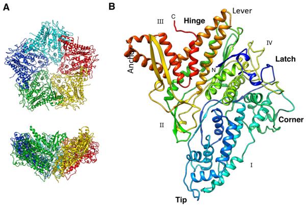
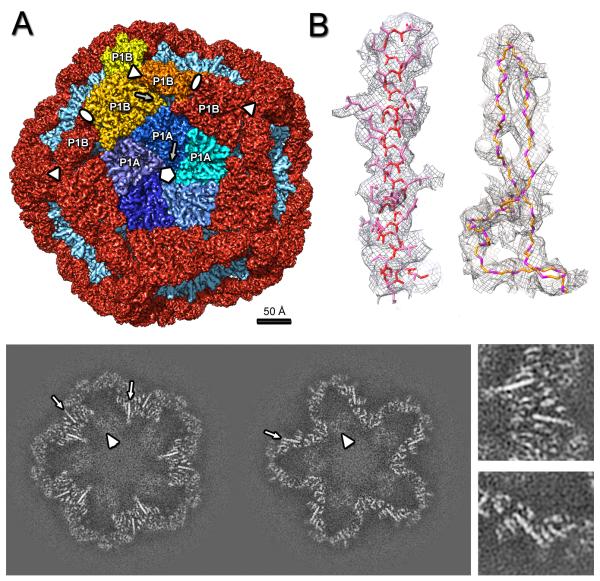
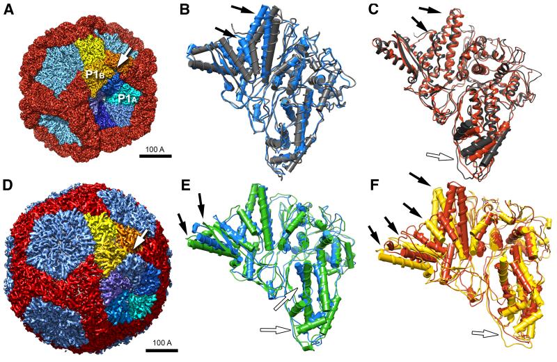
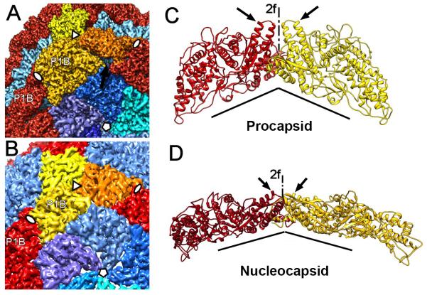
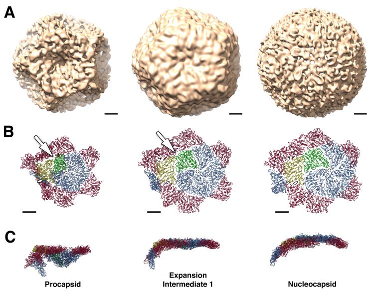
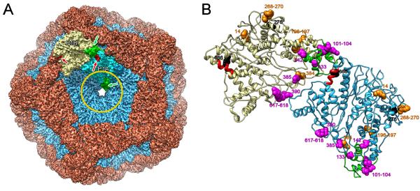
The cystovirus ϕ6 shares several distinct features with other double-stranded RNA (dsRNA) viruses, including the human pathogen, rotavirus: segmented genomes, nonequivalent packing of 120 subunits in its icosahedral capsid, and capsids as compartments for transcription and replication. ϕ6 assembles as a dodecahedral procapsid that undergoes major conformational changes as it matures into the spherical capsid. We determined the crystal structure of the capsid protein, P1, revealing a flattened trapezoid subunit with an α-helical fold. We also solved the procapsid with cryo-electron microscopy to comparable resolution. Fitting the crystal structure into the procapsid disclosed substantial conformational differences between the two P1 conformers. Maturation via two intermediate states involves remodeling on a similar scale, besides huge rigid-body rotations. The capsid structure and its stepwise maturation that is coupled to sequential packaging of three RNA segments sets the cystoviruses apart from other dsRNA viruses as a dynamic molecular machine.


Figures

Figure 1
Figure 1
Crystal structure of P1. (A) Top and side views of the funnel-shaped P1 pentamer (the 5 subunits are in different colors). (B) The P1 subunit (rainbow-colored from blue at the N-terminus to red at the C-terminus) has a trapezoid shape with four edges labeled I - IV. The long helix-turn-helix (in gold) forms a “lever” that rotates during maturation of the procapsid. Five copies of the “tip” line the axial channel through the pentamer. The “corner” of one pentamer subunit fits against the “anchor” of a neighboring subunit. See also Figure S1.
Figure 2
Figure 2
Cryo-EM reconstruction of the ϕ6 procapsid. (A) Segmentation of the outer surface viewed along a 5-fold axis. The 12 inverted 5-fold vertices are occupied by P1A pentamers (the 5 subunits are in shades of blue) set in a dodecahedral frame of sixty P1B subunits (red, except for the three subunits around one 3-fold axis which are in shades of yellow). (B) Density for one α-helix (left) and two β-strands with the corresponding atomic model (side-chains are shown for the α-helix only, for clarity). (C) Slices through the cryoEM reconstruction viewed along the 5-fold axis at 20 Å (left) and 40 Å (right) from the procapsid center. Elongated densities representing α-helices that are approximately in-plane are indicated by arrows and enlarged in the right panels. Scale bar: 100 Å. See also Movie S2.
Figure 3
Figure 3
(A) Cryo-EM reconstruction of the procapsid with subunits color-coded as in Figure 2A but viewed from a different angle. (B) Differences between the P1A subunit in the procapsid (blue) and the P1 crystal structure (dark gray) are localized mainly to the hinge region (black arrows, white arrow in (A)). (C) Comparison of P1A (black) and P1B (red) in the procapsid, showing differences of similar magnitude to those in (B): black arrows point out features most affected and additional differences at the tip (white arrow). (D) Cryo-EM reconstruction of the nucleocapsid (EMD-1206 by Huiskonen et al. (2006) segmented as in (A). (E) Changes in the P1A structure on maturation (procapsid – blue; nucleocapsid – green) localize in the helices next to the hinge region (black arrows, white arrow in (C)). (F) Changes in the P1B structure on maturation (procapsid, red; nucleocapsid, yellow) involve the whole of edge III (black arrows) as well as the tip (white arrow). Scale bar: 100 Å.
Figure 4
Figure 4
Transformation at the hinge. (A) P1B subunits (red and yellow) are tightly connected to P1A subunits (blues) of the inverted vertices in the procapsid. This shell contains cavities between the P1B and P1A subunits (arrows). (B) In the nucleocapsid, the P1B subunits are rotated so that the planes of these flat molecules concide with the tangential plane of the shell, leaving no significant cavities between the subunits. (C) Orientation of P1B subunits (red and yellow ribbons) on either side of a 2-fold icosahedral axis in the procapsid. The subunit planes are almost perpendicular to each other, and their helix-turn-helix motifs are aligned with the 2-fold axis (arrows). (D) Corresponding representation of two P1B subunits in the nucleocapsid. The two subunits are now almost coplanar. (The views shown in C and D are rotated around the 2-fold axis so that the dihedral angles appear considerably larger.) See also Movie S2.
Figure 5
Figure 5
Procapsid expansion. (A) CryoEM reconstructions of three conformational states of the ϕ6 capsid at 16 Å resolution. (B, C) Models of a portion of capsid comprising a pentamer of P1A subunits (blue, green) and surrounding P1B subunits (red, yellow) subunits, viewed from above (B) and from the side (C). Expansion to intermediate 1 is the major transition of the maturing capsid, achieved by rotation of P1B subunits around an axis connecting the 3-fold icosahedral axes (bar in panel A). This rotation appears to stabilize the P1B/P1B interface at the 2-fold axis (Figure 6) and seals gaps between P1A and P1B subunits near the 3-fold axis (arrow). Further expansion to intermediate 2 is achieved by outward movement of the P1A subunits. The final step to the nucleocapsid state is accompanied by local conformational changes in the P1A subunits that correlate with increased outward curvature at the 5-fold axis. Scale bar: 100 Å. See also Movie S3.
Figure 6
Figure 6
(A) Regions of P1 interacting with the s-segment (green) and a monoclonal antibody (red) are shown respectively for the P1A and P1B subunits in the context of the procapsid shell. The circle represents the area covered by the P4 hexamer. (B) Locations of mutations in P1 affecting s- and m-segment binding (magenta) and packaging (orange) are shown for the P1A (cyan) and P1B (yellow) subunits. The proteolytically susceptible sites for factor Xa and trypsin are also shown (black). The subunits are oriented as in the procapsid (panel A) and viewed from the outside. See also Figure S3.

References

    1. Adams PD, Afonine PV, Bunkoczi G, Chen VB, Davis IW, Echols N, Headd JJ, Hung LW, Kapral GJ, Grosse-Kunstleve RW, et al. PHENIX: a comprehensive Python-based system for macromolecular structure solution. Acta crystallographica Section D, Biological crystallography. 2010;66:213–221. - PMC - PubMed
    1. Baker ML, Zhang J, Ludtke SJ, Chiu W. Cryo-EM of macromolecular assemblies at near-atomic resolution. Nature protocols. 2010;5:1697–1708. - PMC - PubMed
    1. Bamford DH, Grimes JM, Stuart DI. What does structure tell us about virus evolution? Curr Opin Struct Biol. 2005;15:655–663. - PubMed
    1. Benevides JM, Juuti JT, Tuma R, Bamford DH, Thomas GJ., Jr. Characterization of subunit-specific interactions in a double-stranded RNA virus: Raman difference spectroscopy of the phi6 procapsid. Biochemistry. 2002;41:11946–11953. - PubMed
    1. Butcher SJ, Dokland T, Ojala PM, Bamford DH, Fuller SD. Intermediates in the assembly pathway of the double-stranded RNA virus phi6. The EMBO journal. 1997;16:4477–4487. - PMC - PubMed

Publication types