Review
. 2013 Nov 29;167(1-2):93-122.
doi: 10.1016/j.vetmic.2013.06.015. Epub 2013 Jul 3.

Equine arteritis virus

Affiliations
Review

Equine arteritis virus

Udeni B R Balasuriya et al. Vet Microbiol. .

Abstract

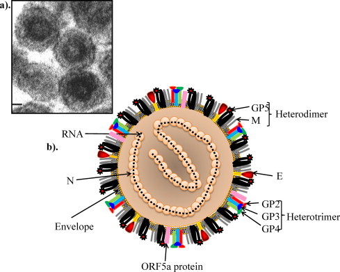
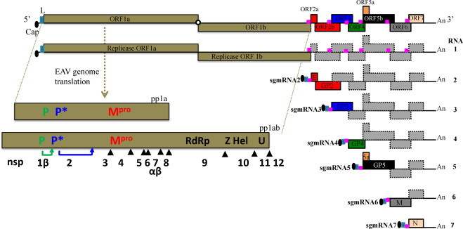
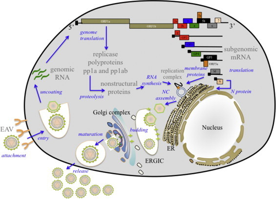
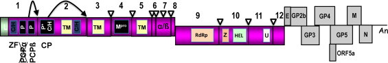
Equine arteritis virus (EAV) is the causative agent of equine viral arteritis (EVA), a respiratory and reproductive disease of equids. There has been significant recent progress in understanding the molecular biology of EAV and the pathogenesis of its infection in horses. In particular, the use of contemporary genomic techniques, along with the development and reverse genetic manipulation of infectious cDNA clones of several strains of EAV, has generated significant novel information regarding the basic molecular biology of the virus. Therefore, the objective of this review is to summarize current understanding of EAV virion architecture, replication, evolution, molecular epidemiology and genetic variation, pathogenesis including the influence of host genetics on disease susceptibility, host immune response, and potential vaccination and treatment strategies.

Keywords: EAV; EVA; Equine arteritis virus.


Figures

Fig. 1
Fig. 1
Equine arteritis virus particle: (a) electron micrograph of EAV (adapted from Snijder et al., Chapter 20, Topley & Wilson's Microbiology and Microbial Infections. London, UK with permission), (b) schematic presentation of EAV particle.
Fig. 2
Fig. 2
The genome organization and polycistronic nature of the EAV genome. The genomic open reading frames (ORFs) are indicated and the names of the corresponding proteins are depicted. The pink boxes represent the body transcription regulatory sequences (TRSs). The nested set of mRNAs that is found in infected cells is depicted below the genome, with RNA1 being identical to the viral genome and sg mRNAs 2–7 being used to express the structural protein genes located in the 3′-proximal quarter of the genome. The light blue box at the 5′ end of each sg mRNA represents the common leader sequence, which is derived from the 5′ end of the genome. With the exception of the bicistronic sg mRNAs 2 and 5, the sg mRNAs are functionally monocistronic. Translation of proteins from sg mRNAs 2 (E and GP2 proteins) and 5 (ORF5a protein and GP5) occurs by leaky scanning of the 5′-proximal end of these sg mRNAs (Firth et al., 2011, Snijder et al., 1999). The ORFs 1a and 1b located at the 5′ end of the genome and are translated into two polyproteins (pp1a and pp1ab) that are further processed into 12–13 nonstructural proteins by three viral proteases (nsps 1, 2, and 4).
Fig. 3
Fig. 3
Schematic overview of EAV life cycle. ER: endoplasmic reticulum; ERGIC: ER–Golgi intermediate compartment; NC: nucleocapsid.
Fig. 4
Fig. 4
Schematic representation of the processing of the EAV replicase polyproteins (pp1a and pp1ab) and generation of individual nsps. The papain-like cysteine protease (PCPβ) and cysteine protease (CP) cleavage sites are indicated by black arrows. The Mpro cleavage sites are indicated by open arrowheads. The genes encoding structural proteins are depicted in gray. P and P* are, PCP and CP, respectively; Mpro, 3C-like main protease; Z, zinc-binding domain; C/H, cysteine/histidine-rich clusters; TM, predicted transmembrane domains; U, nidoviral uridylate-specific endoribonuclease (NendoU). Figure modified from Nidoviruses book chapter on the arterivirus replicase (Van Hemert and Snijder, 2008) with permission from ASM press.
Fig. 5
Fig. 5
Proteolytic processing of EAV replicase polyprotein pp1a through the major and minor pathways. The papain-like cysteine protease (PCPβ) and cysteine protease (CP) cleavage sites are indicated by black arrows. The Mpro cleavage sites are indicated by open arrowheads. P and P* are, PCP and CP, respectively; Mpro, 3C-like main protease; C/H, cysteine/histidine-rich clusters; TM, predicted transmembrane domains.
Fig. 6
Fig. 6
Illustration of the transcription model in nidoviruses. The “discontinuous extension of minus-strand RNA model” proposes that an sg-length negative strand is produced that functions as a template for generation of sg mRNA (Sawicki and Sawicki, 1995). The anti-body TRS in minus-strand may serve as a “jump” signal of the nascent minus strand to leader TRS located at the 5′-end of the plus-strand full-length genome. After the anti-leader (-L) is added to the nascent minus strand, the sg-length minus strand functions as template for positive strand sg mRNA synthesis.
Fig. 7
Fig. 7
Predicted structure of GP5 and M heterodimer. The predicted N-glycosylation sites (Asn-56 and Asn-81) are depicted with an orange circle. The green circle represents the N-glycosylation site (Asn-73) found in isolates of an extensive recent EVA outbreak in North America. The GP5 and M proteins are covalently linked by a disulfide bond (S—S) formed between Cys-34 in the GP5 protein and Cys-8 in the M protein. The major neutralization determinants of EAV that are located in the N-terminal ectodomain of the GP5 major envelope glycoprotein of EAV are depicted. The signal sequence (aa 1–18), four major neutralization sites (A, B, C and D) and putative glycosylation sites (aa positions 56, 73 and 81) are identified.
Fig. 8
Fig. 8
Predicted membrane topology of the minor envelope proteins: E, GP2, GP3, GP4, and ORF5a protein. Two alternative predictions of E protein and GP3 membrane topology are shown. Putative N-glycosylation sites are indicated in yellow circles with corresponding amino acid positions.
Fig. 9
Fig. 9
Two predicted models for the disulfide-bonded structure of the covalently linked GP2—GP3—GP4 heterotrimer. These alternative models differ in membrane topology of GP3, which is currently uncertain. The GP3 protein may be either a class II membrane protein (A) or a class IV membrane protein (B). The intermolecular cystine bridges are depicted arbitrarily with disulfide bonds (S—S) since the Cys positions have not yet been determined. N-glycosylation sites are indicated in orange circles with corresponding amino acid positions.
Fig. 10
Fig. 10
Dual-color immunofluorescence flow cytometry analysis of T cells from carrier and seropositive non-carrier stallions following in vitro infection with EAV. (A) Carrier status was confirmed by repeatedly isolating EAV from semen over an extended period of time. (B) Non-carrier stallions were uninfected but seropositive to EAV following natural infection, not vaccination. Lymphocytes from each stallion that were labeled with both anti-CD3 and anti-nsp1 (12A4) MAbs are shown in the upper right quadrant of each dot plot. Note significant double labeling of lymphocytes from the carrier stallion but not the non-carrier stallion for both CD3 and EAV after in vitro infection.

Similar articles

Cited by

References

    1. Abes S., Moulton H.M., Clair P., Prevot P., Youngblood D.S., Wu R.P., Iversen P.L., Lebleu B. Vectorization of morpholino oligomers by the (R-Ahx-R)4 peptide allows efficient splicing correction in the absence of endosomolytic agents. J. Control. Release. 2006;116:304–313. - PubMed
    1. Anonymous . Equine Viral Arteritis (EVA) and the U.S. Horse Industry. USDA-APHIS, VS, CEAH, National Animal Health Monitoring System; Fort Collins, CO: 2000. NAHMS.
    1. Anonymous . OIE Manual of Diagnostic Tests and Vaccines for Terrestrial Animals (Mammals, Birds and Bees) 2012. 2.5.10. Equine viral arteritis.
    1. Anonymous . Press Release, Inc.; 2012. Equine Arteritis Virus cELISA Correlates Well with SN.
    1. Archambault D., St-Laurent G. Induction of apoptosis by equine arteritis virus infection. Virus Genes. 2000;20:143–147. - PubMed

Publication types

LinkOut - more resources