. 2013 Sep:56:393-403.
doi: 10.1016/j.mcn.2013.07.008. Epub 2013 Jul 26.

FGF14 localization and organization of the axon initial segment

Affiliations

FGF14 localization and organization of the axon initial segment

Maolei Xiao et al. Mol Cell Neurosci. 2013 Sep.

Abstract

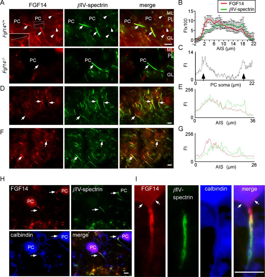
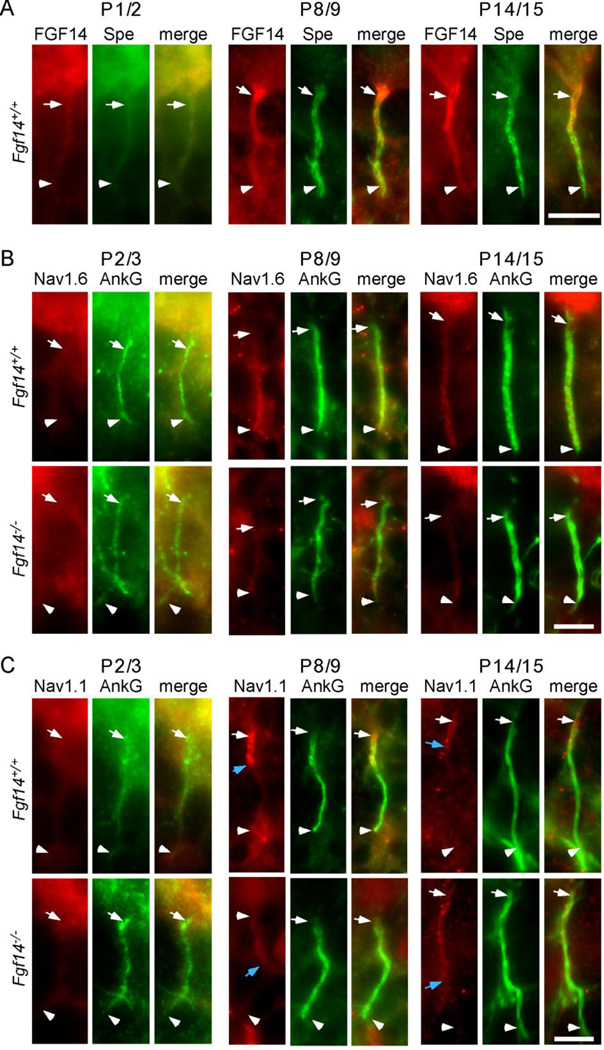
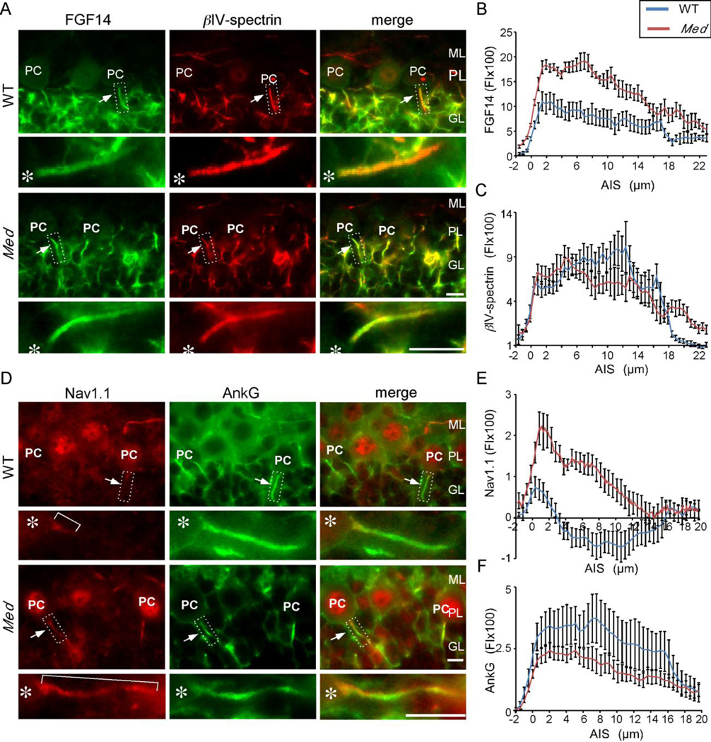
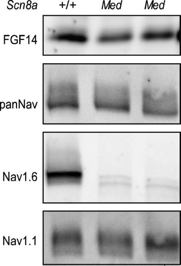
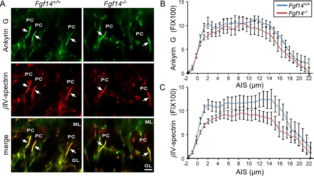
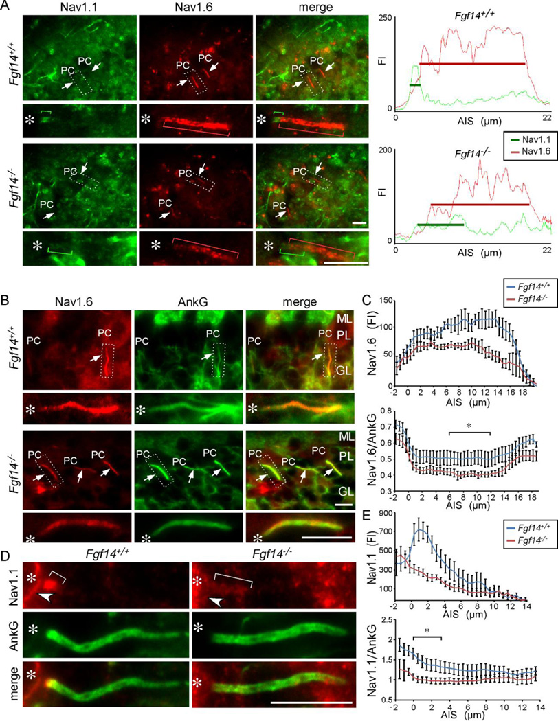
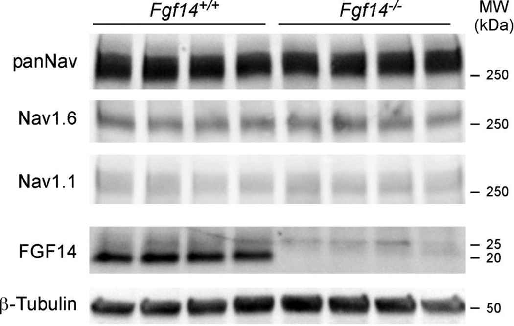
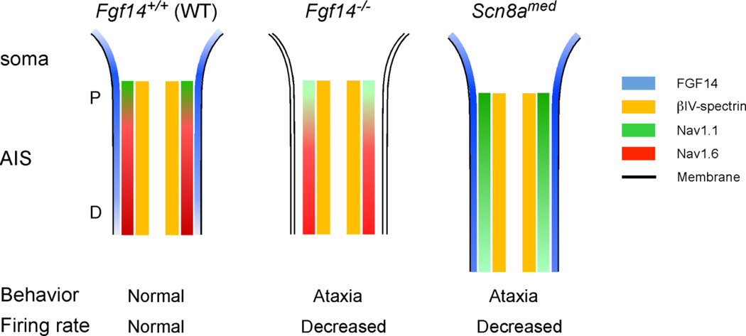
The axon initial segment (AIS) is highly enriched in the structural proteins ankyrin G and βIV-spectrin, the pore-forming (α) subunits of voltage-gated sodium (Nav) channels, and functional Nav channels, and is critical for the initiation of action potentials. We previously reported that FGF14, a member of the intracellular FGF (iFGF) sub-family, is expressed in cerebellar Purkinje neurons and that the targeted inactivation of Fgf14 in mice (Fgf14(-/-)) results in markedly reduced Purkinje neuron excitability. Here, we demonstrate that FGF14 immunoreactivity is high in the AIS of Purkinje neurons and is distributed in a decreasing, proximal to distal, gradient. This pattern is evident early in the postnatal development of Purkinje neurons and is also observed in many other types of central neurons. In (Scn8a(med)) mice, which are deficient in expression of the Nav1.6 α subunit, FGF14 immunoreactivity is markedly increased and expanded in the Purkinje neuron AIS, in parallel with increased expression of the Nav1.1 (Scn1a) α subunit and expanded expression of βIV-spectrin. Although Nav1.1, FGF14, and βIV-spectrin are affected, ankyrin G immunoreactivity at the AIS of Scn8a(med) and wild type (WT) Purkinje neurons was not significantly different. In Fgf14(-/-) Purkinje neurons, βIV-spectrin and ankyrin G immunoreactivity at the AIS were also similar to WT Purkinje neurons, although both the Nav1.1 and Nav1.6 α subunits are modestly, but significantly (p<0.005), reduced within sub-domains of the AIS, changes that may contribute to the reduced excitability of Fgf14(-/-) Purkinje neurons.

Keywords: AIS; Ankyrin G; Axon initial segment; FGF14; Nav; Purkinje neuron; Scn1a; Scn8a; Voltage-gated sodium channel; axon initial segment; fibroblast growth factor 14; iFGF; intracellular fibroblast growth factor; voltage gated sodium channel; βIV-spectrin.


Figures

Figure 1
Figure 1. Proximal to distal gradient in FGF14 immunoreactivity in the AIS of multiple CNS neurons
Cryostat sections of the cerebellum were cut from the brains of adult WT (Fgf14+/+) mice and mice lacking FGF14 (Fgf14−/−) and stained with antibodies against FGF14 and βIV-spectrin as described in Materials and Methods. A. FGF14 immunostaining (red) is prominent at the AIS of Fgf14+/+ Purkinje neurons (arrows) and is higher proximal to the soma and lower distally; immunostaining is also seen in cell bodies of Purkinje neurons (dashed line) and in the AIS of basket/granule neurons (arrowheads). In contrast, no anti-FGF staining is evident in Fgf14−/− cerebellar sections. An antibody against βIV-spectrin (green) was used to define the AIS (arrows). B. Relative anti-FGF14 (red) and anti-βIV-spectrin (green) fluorescence intensities (FI, mean ± SEM), determined from linescan analyses of individual Fgf14+/+ neurons (n = 11) along the AIS. C. Linescan of a single Purkinje neuron along the dashed line shown in (A) demonstrating FGF14 immunofluorescence in the Purkinje neuron soma membrane (arrows). D. Immunostaining of CA1 hippocampal pyramidal neurons reveals a proximal to distal gradient of FGF14 (red) along the AIS (arrows). E. Linescan analysis of anti-FGF14 (red) and anti-βIV-spectrin (green) labeling of the AIS in a representative CA1 hippocampal pyramidal neuron. F. Immunostaining of neurons in layer III of the motor cortex reveal a proximal to distal gradient of FGF14 (red) along the AIS (arrows). G. Linescan analysis of anti-FGF14 (red) and anti-βIV-spectrin (green) labeling of the AIS in a representative neuron in layer III of the motor cortex. H. Isolated Purkinje neurons maintained in culture for 14 days were stained for FGF14 (red) and βIV-spectrin (green), as well as for calbindin (blue; to identify Purkinje neurons). I. High magnification view of an isolated Purkinje neuron showing FGF14 (red) and βIV-spectrin (green) immunoreactivity and a proximal to distal gradient similar to that seen in Purkinje neurons in situ (A). FGF14 immunostaining can also be seen on the soma (arrows). ML, molecular layer, PL, Purkinje neuron layer; GL, granule neuron layer; PC, Purkinje neuron cell body; scale bars are 10 µm.
Figure 2
Figure 2. Localization of AIS-associated proteins during Purkinje neuron maturation in vivo
A. Representative images showing immunostaining for FGF14 (red) and βIV-spectrin (green) in Fgf14+/+ and Fgf14−/− Purkinje neuron AIS at postnatal days 1/2, 8/9, and 14/15. B. Representative images of Nav1.6 (red) and ankyrin G (green) immunostaining in Fgf14+/+ and Fgf14−/− Purkinje neuron AIS at postnatal days 2/3, 8/9 and 14/15. C. Representative images of immunostaining for Nav1.1 (red) and ankyrin G (green) in Fgf14+/+ and Fgf14−/− Purkinje neuron AIS at postnatal days 2/3, 8/9 and 14/15. The white arrows in each panel indicate the positions of the proximal AIS and the white arrowheads, the distal AIS. In C, the blue arrows mark the distal extent of detectable Nav1.1 immunostaining at the AIS. Scale bars are 10 µm.
Figure 3
Figure 3. Changes in the AIS inScn8amed Purkinje neurons
A. Immunostaining showing FGF14 (green) and βIV-spectrin (red) localization in WT and Scn8amed (Med) Purkinje neuron AIS. B, C. Line scan analysis showing increased FGF14 immunostaining (B) and similar βIV-spectrin immunostaining (C) in Scn8amed, compared to WT, Purkinje neuron AIS. Note that the length of the AIS, as demarcated by βIV-spectrin immunostaining, is increased in Scn8amed Purkinje neurons. D. Immunostaining showing Nav1.1 (red) and ankyrin G (green) localization in WT and Scn8amed (Med) Purkinje neuron AIS. E, F. Linescan analysis showing increased Nav1.1 immunostaining (E) and similar ankyrin G immunostaining (F) in Scn8amed, compared to WT, Purkinje neuron AIS. Asterisks mark the locations of the Purkinje neuron soma. FI, Fluorescence intensity; ML, molecular layer, PC, Purkinje neuron soma; PL, Purkinje neuron layer; GL, granule neuron layer. Scale bars are 10 µm. For linescan analyses, n=7–11 values presented are means ± SEM.
Figure 4
Figure 4. FGF14 and Nav α subunit protein levels in cerebellum lacking Nav1.6
Whole cerebellar lysates from WT (+/+) and Scn8amed mice were fractionated and Western blots were probed with anti-FGF14, anti-panNav, anti-Nav1.1, and anti-Nav1.6 antibodies. As expected, Nav1.6 protein expression is deficient in Med mice cerebellum; However, FGF14, total Nav, and Nav1.1 protein levels are not affected.
Figure 5
Figure 5. Structural components of the AIS are localized normally in Purkinje neurons in adult Fgf14−/− mice
A. Immunostaining showing βIV-spectrin (red) and ankyrin G (green) localization in Fgf14+/+ and Fgf14−/− Purkinje neuron AIS. B. Linescan analysis of ankyrin G in the AIS showing similar intensities. C. Linescan analysis revealed reduced βIV-spectrin labeling in the AIS of Fgf14−/− , compared with Fgf14+/+, Purkinje neurons. FI, Fluorescence intensity; ML, molecular layer, PC, Purkinje neuron soma; GL, granule neuron layer. Scale bars are 10 µm. For linescan analysis, n=7–11, error bars equal ± SEM.
Figure 6
Figure 6. Altered distributions of Nav1.1 and Nav1.6 in Fgf14−/− Purkinje neuron AIS
A. Immunostaining showing Nav1.1 (green) and Nav1.6 (red) in the AIS of adult Fgf14+/+ and Fgf14−/− Purkinje neurons. In Fgf14−/− Purkinje neurons, the intensity of anti-Nav1.1 immunostaining is decreased and expanded distally (green bracket), compared with the pattern evident in Fgf14+/+ Purkinje neurons. Similarly, the intensity of anti-Nav1.6 immunostaining is decreased (red bracket) in Fgf14−/−, compared with Fgf14+/+, Purkinje neurons. Right panels: linescans of the AIS of individual Fgf14+/+ and Fgf14−/− Purkinje neurons. B. Immunostaining showing Nav1.6 (red) and ankyrin G (green) distribution in adult Fgf14+/+ and Fgf14−/− Purkinje neuron AIS. C. Linescan analysis showing reduced Nav1.6 immunostaining in the AIS of Fgf14−/−, compared to Fgf14+/+, Purkinje neurons (n = 6𠄷, values presented are means ± SEM). Lower panel: ratio of Nav1.6 to ankyrin G immunofluorescence intensity throughout the AIS. Bracket indicates the region between 6.7 and 10.5 µm that are significantly (p < 0.05) different between Fgf14+/+ and Fgf14−/− Purkinje neurons. D. Immunostaining showing Nav1.1 (red) and ankyrin G (green) localization in adult Fgf14+/+ and Fgf14−/− Purkinje neuron AIS. E. Linescan analysis showing a proximal peak of Nav1.1 immunostaining in Fgf14+/+ compared to reduced immunostaining in Fgf14−/− Purkinje neuron AIS (n=6–10, values presented are means ± SEM). Lower panel: ratio of Nav1.1 to ankyrin G throughout the AIS. Bracket indicates the region between 0 and 3.9 µm that are significantly (p < 0.05) different between Fgf14+/+ and Fgf14−/− Purkinje neurons. Regions in dashed box are enlarged below. Asterisk marks the location of the Purkinje neuron soma. FI, Fluorescence intensity; ML, molecular layer, PC, Purkinje neuron soma; PL, Purkinje neuron layer; GL, granule neuron layer. Scale bars equal 10 µm.
Figure 7
Figure 7. Nav1.1 and 1.6 protein levels in whole cerebellar lysates are not affected by loss of FGF14
Cerebellar lysates from four Fgf14+/+ and four Fgf14−/− adult mice were probed with the anti-FGF14, anti-panNav, anti-Nav1.1, anti-Nav1.6 and anti-β-tubulin antibodies.
Figure 8
Figure 8. Protein distribution patterns in the Purkinje neuron AIS in mice lacking FGF14 or Nav1.6
FGF14 (blue) is distributed in a decreasing proximal (P) to distal (D) gradient along the AIS of Fgf14+/+ cerebellar Purkinje neurons and weakly on the soma membrane. Nav1.1 (green) is distributed in a similar pattern, with high levels proximally and lower levels distally, whereas Nav1.6 (red) is distributed in an inverse pattern with lower levels proximally and higher levels distally. In the absence of FGF14 (Fgf14−/−), the intensity of both the anti-Nav1.1 and the anti-Nav1.6 immunolabeling is decreased. In addition, the distributions of these subunits in the AIS is also shifted such that the Nav1.1 domain is expanded and the Nav1.6 domain is constricted. In Scn8amed mice, which lack Nav1.6, immunostaining for FGF14 and Nav1.1 at the AIS are increased in parallel with an expanded domain for βIV-spectrin, suggesting lengthening of the AIS in Scn8amed Purkinje neurons.

Similar articles

Cited by

References

    1. Ango F, di Cristo G, Higashiyama H, Bennett V, Wu P, Huang ZJ. Ankyrin-Based Subcellular Gradient of Neurofascin, an Immunoglobulin Family Protein, Directs GABAergic Innervation at Purkinje Axon Initial Segment. Cell. 2004;119:257–272. - PubMed
    1. Boiko T, Van Wart A, Caldwell JH, Levinson SR, Trimmer JS, Matthews G. Functional specialization of the axon initial segment by isoform-specific sodium channel targeting. J. Neurosci. 2003;23:2306–2313. - PMC - PubMed
    1. Brusse E, de Koning I, Maat-Kievit A, Oostra BA, Heutink P, van Swieten JC. Spinocerebellar ataxia associated with a mutation in the fibroblast growth factor 14 gene (SCA27): A new phenotype. Mov. Disord. 2006;21:396–401. - PubMed
    1. Burgess DL, Kohrman DC, Galt J, Plummer NW, Jones JM, Spear B, Meisler MH. Mutation of a new sodium channel gene, Scn8a, in the mouse mutant ‘motor endplate disease’. Nat. Genet. 1995;10:461–465. - PubMed
    1. Clark BD, Goldberg EM, Rudy B. Electrogenic tuning of the axon initial segment. Neuroscientist. 2009;15:651–668. - PMC - PubMed

Publication types