

Scorpion venom components that affect ion-channels function
- PMID: 23891887
- PMCID: PMC4089097
- DOI: 10.1016/j.toxicon.2013.07.012
Scorpion venom components that affect ion-channels function
Abstract
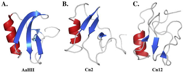
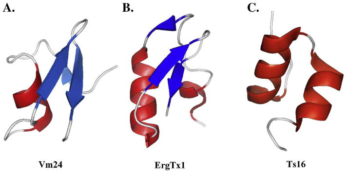
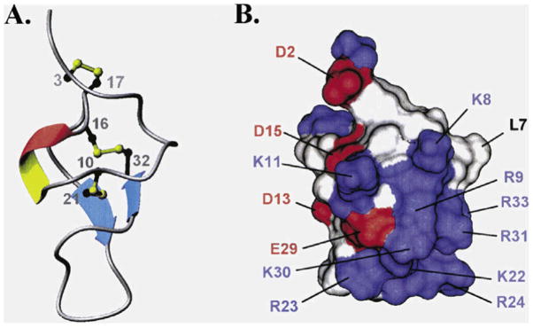
The number and types of venom components that affect ion-channel function are reviewed. These are the most important venom components responsible for human intoxication, deserving medical attention, often requiring the use of specific anti-venoms. Special emphasis is given to peptides that recognize Na(+)-, K(+)- and Ca(++)-channels of excitable cells. Knowledge generated by direct isolation of peptides from venom and components deduced from cloned genes, whose amino acid sequences are deposited into databanks are nowadays in the order of 1.5 thousands, out of an estimate biodiversity closed to 300,000. Here the diversity of components is briefly reviewed with mention to specific references. Structural characteristic are discussed with examples taken from published work. The principal mechanisms of action of the three different types of peptides are also reviewed. Na(+)-channel specific venom components usually are modifier of the open and closing kinetic mechanisms of the ion-channels, whereas peptides affecting K(+)-channels are normally pore blocking agents. The Ryanodine Ca(++)-channel specific peptides are known for causing sub-conducting stages of the channels conductance and some were shown to be able to internalize penetrating inside the muscle cells.
Keywords: Biodiversity; Functional effect; Ion-channel; Scorpion toxin; Structural features.
Copyright © 2013 Elsevier Ltd. All rights reserved.
Figures
References
-
- Abdel-Mottaleb Y, Vandendriessche T, Clynen E, Landuyt B, Jalali Vatanpour H, et al. OdK2, a Kv1.3 channel-selective toxin from the venom Iranian scorpion Odonthobuthus doriae. Toxicon. 2008;51:1424–30. - PubMed
- Acta Trop. 107:71–79.
-
- Almeida DD, Torres TM, Barbosa EG, Lima JPMS, Fernandes-Pedrosa MF. Molecular approaches for structural characterization of a new potassium channel blocker from Tityus stigmurus venom: cDNA cloning, homology modeling, dynamic simulations and docking. Biochemical and Biophysical Research Communications. 2013;430:113–118. - PubMed
-
- Altafaj X, Cheng E, Estève E, Urbani J, Grunwald D, Sabatier J-M, Coronado R, De Waard M, Ronjat M. Maurocalcine and domain A of the II–III loop of the dihydroprydine receptor Cav1.1 subunit share common binding sites on the skeletal ryanodine receptor. J Biol Chem. 2005;280:4013–4016. - PMC - PubMed
Publication types
MeSH terms
Substances
Grants and funding
LinkOut - more resources
Full Text Sources
Other Literature Sources
