. 2013 Jul 15;12(14):2233-40.
doi: 10.4161/cc.25268.

Carboxamide SIRT1 inhibitors block DBC1 binding via an acetylation-independent mechanism

Affiliations

Carboxamide SIRT1 inhibitors block DBC1 binding via an acetylation-independent mechanism

Basil P Hubbard et al. Cell Cycle. .

Abstract

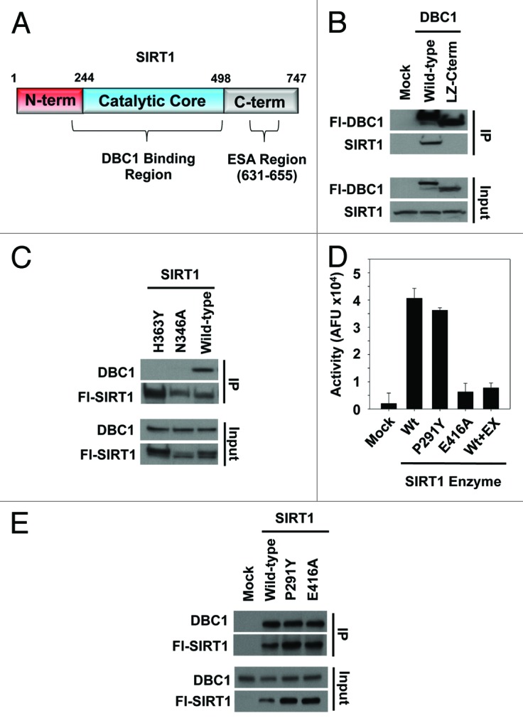
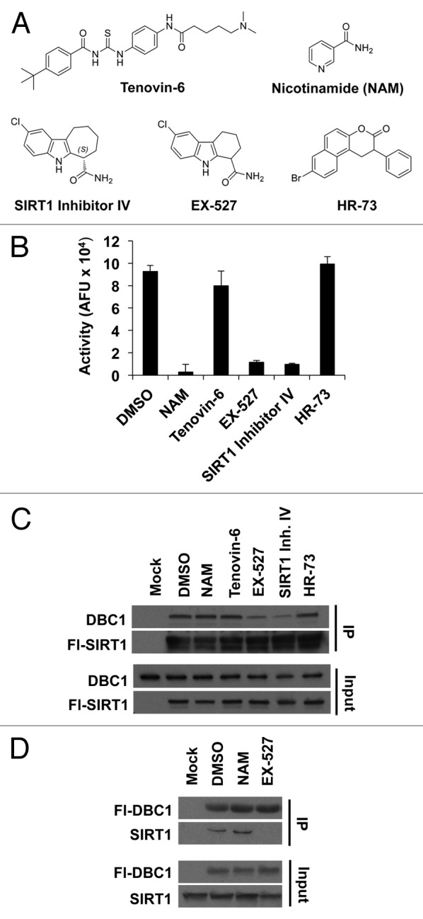
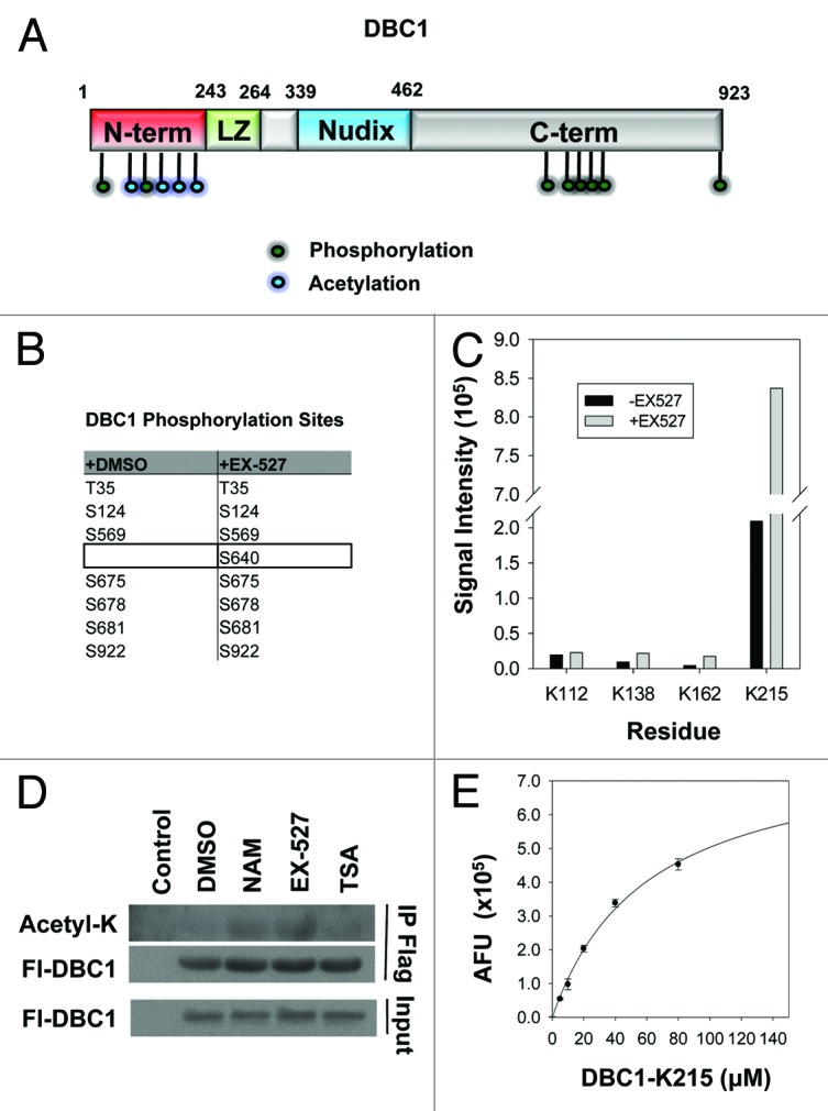
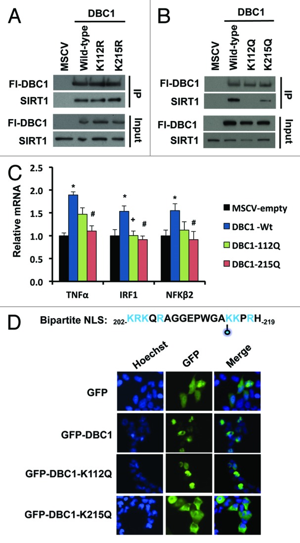
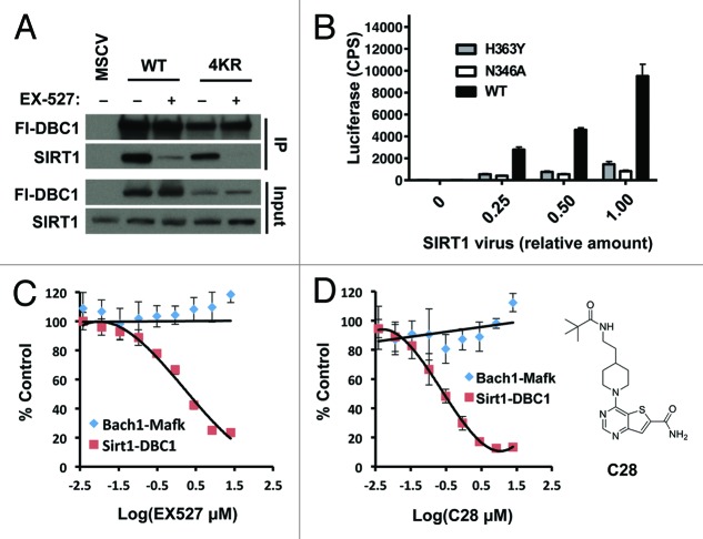
SIRT1 is an NAD (+) -dependent deacetylase that counteracts multiple disease states associated with aging and may underlie some of the health benefits of calorie restriction. Understanding how SIRT1 is regulated in vivo could therefore lead to new strategies to treat age-related diseases. SIRT1 forms a stable complex with DBC1, an endogenous inhibitor. Little is known regarding the biochemical nature of SIRT1-DBC1 complex formation, how it is regulated and whether or not it is possible to block this interaction pharmacologically. In this study, we show that critical residues within the catalytic core of SIRT1 mediate binding to DBC1 via its N-terminal region, and that several carboxamide SIRT1 inhibitors, including EX-527, can completely block this interaction. We identify two acetylation sites on DBC1 that regulate its ability to bind SIRT1 and suppress its activity. Furthermore, we show that DBC1 itself is a substrate for SIRT1. Surprisingly, the effect of EX-527 on SIRT1-DBC1 binding is independent of DBC1 acetylation. Together, these data show that protein acetylation serves as an endogenous regulatory mechanism for SIRT1-DBC1 binding and illuminate a new path to developing small-molecule modulators of SIRT1.

Keywords: DBC1; DBC1 acetylation; DBC1 localization; SIRT1 inhibitors; SIRT1-DBC1 complex regulation.


Figures

None
Figure 1. Regions on SIRT1 and DBC1 involved in complex formation. (A) Schematic of SIRT1 domains illustrating the putative DBC1 binding region and the ESA region. (B) Co-immunoprecipitation of full-length DBC1 or a truncated version lacking amino acids 1–242 with SIRT1 from 293-T cells. (C) Co-immunoprecipitation of wild type SIRT1 and SIRT1 point mutants with DBC1 from 293-T cells. (D) In vitro activity of SIRT1, SIRT1-P291Y and SIRT1-E416A purified from cells using the BIOMOL assay. Ten µM EX-527 was used as a negative control; mean + s.d. shown (n = 3). (E) Co-immunoprecipitation of DBC1 and the corresponding SIRT1 variants from (D) from 293-T cells.
None
Figure 2. Effect of various SIRT1 inhibitors on SIRT1-DBC1 complex formation. (A) Chemical structures of SIRT1 inhibitors. (B) SIRT1 activity in the absence or presence of various inhibitors; mean + s.d. shown (n = 3). (C) Co-immunoprecipitation of Flag-wild-type SIRT1 with DBC1 from 293-T cells following 24 h treatment with the indicated compounds. (D) Co-immunoprecipitation of Flag-DBC1 and SIRT1 from 293-T cells in the presence of the indicated compounds (24 h treatment). Compound doses used in panels (B, C, and D) were NAM (20 mM), Tenovin-6 (25 µM), EX-527 (10 µM), SIRT1 Inhibitor IV (10 µM) and 15 µM HR-73.
None
Figure 3. Post-translational modifications on DBC1 and deacetylation by SIRT1. (A) Schematic of DBC1 domains illustrating the position of acetylation (blue) and phosphorylation (green) sites. (B) Table indicating phosphorylation sites on Flag-DBC1 purified from 293-T cells, identified by LC/MS-MS following 24-h treatment with either DMSO or EX-527 (10 µM). (C) Semi-quantitative LC/MS-MS comparison of acetylation on immunoprecipitated DBC1 from 293-T cells following 24- hour treatment with DMSO or EX-527 (10 µM). (D) Acetylation analysis of immunoprecipitated Flag-DBC1 from 293-T cells treated for 24 h with NAM (20 mM), EX-527 (10 µM) or TSA (1 µM). (E) SIRT1 deacetylation of a native peptide corresponding to Ac-K215 on DBC1, measured using the PNC1-OPT assay; mean ± s.d. is shown for each point (n = 3).
None
Figure 4. Effects of DBC1 acetylation on SIRT1 binding and sub-cellular localization. (A) Co-immunoprecipitation of Flag-wild-type DBC1 or deacetyl-mimetics with endogenous SIRT1 in stably transduced U2OS cells. (B) Co-immunoprecipitation of Flag-wild-type DBC1 or acetyl-mimetics with endogenous SIRT1 in stably transduced U2OS cells. (C) Effect of wild-type DBC1 and acetyl-mimetics on the transcription NFκB- regulated genes in stably transduced U2OS cells; mean + s.e. is shown (n = 3). *Denotes statistical significance (p < 0.05; t-test) for empty vs. DBC1-Wt; +denotes statistical significance (p < 0.05; t-test) for DBC1-Wt vs. DBC1–112Q; and #denotes statistical significance (p < 0.05; t-test) for DBC1-Wt vs. DBC1–215Q. (D) Top: Sequence of putative NLS with K215 indicated with a blue-filled circle, and positively charged residues indicated in blue, Bottom: localization of GFP, wild-type DBC1 and acetyl-mimetics in transiently transfected 293-T cells.
None
Figure 5. Mechanism and dose-dependence of EX-527 and C28. (A) Co-immunoprecipitation of Flag-wild-type DBC1or a non-acetylated DBC1 mutant protein (DBC1–4KR) with endogenous SIRT1 in the presence or absence of EX-527 (10 µM) following 24 h treatment. (B) Validation of the luciferase complementation assay in 293 cells using different titers of SIRT1 virus and non-binding negative control proteins; mean + s.e. is shown (n = 2). Dose-response effect of 24 h treatment with (C) EX-527 or (D) C28 (structure depicted in inset) on the SIRT1-DBC1 interaction and a control protein binding pair (Bach1-Mafk) in 293 cells; mean ± s.e. is shown (n = 2).

References

    1. Picard F, Kurtev M, Chung N, Topark-Ngarm A, Senawong T, Machado De Oliveira R, et al. Sirt1 promotes fat mobilization in white adipocytes by repressing PPAR-gamma. Nature. 2004;429:771–6. doi: 10.1038/nature02583. - DOI - PMC - PubMed
    1. Fulco M, Cen Y, Zhao P, Hoffman EP, McBurney MW, Sauve AA, et al. Glucose restriction inhibits skeletal myoblast differentiation by activating SIRT1 through AMPK-mediated regulation of Nampt. Dev Cell. 2008;14:661–73. doi: 10.1016/j.devcel.2008.02.004. - DOI - PMC - PubMed
    1. Biason-Lauber A, Böni-Schnetzler M, Hubbard BP, Bouzakri K, Brunner A, Cavelti-Weder C, et al. Identification of a SIRT1 mutation in a family with type 1 diabetes. Cell Metab. 2013;17:448–55. doi: 10.1016/j.cmet.2013.02.001. - DOI - PMC - PubMed
    1. Bordone L, Cohen D, Robinson A, Motta MC, van Veen E, Czopik A, et al. SIRT1 transgenic mice show phenotypes resembling calorie restriction. Aging Cell. 2007;6:759–67. doi: 10.1111/j.1474-9726.2007.00335.x. - DOI - PubMed
    1. Hubbard BP, Gomes AP, Dai H, Li J, Case AW, Considine T, et al. Evidence for a common mechanism of SIRT1 regulation by allosteric activators. Science. 2013;339:1216–9. doi: 10.1126/science.1231097. - DOI - PMC - PubMed

Publication types

MeSH terms

LinkOut - more resources