. 2013 Sep;20(9):1085-92.
doi: 10.1038/nsmb.2630. Epub 2013 Jul 28.

Allosteric mechanism of water-channel gating by Ca2+-calmodulin

Affiliations

Allosteric mechanism of water-channel gating by Ca2+-calmodulin

Steve L Reichow et al. Nat Struct Mol Biol. 2013 Sep.

Abstract

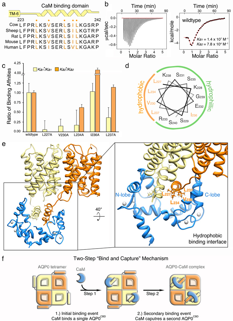
Calmodulin (CaM) is a universal regulatory protein that communicates the presence of calcium to its molecular targets and correspondingly modulates their function. This key signaling protein is important for controlling the activity of hundreds of membrane channels and transporters. However, understanding of the structural mechanisms driving CaM regulation of full-length membrane proteins has remained elusive. In this study, we determined the pseudoatomic structure of full-length mammalian aquaporin-0 (AQP0, Bos taurus) in complex with CaM, using EM to elucidate how this signaling protein modulates water-channel function. Molecular dynamics and functional mutation studies reveal how CaM binding inhibits AQP0 water permeability by allosterically closing the cytoplasmic gate of AQP0. Our mechanistic model provides new insight, only possible in the context of the fully assembled channel, into how CaM regulates multimeric channels by facilitating cooperativity between adjacent subunits.


Conflict of interest statement

The authors declare no financial conflict related to this work.

Figures

Figure 1
Figure 1. Purification and pseudo-atomic model of the aquaporin-0 – calmodulin complex (AQP0–CaM) determined by electron microscopy
(a) Chromatogram showing purification of the AQP0–CaM cross-linked complex from excess free calmodulin (CaM) by size-exclusion chromatography (SEC). (b) Silver stained SDS-PAGE showing fractions from the SEC purification. Lane 1 shows the total starting material. Lane 2, the purified AQP0–CaM cross-linked complex dissociated into three protein bands in SDS migrating at 26kD, 39kD and 65kD. These bands correspond to the AQP0 monomer, the 1:1 AQP0–CaM cross-link and the 2:1 (AQP0)2–CaM cross-link, respectively. Lane 3, is the free CaM migrating as a diffuse band ~13kD. The fully assembled AQP0–CaM complex (Lane 2) dissociates to the 2:1 AQP0–CaM complex, the 1:1 AQP0–CaM complex, and the AQP0 monomer under denaturing SDS conditions due to the stochastic occurrence of peptide bonds made during the cross-linking reaction. (c) Electron micrograph of negatively stained AQP0–CaM particles. Inset, contains symmetrized projection averages of the AQP0–CaM complex (d) Different “views” of the three-dimensional (3D) reconstruction of the AQP0–CaM complex. (e) Fitting of the crystallographic structures of AQP0 (orange and yellow) and CaM (blue) into the 3D reconstruction (grey mesh). (f) Pseudo-atomic model of the AQP0–CaM complex displaying two CaM molecules (A and B) bound to the cytoplasmic C-terminal helices of the AQP0 tetramer.
Figure 2
Figure 2. Hydrophobic interactions involved in AQP0–CaM complex formation
(a) Schematic of the secondary structure (yellow) and primary sequence alignment (blue) of the AQP0 calmodulin binding domain (AQP0CBD). Residues in orange indicate conserved hydrophobic residues within the α-helical AQP0CBD. (b) (left) ITC raw heats of binding Ca2+–CaM to the wildtype AQP0CBD peptide and (right) binding isotherm fit with a two-state binding model. (c) Ratio of binding affinities obtained by ITC for AQP0CBD peptides when each hydrophobic site (• in panel A) was mutated to alanine. (d) Helical wheel analysis identifies a hydrophobic face involved in CaM recognition. (e) Structure of AQP0–CaM showing two AQP0 monomers bound to CaM (colored as in Figure 1). Zoom-view, shows the AQP0CBD (yellow and orange helices) with AQP0 residues forming the proposed hydrophobic interface with CaM shown as stick representations. (f) Schematic illustrating the two-step “bind and capture” mechanism for assembling the 2:1 AQP0–CaM complex, described in the main text.
Figure 3
Figure 3. Calmodulin restricts the dynamics of AQP0
(a) Water density (blue iso-surface) for the equilibrated AQP0–CaM (grey ribbon) in a lipid bilayer (grey stick). (b) Pore profile analysis using the program HOLE showing water permeation pathway (blue surface) in the CaM-bound conformation of the AQP0 protomer (white and grey ribbon). (c) CaM-free per-residue root-mean-square fluctuation (RMSF) values mapped to the AQP0 structure according to color (RMSF = 0.3 Å – blue, 0.6 Å – white and 1.2 Å – red). (d) Zoom view of the cytoplasmic domain of AQP0. (e) Change in protein dynamics (Δ RMSF) of the CaM-bound system compared to the CaM-free system mapped to the AQP0 structure according to color (Δ RMSF. = 0 Å – white, −0.4 Å – light blue, −0.8 Å – dark blue). Asterisks in (d and e) indicate the position of the AQP0 cytoplasmic constriction site II (CSII).
Figure 4
Figure 4. CaM binding closes the AQP0 cytoplasmic constriction site gate II (CSII)
(a–c) Pore profile analysis of AQP0 during the molecular dynamics (MD) simulation. The location of CSII is indicated in (a). Side chains of the CSII residues Tyr149, Phe75 and His66 are displayed as sticks. (a and b, left panel) Tyr149 in a downward “open” conformation. (b, right panel) Tyr149 in an upward “closed” conformation. (c) Plot indicating the pore diameter of the open AQP0 CSII (green) and the closed CSII (red). (d) Plot indicating the population of structures obtained during the CaM–free AQP0 (green) and CaM–bound AQP0 (red) MD simulations clustered according to the displacement of their CSII residues Tyr149 and Phe75. (d, inset) Plot showing the population of structures with CSII displacement < 5 Å and > 8Å. (e) Water channel permeability rates (P; um•s−1) obtained from Oocytes expressing wildtype AQP0 and CSII mutants (Y149G, Y149L, Y149S) obtained under 0mM (blue) and 1.8 mM Ca2+ (green) buffer conditions. The AQP0 Pf is significantly inhibited by Ca2+ (P = 0.012). This effect has previously been shown to be CaM dependent. The water permeability rates of CSII mutant AQP0 channels were deficient in Ca2+ regulation. Each construct was expressed and correctly trafficked to the plasma membrane as demonstrated by immune-blot analysis (e, inset and Supplementary Figure S5). Although expression levels varied among the mutants only WT AQP0 was acutely Ca2+ sensitive and mutation in Y149 generally abolished Ca2+ sensitivity.
Figure 5
Figure 5. Mechanism of Ca2+–CaM regulation of AQP0
The AQP0 water channel exists in equilibrium between an open state (left) and closed state (right). These states are formed by dynamic conformational changes at the cytoplasmic constriction site (CSII) formed by residues Tyr149, Phe75 and His66. Binding of Ca2+–CaM at the C-terminus of AQP0 results in a shift in this equilibrium by allosterically stabilizing the closed CSII state, thereby restricting water channel permeability. The structural models were obtained from the MD simulations. AQP0 is shown as grey ribbon and water molecules in red. Some water molecules were deleted in the figure for display purposes and clarity only. Likewise, The last transmembrane helix of AQP0 is removed for clarity. CSII residues Tyr149 and Phe75 are shown as stick representations.

References

    1. Saimi Y, Ling KY. Calmodulin activation of calcium-dependent sodium channels in excised membrane patches of Paramecium. Science. 1990;249:1441–1444. - PubMed
    1. Zuhlke RD, Pitt GS, Deisseroth K, Tsien RW, Reuter H. Calmodulin supports both inactivation and facilitation of L-type calcium channels. Nature. 1999;399:159–162. - PubMed
    1. Tan HL, et al. A calcium sensor in the sodium channel modulates cardiac excitability. Nature. 2002;415:442–447. - PubMed
    1. Saimi Y, Kung C. Calmodulin as an ion channel subunit. Annu Rev Physiol. 2002;64:289–311. - PubMed
    1. Meissner G. Evidence of a role for calmodulin in the regulation of calcium release from skeletal muscle sarcoplasmic reticulum. Biochemistry. 1986;25:244–251. - PubMed

Methods-only references

    1. Mindell JA, Grigorieff N. Accurate determination of local defocus and specimen tilt in electron microscopy. J Struct Biol. 2003;142:334–347. - PubMed
    1. Emsley P, Cowtan K. Coot: model-building tools for molecular graphics. Acta Crystallogr D Biol Crystallogr. 2004;60:2126–2132. - PubMed
    1. Phillips JC, et al. Scalable molecular dynamics with NAMD. J Comput Chem. 2005;26:1781–1802. - PMC - PubMed
    1. Mackerell AD, et al. All-Atom Empirical Potential for Molecular Modeling and Dynamics Studies of Proteins. J Phys Chem B. 1998;102:3586–3616. - PubMed
    1. Klauda JB, Brooks BR, MacKerell AD, Jr, Venable RM, Pastor RW. An ab initio study on the torsional surface of alkanes and its effect on molecular simulations of alkanes and a DPPC bilayer. J Phys Chem B. 2005;109:5300–5311. - PubMed

Publication types

MeSH terms

Associated data