. 2013 Oct 1;305(7):H1031-40.
doi: 10.1152/ajpheart.00434.2013. Epub 2013 Jul 26.

Focal myocardial infarction induces global remodeling of cardiac sympathetic innervation: neural remodeling in a spatial context

Affiliations

Focal myocardial infarction induces global remodeling of cardiac sympathetic innervation: neural remodeling in a spatial context

Olujimi A Ajijola et al. Am J Physiol Heart Circ Physiol. .

Abstract

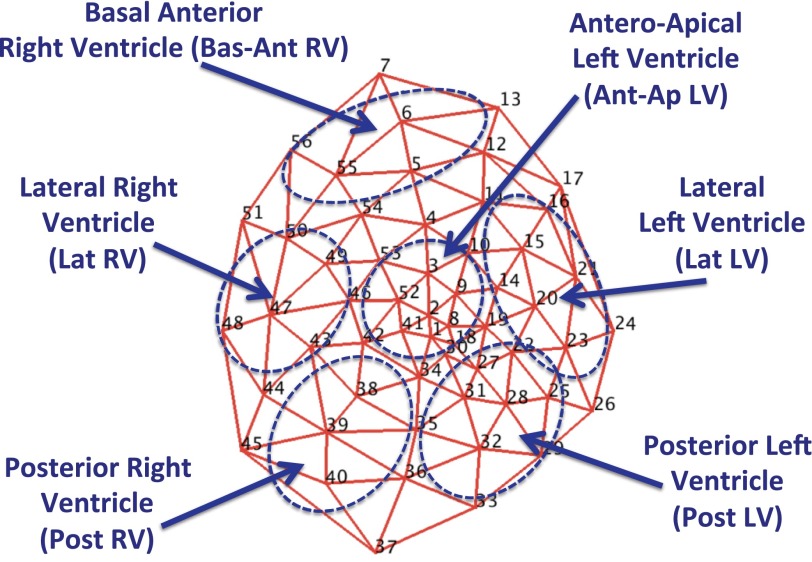
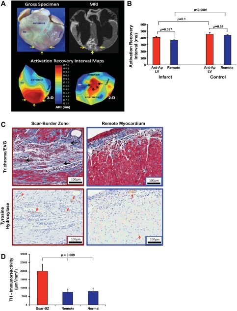
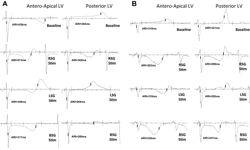
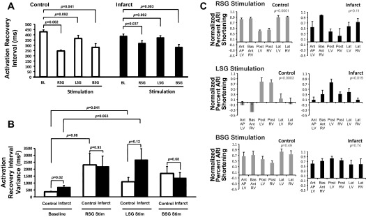
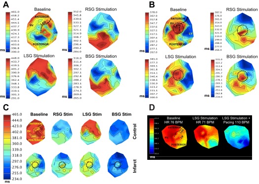
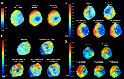
Myocardial infarction (MI) induces neural and electrical remodeling at scar border zones. The impact of focal MI on global functional neural remodeling is not well understood. Sympathetic stimulation was performed in swine with anteroapical infarcts (MI; n = 9) and control swine (n = 9). A 56-electrode sock was placed over both ventricles to record electrograms at baseline and during left, right, and bilateral stellate ganglion stimulation. Activation recovery intervals (ARIs) were measured from electrograms. Global and regional ARI shortening, dispersion of repolarization, and activation propagation were assessed before and during sympathetic stimulation. At baseline, mean ARI was shorter in MI hearts than control hearts (365 ± 8 vs. 436 ± 9 ms, P < 0.0001), dispersion of repolarization was greater in MI versus control hearts (734 ± 123 vs. 362 ± 32 ms(2), P = 0.02), and the infarcted region in MI hearts showed longer ARIs than noninfarcted regions (406 ± 14 vs. 365 ± 8 ms, P = 0.027). In control animals, percent ARI shortening was greater on anterior than posterior walls during right stellate ganglion stimulation (P = 0.0001), whereas left stellate ganglion stimulation showed the reverse (P = 0.0003). In infarcted animals, this pattern was completely lost. In 50% of the animals studied, sympathetic stimulation, compared with baseline, significantly altered the direction of activation propagation emanating from the intramyocardial scar during pacing. In conclusion, focal distal anterior MI alters regional and global pattern of sympathetic innervation, resulting in shorter ARIs in infarcted hearts, greater repolarization dispersion, and altered activation propagation. These conditions may underlie the mechanisms by which arrhythmias are initiated when sympathetic tone is enhanced.

Keywords: autonomic nervous system; cardiac innervation; neural remodeling; sympathetic nerves.


Figures

Fig. 1.
Fig. 1.
Polar map of 56-electrode sock and prespecified regions. The mesh represents the locations of all 56 electrodes (nos. 1–56) contained in the multielectrode sock and the 6 prespecified regions (with abbreviations) used in the study.
Fig. 2.
Fig. 2.
Myocardial infarct model: structure-function correlation. A, top: representative gross tissue (left) and MRI (right) images of the patchy anteroapical myocardial infarct. Bottom, three-dimensional (3-D; left) and two-dimensional (2-D; right) activation recovery interval (ARI) maps. Yellow arrows show the myocardial infarct (MI) location. B: graphic representation of ARI in the infarcted region versus all other locations in infarcted and control animals. Data are means ± SE. C: representative histological slides showing the patchy intramyocardial scar (black arrows) after trichrome/elastic von Giessen (EVG) staining and diaminobenzidine detection of anti-tyrosine hydroxylase (TH) antibody (red arrows) from the peri-infarcted (red box) and remote (blue box) myocardium. Magnification: ×20. Scale bars = 100 μm. Scant intramyocardial staining and a subepicardial nerve bundle are labeled (red arrow) in the remote myocardium compared with extensive staining in the infarct scar-border zone (BZ). D: graphic representation (means ± SE) of TH immunoreactivity expressed as the area of positive staining (in μm2) divided by the total area (in mm2) in the scar-BZ region, remote regions from the scar, and in normal control animals. The P value represents ANOVA of the three groups.
Fig. 3.
Fig. 3.
Electrogram responses to sympathetic stimulation. A and B: representative electrograms from control animals (A) and infarcted animals (B) showing two sampled regions: the Ant-Ap LV (location of the focal scar in infarcted animals) and Post LV (a location disparate from the infarct bed, with a separate coronary artery supply). Representative responses to right, left, and bilateral stellate ganglia (RSG, LSG, and BSG, respectively) stimulation are shown.
Fig. 4.
Fig. 4.
Effects of sympathetic stimulation on ARIs and repolarization heterogeneity. A: ARI shortening at baseline (BL) and during RSG, LSG, and BSG stimulation for control and infarcted animals (n = 8 for control RSG stimulation and n = 9 for all others). Note the compressed scales for RSG and BSG stimulation; the pattern of ARI shortening during BSG stimulation is more even, as emphasized in C and in Fig. 5C. B: global dispersion of repolarization (DOR) at BL and during RSG, LSG, and BSG stimulation in control and infarcted hearts. C: percent ARI shortening in each region normalized to the maximum in each animal during RSG, LSG, and BSG stimulation in control and infarcted hearts (n = 8 for control RSG stimulation and n = 9 for all others). All graphic data shown in A–C are means ± SE. P values by ANOVA are shown.
Fig. 5.
Fig. 5.
2-D ARI maps in control and infarcted hearts. A and B: representative 2-D polar maps of ARIs of control animals (A) and infarcted animals (B) at BL and during RSG, LSG, and BSG stimulation. Myocardial regions are shown in the BL control map (and in Fig. 1). The black dashed circle represents the anteroapical location of the infarct. Note the more even pattern of ARI shortening during BSG stimulation in control animals (A, bottom right), as indicated by the smaller ARI scale. C: polar maps from A and B on a synchronized scale. The top and bottom maps represent control and infarcted animals, respectively, at BL and during RSG, LSG, and BSG stimulation. Myocardial regions are shown in the BL control map. The black dashed circle represents the anteroapical location of the infarct. D: representative 2-D ARI map of a normal animal at BL, during LSG stimulation, and during LSG stimulation with simultaneous atrial pacing at 110 beats/min (BPM). The posterior wall of the heart exhibited shorter ARI during LSG stimulation; this was unchanged when the heart rate was increased. LAD, left anterior descending coronary artery.
Fig. 6.
Fig. 6.
Impact of sympathetic stimulation on impulse propagation from the myocardial scar. A: 2-D activation maps showing pacing in a control animal. B and C: 2-D activation maps for two animals at baseline (sinus rhythm), during scar pacing, and during scar pacing simultaneous with RSG, LSG, and BSG stimulation. The direction of activation propagation was modulated by sympathetic stimulation in both animals. D: representative example of an animal where sympathetic stimulation did not result in modulation of activation propagation. For B–D, white dashed arrows show the region of activation propagation, whereas the white equal signs indicate regions of functional block during. The white electrical pulse symbols represent pacing sites.

References

    1. Ajijola OA, Lellouche N, Bourke T, Tung R, Ahn S, Mahajan A, Shivkumar K. Bilateral cardiac sympathetic denervation for the management of electrical storm. J Am Coll Cardiol 59: 91–92, 2012 - PMC - PubMed
    1. Ajijola OA, Shivkumar K. Neural remodeling and myocardial infarction: the stellate ganglion as a double agent. J Am Coll Cardiol 59: 962–964, 2012 - PMC - PubMed
    1. Ajijola OA, Vaseghi M, Zhou W, Yamakawa K, Benharash P, Hadaya J, Lux RL, Mahajan A, Shivkumar K. Functional differences between junctional and extrajunctional adrenergic receptor activation in mammalian ventricle. Am J Physiol Heart Circ Physiol 304: H579–H588, 2013 - PMC - PubMed
    1. Ajijola OA, Wisco JJ, Lambert HW, Mahajan A, Stark E, Fishbein MC, Shivkumar K. Extracardiac neural remodeling in humans with cardiomyopathy. Circ Arrhythm Electrophysiol 5: 1010–1116, 2012 - PMC - PubMed
    1. Armour JA. Functional anatomy of intrathoracic neurons innervating the atria and ventricles. Heart Rhythm 7: 994–996, 2010 - PubMed

Publication types

MeSH terms

LinkOut - more resources