. 2013 Oct;33(7):983-90.
doi: 10.1007/s10571-013-9966-z. Epub 2013 Jul 28.

Conditioned medium of Wnt/β-catenin signaling-activated olfactory ensheathing cells promotes synaptogenesis and neurite growth in vitro

Affiliations

Conditioned medium of Wnt/β-catenin signaling-activated olfactory ensheathing cells promotes synaptogenesis and neurite growth in vitro

Zhenyu Yang et al. Cell Mol Neurobiol. 2013 Oct.

Abstract

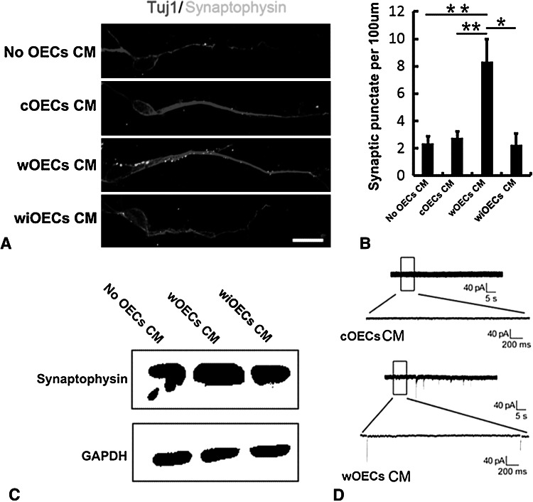
Olfactory ensheathing cells (OECs), the major glia cells in the olfactory system, have been extensively studied because of their ability to promote axonal growth and regeneration. Whether it could facilitate synaptogenesis is an important, but remains as yet an unanswered question. We have identified a subgroup of Wnt signaling-activated OECs, spatiotemporal distribution of which in the olfactory bulb suggests a role for these cells in both axonal growth and synaptogenesis. In the present study, we explored this possibility in vitro. OECs were primarily cultured, in which Wnt signaling was activated by overexpressing β-catenin, and inhibited by dominant negative TCF4. Neurite growth and synaptogenesis were assessed by co-culturing neurons with conditioned medium from control OECs (cOECs CM), Wnt/β-catenin signaling-activated OECs (wOECs CM), or Wnt signaling-inhibited OECs (wiOECs). The results showed that although cOECs CM enhances axonal growth, wOECs CM exhibited a stronger axonal growth-promoting effect, than cOECs CM. More importantly, wOECs CM stimulates synatpogenesis, demonstrated by the expression of Synaptophysin and whole-cell patch clamp recording. In contrast, both cOECs CM and wiOECs CM do not affect synaptogenesis. Our data, for the first time, demonstrated that, in comparison with regularly cultured OECs, wOECs CM are more effective in enhancing axonal growth, and can promote synaptogenesis, probably by secreting factors. These results suggest a potential application of wOECs for treating spinal cord injury.


Conflict of interest statement

The authors claim no conflict of interests.

Figures

Fig. 1
Fig. 1
Phenotypes of primary cultured OECs. a Immunocytochemistry of BLBP. b Immunocytochemistry of P75. c Percentages of BLBP positive and P75 positive cells in culture. Bar 50 μm
Fig. 2
Fig. 2
Activation of Wnt/β-catenin signaling in OECs. a, b Immunocytochemistry of PY489 in cOECs and wOECs. Bar 100 μm. c Percentages of PY489 positive cells in cOECs and wOECs. EbC increases the percentages of PY489 positive cells. d, e Western blot and gray scale analysis of β-catenin in cOECs and wOECs. EbC increases the expression of nuclear β-catenin. f Real-time RT-PCR of Axin2 in cOECs, wOECs, and wiOECs. Dominant negative TCF4 inhibits the β-catenin-induced Wnt activation. *p < 0.05, **p < 0.01, n = 3
Fig. 3
Fig. 3
wOECs CM promotes neurite growth. a Strategy of OEC-neuron co-culture. Neurons were cultured in the conditioned medium of OECs. be Immunocytochemistry of Tuj1 in neurons cultured without OECs, with cOECs CM, wOECs CM, or wiOECs CM. Bar 120 μm. f, g Quantification of the length of the longest neurite and the average length of primary neuritis in neurons cultured without OECs, with cOECs CM, wOECs CM, or wiOECs CM. Although cOECs CM enhances neurite outgrowth, wOECs CM can further promote neurite outgrowth. wiOECs CM does not promote neurite growth as do wOECs CM does. *p < 0.05, **p < 0.01, n = 3
Fig. 4
Fig. 4
wOECs CM promotes synaptogensis. a, b Double-staining of Tuj1 and Synaptophysin, and quantification of Synaptophysin positive punctuates in neurons cultured without OECs, with cOECs CM, wOECs CM, or wiOECs CM. Bar 100 μm. c Western blot of Synaptophysin in neurons cultured with cOECs CM, wOECs CM, or wiOECs CM. cOECs CM and wiOECs CM have no effects on, while wOECs stimulate the expression of synaptophysin. d Whole-cell patch clamp recording of mEPSCs in neurons cultured with cOECs CM or wOECs CM. wOECs CM promotes the functional activity of synapse. *p < 0.05, **p < 0.01, n = 3

Similar articles

Cited by

References

    1. Au E, Roskams AJ (2003) Olfactory ensheathing cells of the lamina propria in vivo and in vitro. Glia 41(3):224–236. doi:10.1002/glia.10160 - PubMed
    1. Au WW, Treloar HB, Greer CA (2002) Sublaminar organization of the mouse olfactory bulb nerve layer. J Comp Neurol 446(1):68–80 - PubMed
    1. Booker-Dwyer T, Hirsh S, Zhao H (2008) A unique cell population in the mouse olfactory bulb displays nuclear beta-catenin signaling during development and olfactory sensory neuron regeneration. Dev Neurobiol 68(7):859–869. doi:10.1002/dneu.20606 - PubMed
    1. Budnik V, Salinas PC (2011) Wnt signaling during synaptic development and plasticity. Curr Opin Neurobiol 21(1):151–159. doi:10.1016/j.conb.2010.12.002 - PMC - PubMed
    1. Chung RS, Woodhouse A, Fung S, Dickson TC, West AK, Vickers JC, Chuah MI (2004) Olfactory ensheathing cells promote neurite sprouting of injured axons in vitro by direct cellular contact and secretion of soluble factors. Cell Mol Life Sci 61(10):1238–1245. doi:10.1007/s00018-004-4026-y - PMC - PubMed

Publication types

Substances

LinkOut - more resources