. 2013 Oct;33(7):991-1001.
doi: 10.1007/s10571-013-9967-y. Epub 2013 Jul 28.

Neuronal damage using fluoro-Jade B histofluorescence and gliosis in the gerbil septum submitted to various durations of cerebral ischemia

Affiliations

Neuronal damage using fluoro-Jade B histofluorescence and gliosis in the gerbil septum submitted to various durations of cerebral ischemia

Chan Woo Park et al. Cell Mol Neurobiol. 2013 Oct.

Abstract

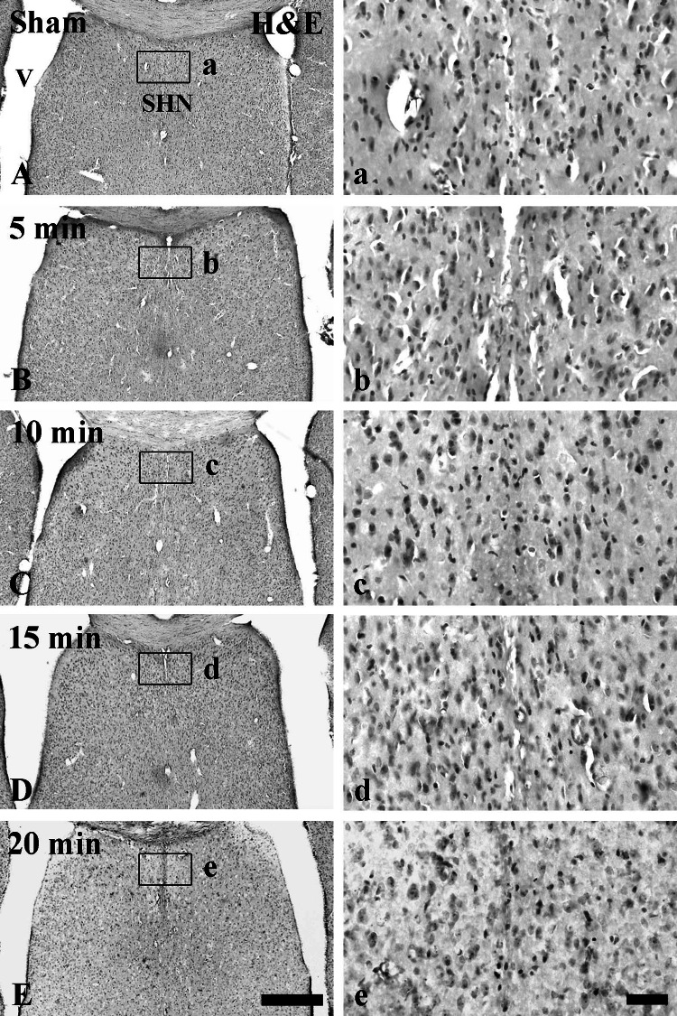
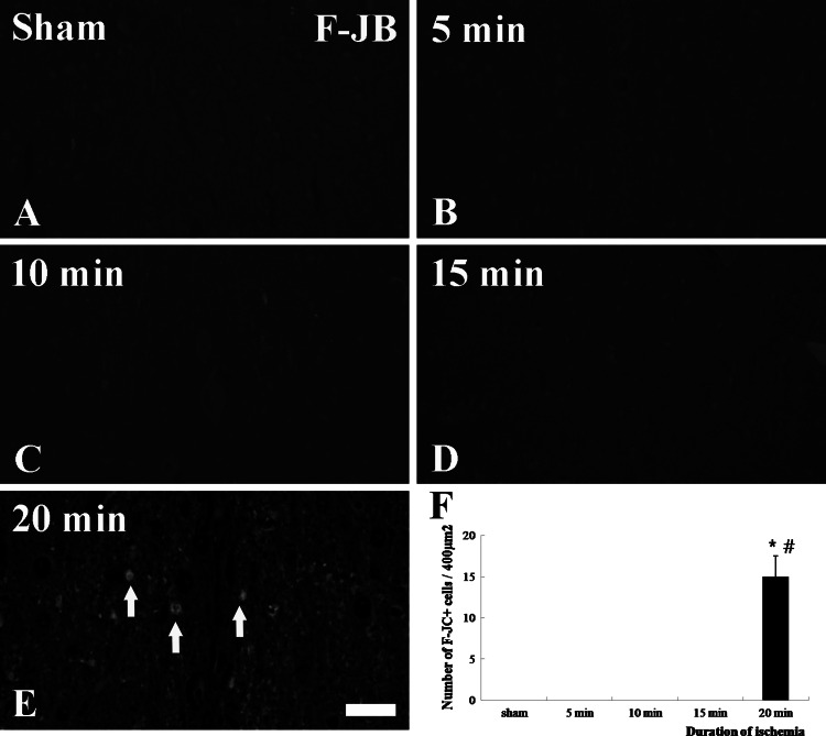
The extent of neuronal damage/death in some brain regions is highly correlated to duration time of transient ischemia. In the present study, we carried out neuronal degeneration/death and glial changes in the septum 4 days after 5, 10, 15, and 20 min of transient cerebral ischemia using gerbils. To examine neuronal damage, Fluoro-Jade B (F-J B, a marker for neuronal degeneration) histofluorescence staining was used. F-J B positive ((+)) cells were detected in the septo-hippocampal nucleus (SHN) of the septum only in the 20 min ischemia-group; the mean number of F-J B(+) neurons was 14.9 ± 2.5/400 μm(2) in a section. Gliosis of astrocytes and microglia was examined using anti-glial fibrillary acidic protein (GFAP) and anti-ionized calcium-binding adapter molecule 1 (Iba-1), respectively. In all the ischemia-groups, GFAP- and Iba-1-immunoreactive astrocytes and microglia, respectively, were increased in number, and apparently tended to be increased in their immunoreactivity. Especially, in the 20 min ischemia-group, the number and immunoreactivity of Iba-immunoreactive microglia was highest and strongest in the ischemic SHN 4 days after ischemia-reperfusion. In brief, our findings showed that neuronal damage/death in the SHN occurred and gliosis was apparently increased in the 20 min ischemia-group at 4 days after ischemia-reperfusion.


Conflict of interest statement

The authors have declared that there is no conflict of interest.

Figures

Fig. 1
Fig. 1
H–E staining in the septum of the sham-group (A, a) and ischemia-groups (5 min (B, b), 10 min (C, c), 15 min (D, d), and 20 min (E, e)) 4 days after ischemia–reperfusion. In all the ischemia-groups, H–E staining is not apparently changed compared to that in the sham-group. However, H–E positive small cells tend to be increased with duration time of ischemia in the septo-hippocampal nucleus (SHN) of the septum. ae High magnification framed in AE. Scale bar 200 μm (AE), 50 μm (ae)
Fig. 2
Fig. 2
CV staining (ae) and immunohistochemistry for NeuN (fJ) in the SHN of the sham-group (a, f) and ischemia-groups (5 min (b, g), 10 min (c, h), 15 min (d, i), and 20 min (e, j)) 4 days after ischemia–reperfusion. In all the ischemia-groups, numbers of CV positive neurons are not apparently changed in the ischemic SHN; however, NeuN-immunoreactive neurons tend to be slightly decreased in number. Scale bar 50 μm
Fig. 3
Fig. 3
F-J B staining of the SHN of the sham-group (a) and ischemia-groups (5 min (b), 10 min (c), 15 min (d), and 20 min (e)) 4 days after ischemia–reperfusion. No F-J B positive neurons are observed in the SHN of the sham-group and 5–15 min ischemia-group. However, in the 20 min ischemia-group, many F-J B positive cells (arrows) are observed in the SHN. Scale bar 50 μm. f Mean number of F-J B positive neurons/40 mm2 in a section of the SHN 4 days after ischemia–reperfusion (n = 10 per each group; *P < 0.05, significantly different from the sham-group; # P < 0.05, significantly different from the preceding-group). The bars indicate the mean ± SEM
Fig. 4
Fig. 4
Immunohistochemistry for GFAP (ae) and Iba-1 (fj) in the SHN of the sham-group (a) and ischemia-groups (5 min (b), 10 min (c), 15 min (d), and 20 min (e)) 4 days after ischemia–reperfusion. In the sham-group, typical GFAP-immunoreactive astrocytes (arrows) and Iba-1-immunoreactive microglia (arrows) are easily detected. In all the ischemia-groups, GFAP and Iba-1 immunoreactivity tends to be increased according to the duration of ischemia. Scale bar 50 μm
Fig. 5
Fig. 5
Relative optical density (ROD) as % of GFAP (a) and Iba-1 (b) immunoreactive structures/400 μm2 of the SHN 4 days after ischemia–reperfusion (n = 10 per each group; + P < 0.05, significantly different from the sham-group; # P < 0.05, significantly different from the preceding-group). The bars indicate the mean ± SEM

Similar articles

Cited by

References

    1. Barron KD (1995) The microglial cell. A historical review. J Neurol Sci 134(Suppl):57–68 - PubMed
    1. Bartus RT, Dean RL, Cavanaugh K, Eveleth D, Carriero DL, Lynch G (1995) Time-related neuronal changes following middle cerebral artery occlusion: implications for therapeutic intervention and the role of calpain. J Cereb Blood Flow Metab 15(6):969–979 - PubMed
    1. Brashear HR, Zaborszky L, Heimer L (1986) Distribution of GABAergic and cholinergic neurons in the rat diagonal band. Neuroscience 17(2):439–451 - PubMed
    1. Buchan AM, Pulsinelli WA (1990) Septo-hippocampal deafferentation protects CA1 neurons against ischemic injury. Brain Res 512(1):7–14 - PubMed
    1. Candelario-Jalil E, Alvarez D, Merino N, Leon OS (2003) Delayed treatment with nimesulide reduces measures of oxidative stress following global ischemic brain injury in gerbils. Neurosci Res 47(2):245–253 - PubMed

Publication types

MeSH terms

LinkOut - more resources