. 2013 Sep 6;288(36):25701-25716.
doi: 10.1074/jbc.M113.502674. Epub 2013 Jul 26.

A novel p38 mitogen-activated protein kinase/Elk-1 transcription factor-dependent molecular mechanism underlying abnormal endothelial cell proliferation in plexogenic pulmonary arterial hypertension

Affiliations

A novel p38 mitogen-activated protein kinase/Elk-1 transcription factor-dependent molecular mechanism underlying abnormal endothelial cell proliferation in plexogenic pulmonary arterial hypertension

Monal Patel et al. J Biol Chem. .

Erratum in

  • J Biol Chem. 2013 Dec 27;288(52):36855. Ryan-Hemnes, Anna [corrected to Hemnes, Anna Ryan]

Abstract

Plexiform lesions (PLs), the hallmark of plexogenic pulmonary arterial hypertension (PAH), contain phenotypically altered, proliferative endothelial cells (ECs). The molecular mechanism that contributes to EC proliferation and formation of PLs is poorly understood. We now show that a decrease in intersectin-1s (ITSN-1s) expression due to granzyme B (GrB) cleavage during inflammation associated with PAH and the high p38/Erk1/2(MAPK) activity ratio caused by the GrB/ITSN cleavage products lead to EC proliferation and selection of a proliferative/plexiform EC phenotype. We used human pulmonary artery ECs of PAH subjects (EC(PAH)), paraffin-embedded and frozen human lung tissue, and animal models of PAH in conjunction with microscopy imaging, biochemical, and molecular biology approaches to demonstrate that GrB cleaves ITSN-1s, a prosurvival protein of lung ECs, and generates two biologically active fragments, an N-terminal fragment (GrB-EH(ITSN)) with EC proliferative potential and a C-terminal product with dominant negative effects on Ras/Erk1/2. The proliferative potential of GrB-EH(ITSN) is mediated via sustained phosphorylation of p38(MAPK) and Elk-1 transcription factor and abolished by chemical inhibition of p38(MAPK). Moreover, lung tissue of PAH animal models and human specimens and EC(PAH) express lower levels of ITSN-1s compared with controls and the GrB-EH(ITSN) cleavage product. Moreover, GrB immunoreactivity is associated with PLs in PAH lungs. The concurrent expression of the two cleavage products results in a high p38/Erk1/2(MAPK) activity ratio, which is critical for EC proliferation. Our findings identify a novel GrB-EH(ITSN)-dependent pathogenic p38(MAPK)/Elk-1 signaling pathway involved in the poorly understood process of PL formation in severe PAH.

Keywords: Endothelium; Proliferation; Pulmonary Hypertension; Vascular Biology; p38 MAPK.


Figures

FIGURE 1.
FIGURE 1.
GrB cleaves ITSN in vitro and in vivo. A, in vitro cleavage of affinity-purified ITSN(1–440)-GST (lane a) by 500 nm hGrB followed by SDS-PAGE/Coomassie staining results in GrB-EHITSN and a 50-kDa C-terminal fragment, ITSN(271–440)-GST (lane b). The mutant ITSN(1–440)-GST (D271E) is not cleaved by the same amount of GrB (lanes c and d). Hsp90 was used as control (lanes e and f); arrows in lane f indicate the GrB cleavage products. Western blot of lung lysates of LPS-treated mice (4-h LPS exposure) with ITSN Ab (N-terminal epitope) shows loss of full-length ITSN-1s (lane h versus lane g) and the appearance of GrB-EHITSN cleavage product (arrow). GrB is detected by Western blot in mouse lung lysates at 4 h after 90 μg of LPS/mouse (lane l). No GrB immunoreactivity was detected in untreated mice (lane k). PAH was induced in CD1 mice (females (F) in lane i and males (M) in lane j). Western blot revealed decreased ITSN-1 expression and presence of GrB-EHITSN cleavage product (lanes i and j versus lane g) as well as GrB expression (lanes m and n). n = 3. The control (lane g) shows the level of ITSN-1s in a 3-month-old CD1 mouse. It is a suitable control for both LPS- and MCT-treated mice because studies indicated that 3–6-month-old CD1 mice show similar levels of ITSN. Sequence alignment shows a conserved GrB cleavage site in rodents and humans (blast.ncbi.nlm.nih.gov). B, male Sprague-Dawley rats (200–250 g) were injected with one dose of MCT (60 mg/kg intraperitoneally). Four weeks after treatment, the lungs were perfused with PBS, harvested, and used to prepare tissue lysates that were subjected to Western blot analyses using ITSN-1 Ab. Actin was used as a loading control. C, human lung lysates (70 μg of protein) of FD1-Ctrl and PAH tissue (two different locations: PAH I and PAH II) were subjected to SDS-PAGE and electrotransfer followed by Western blot using ITSN-1 and GrB Abs. D, decrease of full-length ITSN-1s was also detected in ECPAH lysates (lanes b, c, and d) by reference to a representative ECCtrl (lane a; the studies were performed on three different batches/donors and Lonza ECCtrl with similar results). Data are representative for three different ECPAH preparations: ECPAH-B397 (lane b), ECPAH-CC-005 (lane c), and ECPAH-CC016 (lane d). ITSN-1s down-regulation quantified by densitometry was different among the three EC lines used. E, RT-PCR and densitometry of ITSN-1s mRNA levels in ECPAH (lanes b, c, and d) by reference to ECCtrl (lane a). Data are representative for three independent experiments applied on ECCtrl and three different ECPAH lines: B397 (lane b), CC-005 (lane c), and CC016 (lane d). Results are expressed as mean ± S.E. (error bars). *, p < 0.001 versus control. All data are representative of three independent experiments. LPS and MCT treatments were applied to at least three mice per experimental condition.
FIGURE 2.
FIGURE 2.
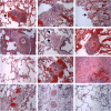
Pathological findings in PAH patients. PA vasculopathy with PL (brown arrow) and associated angiomatoid dilation (black arrow) (a), eccentric intimal fibrosis in lumen of artery with near occlusion of smaller adjacent branch (b), muscularization of arteriole without intimal fibrosis (c, arrow), medial hypertrophy and concentric medial fibrosis (d), media hypertrophy and cellular occlusion of lumen of the artery with adjacent angiomatoid dilation of vessel (arrows) likely with an associated PL in another plane (e), PL with associated angiomatoid dilation (f), muscularization of arteriole (g), PL and associated angiomatoid dilation (h), intimal fibrosis and medial proliferation of collapsed PA (i), concentric intimal fibrosis and medial hypertrophy and adjacent dilation lesion (j), intimal fibrosis and medial hypertrophy (k), and normal control (l). Scale bar, 100 μm (a–l).
FIGURE 3.
FIGURE 3.
ITSN is down-regulated in human PAH specimens. Lung tissue sections of PAH (A, B, and C) and FD2-Ctrl (D) were subjected to IHC using ITSN Ab and CD31 Ab. Panel a1 illustrates that CD31 immunoreactivity of complex lesion with focal proliferation of several endothelial channels and partial destruction of the arterial wall. ITSN-1s pAb/anti-rabbit IgG-Alexa Fluor 594 staining (panel a2) is barely detected. DAPI staining (panels a3 and b2) of the nuclei documents the hypercellularity and the concentric thickening/distribution of intimal cells. The merged image (panel a4) illustrates co-localization of CD31 with ITSN-1s remnants (arrow) in several ECs of the damaged arterial wall as well as ITSN-1s immunoreactivity associated with pulmonary epithelial cells (arrowheads). ITSN-1s immunoreactivity is barely detected at the level of a thin walled lymphatic in close proximity of the PL (A, panels a1 and a4, asterisk). Clusters of proliferative ECs (B, panel b1, circled area) lacking ITSN-1s (panels b2 and b2.1) surrounded by concentric intimal thickening are seen in the lumen of a small PA. C, inset, ITSN immunoreactivity is associated with ECs lining the blood vessels in FD2-Ctrl. D, inset, low ITSN-1s staining in ECs lining the small blood vessels not yet detectably affected by remodeling in PAH specimen. The results are representative for 12 PAH cases. Scale bars, 50 (A, B, C, and D) and 25 μm (panel b2.1).
FIGURE 4.
FIGURE 4.
GrB immunoreactivity in the PAH human lungs and the microenvironment of PLs. Representative GrB/Alexa Fluor 594-CD31/Alexa Fluor 488 IHC of PAH human lung tissue sections illustrates close proximity between ECs and GrB immunoreactivity (A, C, and D); frequently GrB co-localizes with CD31 (C, panel c1) or was detected inside ECs labeled by CD31 (panels d4 and d4.1). B, large PA from PAH specimen does not show GrB staining. E, FD2-Ctrl lung sections do not show detectable GrB immunoreactivity. F, representative H&E staining of a concentric (onion skin) obliterative lesion in human PAH lungs. G, GrB (arrows) within the same obliterative lesion. Arrowheads in G and insets g1 and g2 (boxed areas in G) illustrate GrB immunoreactivity in close proximity of ECs. H, GrB/Alexa Fluor 594-CD31/Alexa Fluor 488 IHC of a large PL shows GrB immunoreactivity (panel h1), the high EC content (panel h2), and frequent presence of GrB in close proximity of CD31-labeled ECs (panel h4, arrows). DAPI staining of the nuclei is shown (panel h3). The results are representative for 12 PAH cases. Three independent IHC experiments were performed. Scale bars, 25 (A, B, and C), 50 (D and E), 10 (panels c1 and d4.1), 40 (F and G), 20 (panels g1 and g2), and 50 μm (H).
FIGURE 5.
FIGURE 5.
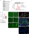
A, Myc-GrB-EHITSN expression causes human PAEC proliferation. Lysates (70 μg of total protein/lane) of control (lane a) and myc-GrB-EHITSN-transfected ECs (lane b) were assessed for myc-GrB-EHITSN protein expression by Western blotting (WB) using myc Ab. Immunofluorescent staining using myc pAb followed by anti-rabbit IgG-Alexa Fluor 594 of control (B) and myc-GrB-EHITSN-transfected cells (C, panel c1). Control (D) and myc-GrB-EHITSN-transfected (E) ECs 48 h post-transfection were incubated with BrdU reagent for 6 h at 37 °C. BrdU mAb/Alexa Fluor 488 were used to detect proliferating cells. F, quantification of BrdU-positive (BrdU+) cells. The results are expressed as BrdU+ ECs/high power field (50 power field images/experimental condition). Data are representative of three different experiments and were normalized per total number of cells counted. G, MTT assay applied on control and myc-GrB-EHITSN-transfected ECs 48 h post-transfection (w, well). H, quantification of the extent of myc-GrB-EHITSN-transfected EC proliferation. Data are presented as mean ± S.E. (error bars) from three to five independent experiments. *, p < 0.05 compared with the control. Scale bars, 20 (B and C), 10 (panel c1), and 50 μm (D and E).
FIGURE 6.
FIGURE 6.
Myc-GrB-EHITSN specifically activates p38MAPK. A, lysates of control (lane a1) and myc-GrB-EHITSN-transfected ECs (lane a2) were assessed for Erk1/2MAPK, JNK, PI3K/Akt, and p38 phosphorylation. B, time course of p38 activity in myc-GrB-EHITSN-expressing ECs (lanes c–e, 48, 72, and 96 h post-transfection) followed by densitometry. Non-transfected ECs (lane a) and myc-ITSN-transfected ECs 48 h post-transfection (panel b) were used for comparison. Densitometric values ±S.E. (error bars) are representative for three independent experiments. C, SB203580 abolishes the proliferative response in myc-GrB-EHITSN-expressing ECs. Data are shown as mean ± S.E. (error bars). *, p < 0.01; **, p < 0.05 compared with the control. D, immunofluorescent staining showing diffuse p38MAPK staining both in control (panel d1) and myc-GrB-EHITSN-transfected ECs (panel d2). E, immunofluorescent staining for phospho-p38MAPK in control (panel e1), myc-GrB-EHITSN-expressing ECs (panel e2), and ECPAH (B397; panel e3). F and G, phospho-p38/CD31 IHC on PAH human lung tissue sections, cases 10 and 3, respectively. Arrowheads point to phospho-p38 immunoreactivity within CD31-labeled EC profiles. H, phospho-p38/CD31 IHC on FD2-Ctrl human lung tissue. Scale bars, 20 (D and E), 10 (F), and 25 μm (G and H). DU, densitometry units.
FIGURE 7.
FIGURE 7.
Concurrent expression of GrB/ITSN-1s cleavage products results in a high p38/Erk1/2 activity ratio. A, lysates of MCT-treated mouse lungs (70 μg of total protein) were resolved by SDS-PAGE, electrotransferred to nitrocellulose membranes, and further analyzed by Western blotting for phospho-p38 and phospho-Erk1/2 immunoreactivity. Total kinase was used as a loading control. Representative blots and corresponding densitometry (phosphokinase (P-kinase)/total kinase (T-kinase) ratio) are shown. The results of three independent experiments (three male (M) and three female (F) mice; 8-week MCT treatment) are expressed as mean ± S.E. (error bars). *, p < 0.05 versus control. B, human lung lysates (70 μg of protein) of FD1-Ctrl and PAH tissue sampled from two different locations (PAH I and PAH II) were analyzed as above. Total kinase was used as a loading control. Given the decreased expression of p38 in PAH lung lysates, actin was further used to confirm equal protein loading. Representative blots and corresponding densitometric analyses of p38 and Erk1/2 phosphorylation are shown. n = 3. Data are expressed as mean ± S.E. (error bars). **, p < 0.01 compared with the control.
FIGURE 8.
FIGURE 8.
Myc-GrB-EH activates Elk-1 transcription factor and cellular immediate early response gene c-fos. A, nuclear extracts (three different concentrations) prepared from control, myc-ITSN-, and myc-GrB-EHITSN-transfected ECs were assayed for Elk-1 activity using an ELISA kit designed to detect only active Elk-1. Data are representative of three independent experiments performed in triplicates and are expressed as mean ± S.E. (error bars). *, p < 0.05 versus control. B, Western blot of nuclear extracts (45 μg/lane) from control, myc-GrB-EHITSN-, and myc-ITSN-transfected ECs using c-Fos Ab and consequent densitometric analysis. Data were normalized to β-actin. Data represent mean ± S.E. (error bars) from three different experiments. *, p < 0.05 versus control. C, a gel shift assay was used to analyze the c-fos SRE binding activity in control ECs and myc-GrB-EHITSN-transfected cells. All lanes have biotin-labeled c-fos probe containing the Elk-1 binding sequence. Lane a, biotin-labeled c-fos probe alone. Nuclear extract from transfected cells shows a shift of the c-fos·Elk-1 complex (lanes c, d, and f). This DNA·protein complex is not detected in control ECs (lane b) or in the presence of a 100-fold excess of unlabeled probe (lane e). In the presence of an Ab specific for Elk-1 protein, a supershift is detected in myc-GrB-EHITSN-transfected ECs (lane g). However, this supershifted complex is not detected in the presence of Sap-1a Ab (lane f). D, ChIP assay applied to control and myc-GrB-EHITSN-transfected ECs using Elk-1 Ab and rabbit IgG. Immunoprecipitated DNA samples were further amplified by quantitative RT-PCR. Data represent mean ± S.E. (error bars) from three different experiments. *, p < 0.01 versus control; ns, not statistically significant. DU, densitometry units.
FIGURE 9.
FIGURE 9.
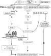
Diagram of the proposed GrB-EHITSN/p38MAPK/Elk-1-mediated molecular mechanism resulting in the development of a proliferative/plexiform EC phenotype in severe PAH. The domain structure of full-length ITSN is shown. KLERQ, coiled coil domain. CD8+ T-lymphocytes and increased GrB expression cause cleavage of ITSN at IDQD271GK, generating an N-terminal cleavage product (GrB-EHITSN) and a C-terminal cleavage product (GrB-SH3A-EITSN). Because ITSN has only one cleavage site for GrB and is not a substrate for any caspases (PeptideCutter), the two cleavage products are generated in equimolar amounts. Both GrB/ITSN cleavage products are biologically active and interfere with the activity of two mitogen-activated protein kinases: GrB-EHITSN activates p38, whereas GrB-SH3A-EITSN has dominant negative effects on Ras/Erk1/2 survival signaling (21). In the context of full-length ITSN, EH domains are under the inhibitory control of SH3A–E (20). ITSN binds mSos and regulates GTP-Ras levels and thus downstream Erk1/2 survival signaling (green font). GrB-EHITSN activates Elk-1 transcription factor via p38MAPK. Sustained p38 activation and Elk-1 positive regulation of c-fos promoter favor the selection of a proliferative/plexiform EC phenotype. A negative cross-talk from p38 to Erk1/2 occurs because p38 can enhance MEK dephosphorylation or up-regulate protein phosphatase 2A (44). The C-terminal cleavage product (GrB-SH3A-EITSN) may perhaps interact with Smad2, recently shown to bind SH3 domains (49), causing ineffective assembly of TGFβ receptor 1 (TGFβR1)·Smad2·SARA (Smad anchor for receptor activation) signaling complex; deficient Smad2-dependent TGFβ signaling results in inhibition of its antiproliferative activity. SRF, serum response factor.

Similar articles

Cited by

References

    1. Voelkel N. F., Quaife R. A., Leinwand L. A., Barst R. J., McGoon M. D., Meldrum D. R., Dupuis J., Long C. S., Rubin L. J., Smart F. W., Suzuki Y. J., Gladwin M., Denholm E. M., Gail D. B. (2006) Right ventricular function and failure: report of a National Heart, Lung, and Blood Institute working group on cellular and molecular mechanisms of right heart failure. Circulation 114, 1883–1891 - PubMed
    1. Tuder R. M., Marecki J. C., Richter A., Fijalkowska I., Flores S. (2007) Pathology of pulmonary hypertension. Clin. Chest Med. 28, 23–42 - PMC - PubMed
    1. Masri F. A., Xu W., Comhair S. A., Asosingh K., Koo M., Vasanji A., Drazba J., Anand-Apte B., Erzurum S. C. (2007) Hyperproliferative apoptosis-resistant endothelial cells in idiopathic pulmonary arterial hypertension. Am. J. Physiol. Lung Cell Mol. Physiol. 293, L548–L554 - PubMed
    1. Singla S., Predescu D., Bardita C., Wang M., Zhang J., Balk R. A., Predescu S. (2011) Pro-inflammatory endothelial cell dysfunction is associated with intersectin-1s down-regulation. Respir. Res. 12, 46. - PMC - PubMed
    1. Tu L., Dewachter L., Gore B., Fadel E., Dartevelle P., Simonneau G., Humbert M., Eddahibi S., Guignabert C. (2011) Autocrine fibroblast growth factor-2 signaling contributes to altered endothelial phenotype in pulmonary hypertension. Am. J. Respir. Cell Mol. Biol. 45, 311–322 - PMC - PubMed

Publication types

MeSH terms