. 2013 Sep 20;288(38):27646-27657.
doi: 10.1074/jbc.M113.463067. Epub 2013 Jul 26.

Ras-mutant cancer cells display B-Raf binding to Ras that activates extracellular signal-regulated kinase and is inhibited by protein kinase A phosphorylation

Affiliations

Ras-mutant cancer cells display B-Raf binding to Ras that activates extracellular signal-regulated kinase and is inhibited by protein kinase A phosphorylation

Yanping Li et al. J Biol Chem. .

Abstract

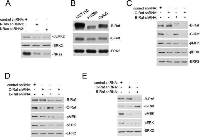
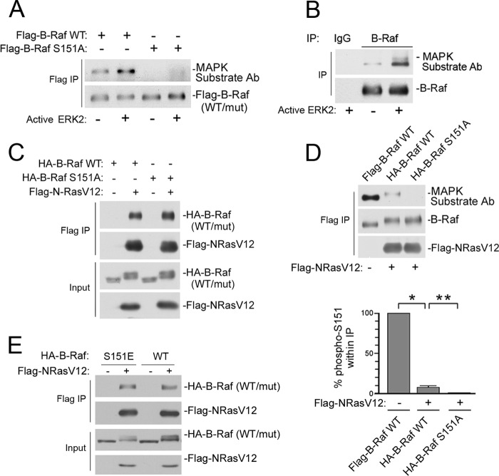
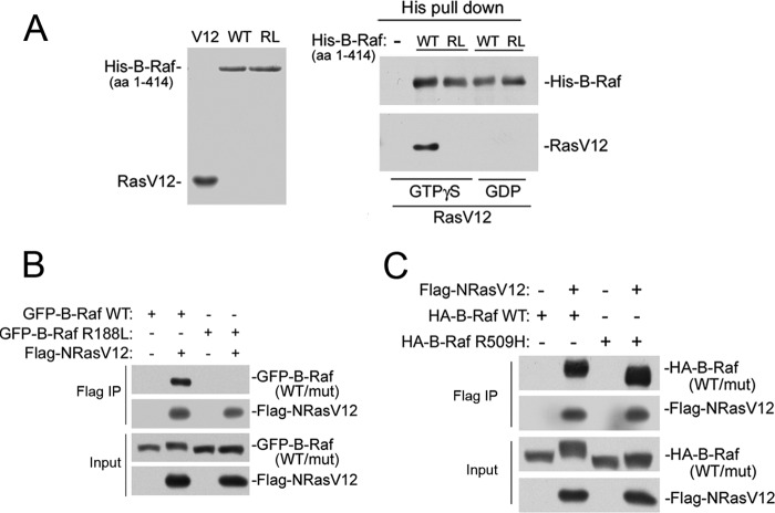
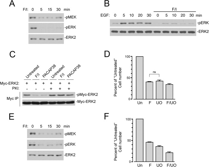
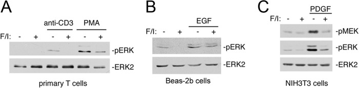
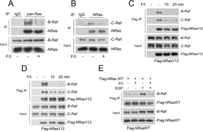
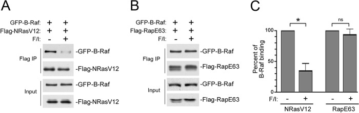
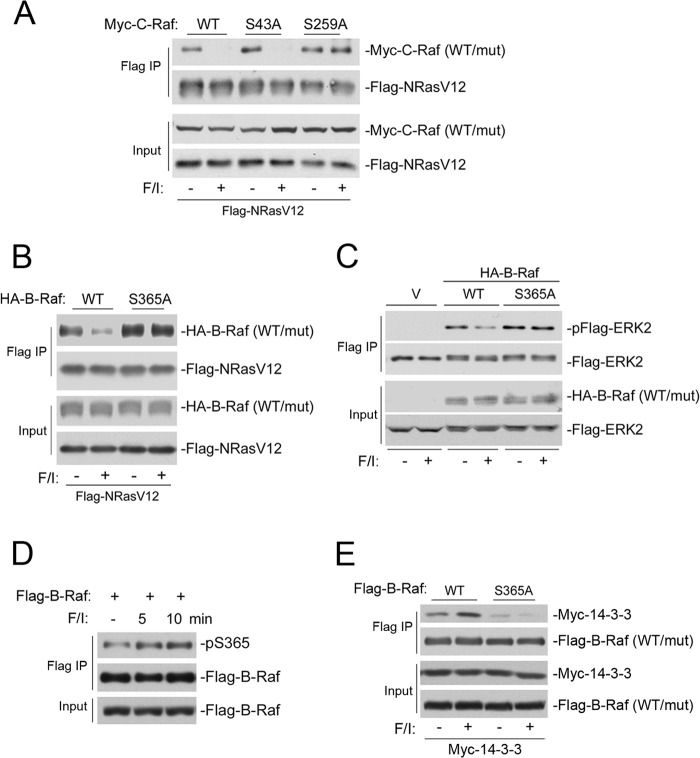
The small G protein Ras regulates proliferation through activation of the mitogen-activated protein (MAP) kinase (ERK) cascade. The first step of Ras-dependent activation of ERK signaling is Ras binding to members of the Raf family of MAP kinase kinase kinases, C-Raf and B-Raf. Recently, it has been reported that in melanoma cells harboring oncogenic Ras mutations, B-Raf does not bind to Ras and does not contribute to basal ERK activation. For other types of Ras-mutant tumors, the relative contributions of C-Raf and B-Raf are not known. We examined non-melanoma cancer cell lines containing oncogenic Ras mutations and express both C-Raf and B-Raf isoforms, including the lung cancer cell line H1299 cells. Both B-Raf and C-Raf were constitutively bound to oncogenic Ras and contributed to Ras-dependent ERK activation. Ras binding to B-Raf and C-Raf were both subject to inhibition by the cAMP-dependent protein kinase PKA. cAMP inhibited the growth of H1299 cells and Ras-dependent ERK activation via PKA. PKA inhibited the binding of Ras to both C-Raf and B-Raf through phosphorylations of C-Raf at Ser-259 and B-Raf at Ser-365, respectively. These studies demonstrate that in non-melanocytic Ras-mutant cancer cells, Ras signaling to B-Raf is a significant contributor to ERK activation and that the B-Raf pathway, like that of C-Raf, is a target for inhibition by PKA. We suggest that cAMP and hormones coupled to cAMP may prove useful in dampening the effects of oncogenic Ras in non-melanocytic cancer cells through PKA-dependent actions on B-Raf as well as C-Raf.

Keywords: B-Raf; C-Raf; ERK; MAP Kinases (MAPKs); Protein Kinase A (PKA); Raf; Ras.


Figures

FIGURE 1.
FIGURE 1.
Both B-Raf and C-Raf contribute to the basal ERK activation in Ras-mutant cancer cells. A, basal ERK activation in H1299 cells is dependent on NRas. H1299 cells were transfected with one of two shRNAs for NRas (shRNA1, shRNA2) or control shRNA. Cells were harvested after 48 h, and ERK activation was measured by Western blot using phosphorylation-specific antibodies against phosphorylated/activated ERK (pERK) (first panel). Total endogenous ERKs are shown as a loading control in the second panel. The efficiencies of knockdown of endogenous NRas by each shRNA are shown in the third panel. B, the expression levels of endogenous B-Raf (first panel) and C-Raf (second panel) in three Ras-mutant cancer cells (HCT116, H1299, and Calu-6 cells) are shown in the left, middle, and right lanes, respectively. Total endogenous ERK2 levels are shown in the third panel as a loading control. C–E, both B-Raf and C-Raf contribute to the basal ERK activation in H1299 cells (C), HCT116 cells (D), and Calu-6 cells (E). Cells were transfected with control shRNA, B-Raf shRNA, or C-Raf shRNA as indicated. Cells were harvested, and endogenous protein levels were assayed by Western blot. The efficiencies of knockdown of endogenous B-Raf and C-Raf are shown in the first and second panels, respectively. MEK activation was measured using phosphorylation-specific antibodies against pMEK (third panel). ERK activation was measured using phosphorylation-specific antibodies against pERK (fourth panel). Total endogenous ERK2 levels are shown in the fifth panel.
FIGURE 2.
FIGURE 2.
The serine 151 site in B-Raf is largely unphosphorylated in H1299 cells. A, MAPK substrate antibody is selective for phosphorylation of Ser-151 on B-Raf. Cells were transfected with FLAG-B-Raf wild type (WT) or the FLAG-B-Raf S151A mutant (mut) as indicated. Cells were harvested, and FLAG-B-Raf was immunoprecipitated and incubated in kinase buffer in the absence (−) or presence (+) of active recombinant ERK2. The phosphorylations of both WT and mutant B-Raf were examined by Western blotting with the MAP kinase substrate antibody (MAPK substrate antibody (top panel). The levels of FLAG-B-Raf (WT or mut) within the immunoprecipitates are shown in the bottom panel. B, Ser-151 was largely unphosphorylated in H1299 cells. Cells were lysed, and endogenous B-Raf proteins were enriched by immunoprecipitation. Each sample was incubated in the absence (−) or presence (+) of active recombinant ERK2, and the Ser(P)-151 level was examined using the MAPK substrate Ab (top panel). The levels of endogenous B-Raf within the immunoprecipitates are shown in the bottom panel. C, B-Raf WT and B-Raf S151A bind equally well to FLAG-NRasV12. H1299 cells were transfected with HA-B-Raf wild type or HA-B-Raf S151A mutant in the presence or absence of FLAG-NRasV12, as indicated. Cells were harvested for IP with FLAG antibody, and the amount of HA-B-Raf (WT) or HA-B-Raf S151A (mut) protein bound to FLAG-NRasV12 was determined by Western blot using HA Ab (first panel). The levels of FLAG proteins within the FLAG IP are shown in the second panel, and the input levels of HA-B-Raf and FLAG-NRasV12 are shown in the third and fourth panels, respectively. D, phosphorylation of B-Raf on Ser-151 inhibits its association with active Ras. H1299 cells were transfected with either FLAG-B-Raf (first lane), HA-B-Raf WT and FLAG-NRasV12 (second lane), or HA-B-Raf S151A and FLAG-NRasV12 (third lane). All cells were subjected to IP using FLAG Ab (FLAG-B-Raf; first lane, FLAG-NRasV12; second and third lanes). The volumes of the protein loaded were adjusted to allow examination of equivalent amounts of B-Raf in each condition. The presence of Ser-151 phosphorylation within each IP was examined using the MAPK Substrate Ab (first panel). The third lane of this panel shows the control using B-Raf S151A, demonstrating the specificity of the MAPK substrate Ab for Ser(P)-151. The levels of B-Raf within each IP are shown using B-Raf Ab (second panel). The levels of FLAG-NRavV12 within each IP are shown in the third panel. Note that much less Ser(P)-151 was detected by the MAPK Substrate Ab in FLAG-NRavV12 IP (second lane) compared with the FLAG-B-Raf IP (first lane). Quantitation of binding was performed by ImageJ and normalized to total FLAG protein within the IP. The normalized data are represented as the mean ± S.E of four independent experiments. The bar graph shows the percentage of phosphorylated Ser-151, normalized for the level of B-Raf within each IP, compared with that seen in the first lane (100%). *, statistical significance is <0.0001. **, statistical significance is <0.05. E, B-Raf WT and B-Raf S151E bind equally well to FLAG-NRasV12. H1299 cells were transfected with HA-B-Raf wild type or HA-B-Raf S151E mutant in the presence or absence of FLAG-NRasV12 as indicated. Cells were harvested for IP with FLAG antibody, and the amount of HA-B-Raf (WT) or HA-B-Raf S151E (mut) protein bound to FLAG-NRasV12 was determined by Western blot using HA Ab (first panel). The levels of FLAG proteins within the FLAG IP are shown in the second panel, and the input levels of HA-B-Raf (WT/mut) and FLAG-NRasV12 are shown in the third and fourth panels, respectively.
FIGURE 3.
FIGURE 3.
Basal B-Raf binding to active Ras is direct. A, the B-Raf R188L mutant prevents binding to RasV12 in vitro. The Ras binding domain of B-Raf (amino acids 1–414) and the corresponding R188L mutant were purified as His-tagged proteins and examined for their ability to bind GTP-loaded RasV12 in vitro. Proteins were purified and prepared as described under “Experimental Procedures.” Left panel, the bands corresponding to purified RasV12 (V12) and His-B-Raf wild type fragment (WT) and the His-B-Raf R188L fragment (RL) are shown by Coomassie staining after SDS-PAGE. Right panel, RasV12, loaded with either GTPγS (first through third lanes) or GDP (fourth and fifth lanes), were incubated alone (first lane), with the His-tagged WT B-Raf fragment (second and fourth lanes), or with the RL mutant (third and fifth lanes) as indicated. The levels of His-B-Raf WT or RL fragments within the His pulldown assay are shown in the upper panel. The presence of RasV12 within the His pulldown assay is shown in the lower panel. B, the binding of B-Raf to NRasV12 is direct. H1299 cells were transfected with wild type GFP-B-Raf or the GFP-B-Raf R188L mutant in the presence or absence of FLAG-NRasV12 as indicated. The level of GFP-B-Raf associated with FLAG-NRasV12 is shown by Western blotting with GFP Ab after FLAG IP (first panel). The levels of FLAG proteins within the FLAG IP are shown in the second panel. The input levels of GFP proteins and FLAG-NRasV12 are shown in the third and fourth panels, respectively. C, binding of B-Raf to NRasV12 is independent of B-Raf dimerization. H1299 cells were transfected with wild type HA-B-Raf or the HA-B-Raf R509H mutant in the presence or absence of FLAG-NRasV12 as indicated. The level of HA-B-Raf (WT) or HA-B-Raf R509H (mut) associated with FLAG-NRasV12 was examined after FLAG IP by Western blotting with HA Ab (first panel). The FLAG-NRasV12 within the IP is also shown using Western blotting with FLAG Ab (second panel). The input levels of HA proteins (WT or mut) and FLAG-NRasV12 are shown (third and fourth panels).
FIGURE 4.
FIGURE 4.
cAMP inhibits ERK activation and cellular proliferation in H1299 and HCT116 cells. A, cAMP inhibits the basal ERK activation in H1299. H1299 cells were serum-starved overnight and treated with F/I for the indicated times. MEK activation was measured using phosphorylation-specific antibodies against activated MEK (pMEK) (first panel). ERK activation was detected by Western blot using phosphorylation-specific antibodies (pERK) (second panel). Total levels of ERK2 are shown in the third panel. B, cAMP inhibits growth factor stimulated ERK activation in H1299 cells. Cells were serum-starved overnight and treated with EGF (1 ng/ml) for the indicated times in the presence and absence of 10 min F/I pretreatment. ERK activation (pERK) was detected by Western blot using phosphorylation-specific antibodies (pERK). pERK levels are shown in the top panel. Total levels of ERK2 are shown in the bottom panel. C, PACAP38 inhibits the basal ERK activation in H1299 in a PKA-dependent manner. H1299 cells were transfected with Myc-ERK2 in the presence or absence of protein kinase inhibitor (PKI), a selective peptide inhibitor of PKA. After serum starvation, cells were treated with F/I or PACAP38. Myc-ERK2 activation (pMyc-ERK2) was measured within the Myc IP by Western blot using phosphorylation-specific antibodies. D, cAMP inhibits the proliferation of H1299 cells. H1299 cells were plated, serum-starved, and treated with F/I (F) and/or UO126 (UO) for 3 days, and cell numbers were assessed by MTT assay. Relative cell numbers after 3 days are shown as the percent of cell number compared with the untreated group. Normalized averages of the three different experiments are shown ± S.E. There was statistical significance between all conditions (p < 0.05) except those marked with ns (not significant). E, cAMP inhibits the basal ERK activation in HCT116 cells. Cells were serum-starved overnight and treated with F/I for the indicated times. MEK activation was measured using phosphorylation-specific antibodies against activated MEK (pMEK) (first panel). ERK activation was detected by Western blot using phosphorylation-specific antibodies (pERK) (second panel). Total levels of ERK2 are shown in the third panel. F, cAMP inhibits cellular proliferation in HCT116 cells. HCT116 cells were plated, serum-starved, and treated with F/I (F), UO126 (UO), or both (F/UO) for 3 days, and cell numbers were assessed by MTT assay. Relative cell numbers after 3 days are shown as the percent of cell number compared with the untreated group. Normalized averages of the three different experiments are shown ± S.E. There was statistical significance between all conditions (p < 0.05).
FIGURE 5.
FIGURE 5.
cAMP inhibits ERK activation in multiple non-cancerous cell types. A, cAMP inhibits the stimulated ERK activation in primary T cells. CD4-positive T cells were harvested from mouse spleen. Cells were plated and stimulated with either anti-CD3 cross-linking or phorbol-12-myristate-13-acetate in the presence and absence of 10 min of F/I pretreatment. ERK activation was detected by Western blot using phosphorylation-specific antibodies (pERK). Total levels of ERK2 are shown as controls. B, cAMP inhibits both the basal and stimulated ERK activation in Beas-2b lung epithelial cells. Cells were plated and stimulated with EGF for 20 min in the presence and absence of 10 min F/I pretreatment. ERK activation was detected by Western blot using phosphorylation-specific antibodies (pERK). Total levels of ERK2 are shown as controls. C, cAMP inhibits the stimulated ERK activation in NIH3T3 cells. Cells were plated and stimulated with PDGF for 5 min in the presence and absence of 10 min F/I pretreatment. MEK activation was measured using phosphorylation-specific antibodies against activated MEK (pMEK) (first panel). ERK activation was detected by Western blot using phosphorylation-specific antibodies (pERK) (second panel). Total levels of ERK2 are shown in the third panel.
FIGURE 6.
FIGURE 6.
cAMP inhibits the binding between B-Raf and Ras in H1299 and HCT116 cells. A, cAMP inhibits the binding between endogenous B-Raf and NRas in H1299 cells. H1299 cells were treated with F/I or left untreated as indicated. Cell lysates were subjected to IP with a control IgG (first lane) or a pan-Ras antibody (second and third lanes). The levels of endogenous B-Raf within each IP are shown in the first panel, and the levels of endogenous NRas within each IP are shown in the second panel. The input levels of B-Raf and NRas are shown in the third and fourth panels, respectively. B, cAMP inhibits the binding between endogenous C-Raf and NRas in H1299 cells. H1299 cells were treated with F/I or left untreated as indicated. Cell lysates were subjected to IP with a control IgG (first panel) or an NRas-specific antibody (second and third panels). The levels of endogenous C-Raf within each IP are shown in the first panel, and the levels of endogenous NRas within each IP are shown in the second panel. The input levels of C-Raf and NRas are shown in the third and fourth panels, respectively. C, cAMP inhibits the binding of both endogenous B-Raf and C-Raf to FLAG-NRasV12 in H1299 cells. H1299 cells were transfected with empty vector (first panel) or FLAG-NRasV12 (second through fourth lanes). After serum starvation, cells were treated with F/I as indicated, and lysates were subjected to FLAG IP. The levels of B-Raf and C-Raf within the IPs are shown in the first and second panels, respectively, and the levels of FLAG-NRasV12 within each IP are shown in the third panel. The fourth and fifth panels show the input levels of endogenous B-Raf and C-Raf, respectively. The sixth panel shows the input level of transfected FLAG-NRasV12. D, cAMP inhibits the binding of both endogenous B-Raf and C-Raf to FLAG-NRasV12 in HCT116 cells. HCT116 cells were transfected with empty vector (first panel) or FLAG-NRasV12 (second through fourth lanes). After serum starvation, cells were treated with F/I as indicated, and lysates were subjected to FLAG IP. The levels of B-Raf and C-Raf within the IPs are shown in the first and second panels, respectively, and the levels of FLAG-NRasV12 within each IP are shown in the third panel. The fourth and fifth panels show the input levels of endogenous B-Raf and C-Raf, respectively. The sixth panel shows the input level of transfected FLAG-NRasV12. E, cAMP blocks EGF stimulation of B-Raf binding to wild type FLAG-NRas (WT) in Hek293 cells. All cells were transfected with FLAG-NRas WT. After serum starvation, cells were treated with F/I or EGF as indicated and subjected to FLAG IP. The level of endogenous B-Raf within the IP is shown in the first panel, and the levels of WT FLAG-NRas within each IP are shown in the second panel. The input levels of endogenous B-Raf and transfected WT FLAG-NRas are shown in the third and fourth panels, respectively.
FIGURE 7.
FIGURE 7.
cAMP does not inhibit the binding between B-Raf and Rap1. A, cAMP inhibits the binding of B-Raf to NRasV12 in Hek293 cells. Hek293 cells were transfected with FLAG-NRasV12 and GFP-B-Raf. After serum starvation, cells were treated with F/I as indicated and subjected to FLAG IP. The levels of GFP-B-Raf within each IP are shown in the first panel, and the levels of FLAG-NRasV12 within each IP are shown in the second panel. The input levels of GFP-B-Raf (GFP) and transfected FLAG-NRasV12 (FLAG) are shown in the third and fourth panels, respectively. B, cAMP does not inhibit the binding of B-Raf to RapE63 in Hek293 cells. Hek293 cells were transfected with FLAG-RapE63 and GFP-B-Raf. After serum starvation, cells were treated with F/I as indicated and subjected to FLAG IP. The levels of GFP-B-Raf within each IP are shown in the first panel, and the levels of FLAG-RapE63 within each IP are shown in the second panel. The input levels of GFP-B-Raf (GFP) and transfected FLAG-RapE63 (FLAG) are shown in the third and fourth panels, respectively. C, the results of A and B are presented as the percent of B-Raf associating with NRasV12 or RapE63 treated with F/I (+) or left untreated (−). Untreated (−) is 100%. Quantitation of binding was performed by ImageJ and normalized to total FLAG protein within the IP. The normalized data are represented as the mean ± S.E from three independent experiments. ns, not significant. *, statistical significance is <0.05.
FIGURE 8.
FIGURE 8.
The ability of cAMP to block Raf association with Ras is regulated by phosphorylation of serine 259 in C-Raf and serine 365 in B-Raf. A, cAMP decreases the interaction between NRasV12 and C-Raf WT but not the interaction between NRasV12 and C-Raf S259A. H1299 cells were transfected with FLAG-NRasV12 and Myc-C-Raf (first and second lanes), Myc-C-Raf S43A (third and fourth lanes), or Myc-C-Raf S259A (fifth and sixth lanes). After serum starvation, the cells were treated with F/I or left untreated as indicated, and lysates were subjected to FLAG IP. The presence of Myc-C-Raf within each IP is shown in the first panel. The presence of FLAG-NRasV12 within the FLAG IP is shown in the second panel. The third and fourth panels show the input levels of Myc-C-Raf (WT or mut) and FLAG-NRasV12, respectively. B, cAMP decreases the interaction between FLAG-NRasV12 and HA-B-Raf wild type but not HA-B-Raf S365A. H1299 cells were transfected with FLAG-NRasV12 with either wild type (WT) HA-B-Raf (first and second lanes) or with HA-B-Raf S365A (third and fourth lanes). After serum starvation, the cells were treated with F/I or left untreated as indicated, and lysates were subjected to FLAG IP. The presence of HA-B-Raf within each IP is shown in the first panel. The levels of FLAG-NRasV12 with each IP are shown in the second panel. The third and fourth panels show the input levels of HA-B-Raf (WT or mut) and FLAG-NRasV12, respectively. C, cAMP decreases the activation of ERK induced by HA-B-Raf wild type but not S365A mutant. H1299 cells were transfected with vector, HA-B-Raf, or HA-B-Raf S365A along with FLAG-ERK2. After serum starvation, cells were treated with F/I or left untreated as indicated, and lysates were subjected to FLAG IP. FLAG-ERK2 activation within the IP was measured by Western blot using pERK antibodies (top panel). The levels of FLAG-ERK2 within each IP are shown in the second panel. The input levels of transfected HA-B-Raf (WT or mut) and FLAG-ERK2 are shown in the third and fourth panels, respectively. D, B-Raf was basally phosphorylated on Ser-365 and further phosphorylated on Ser-365 after cAMP treatment. H1299 cells were transfected with FLAG-B-Raf wild type. After serum starvation, cells were treated with F/I for the indicated times, and lysates were subjected to FLAG IP. The phosphorylation of Ser-365 (pSer365) was detected in the IP using a phospho-specific Ab recognizing B-Raf Ser(P)-365 (first panel). The levels of FLAG-B-Raf within each IP are shown in the second panel. The levels of FLAG-B-Raf within the total lysates (Input) are shown in the third panel. E, cAMP increases the interaction between FLAG-B-Raf and Myc-14-3-3 but not the interaction between FLAG-B-Raf (S365A) and Myc-14-3-3. H1299 cells were transfected with Myc-14-3-3 with either wild type FLAG-B-Raf (first and second lanes) or FLAG-B-Raf S365A (third and fourth lanes). After serum starvation, cells were treated with F/I or left untreated as indicated, and lysates were subjected to FLAG IP. The levels of Myc-14-3-3 within the IP are shown in the first panel, and the levels of FLAG-B-Raf (WT or mut) within the IP are shown in the second panel. The third and fourth panels show the input levels of Myc-14-3-3 and FLAG-B-Raf (WT or mut), respectively.

Similar articles

Cited by

References

    1. Prior I. A., Lewis P. D., Mattos C. (2012) A comprehensive survey of Ras mutations in cancer. Cancer Res. 72, 2457–2467 - PMC - PubMed
    1. Bos J. L. (1989) ras oncogenes in human cancer. A review. Cancer Res. 49, 4682–4689 - PubMed
    1. Ji Z., Flaherty K. T., Tsao H. (2012) Targeting the RAS pathway in melanoma. Trends Mol. Med. 18, 27–35 - PMC - PubMed
    1. Rinehart J., Adjei A. A., Lorusso P. M., Waterhouse D., Hecht J. R., Natale R. B., Hamid O., Varterasian M., Asbury P., Kaldjian E. P., Gulyas S., Mitchell D. Y., Herrera R., Sebolt-Leopold J. S., Meyer M. B. (2004) Multicenter phase II study of the oral MEK inhibitor, CI-1040, in patients with advanced non-small-cell lung, breast, colon, and pancreatic cancer. J. Clin. Oncol. 22, 4456–4462 - PubMed
    1. Klein P. J., Schmidt C. M., Wiesenauer C. A., Choi J. N., Gage E. A., Yip-Schneider M. T., Wiebke E. A., Wang Y., Omer C., Sebolt-Leopold J. S. (2006) The effects of a novel MEK inhibitor PD184161 on MEK-ERK signaling and growth in human liver cancer. Neoplasia 8, 1–8 - PMC - PubMed

Publication types

MeSH terms

Substances