

Emerging common molecular pathways for primary dystonia
- PMID: 23893453
- PMCID: PMC3838975
- DOI: 10.1002/mds.25547
Emerging common molecular pathways for primary dystonia
Abstract
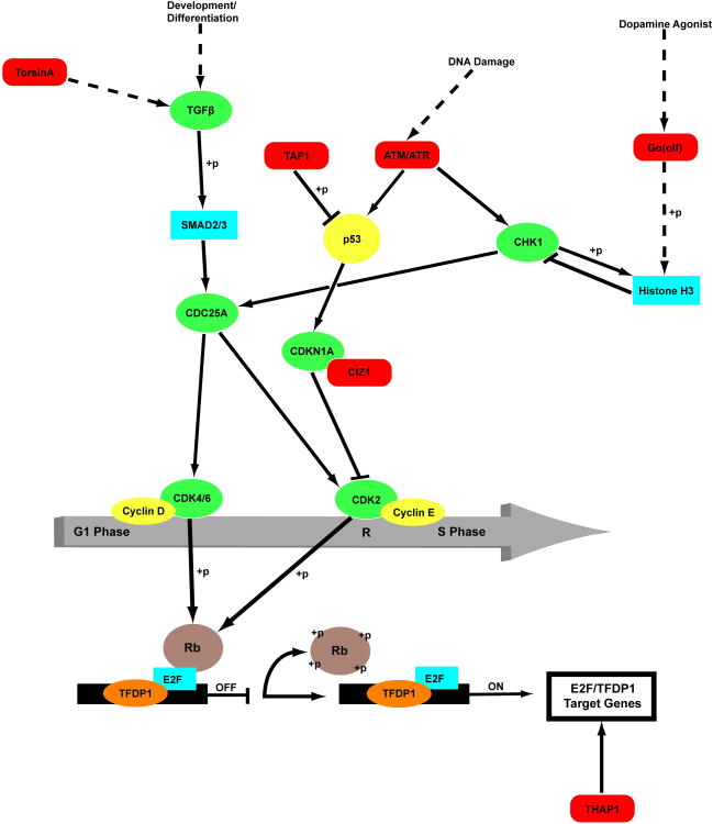
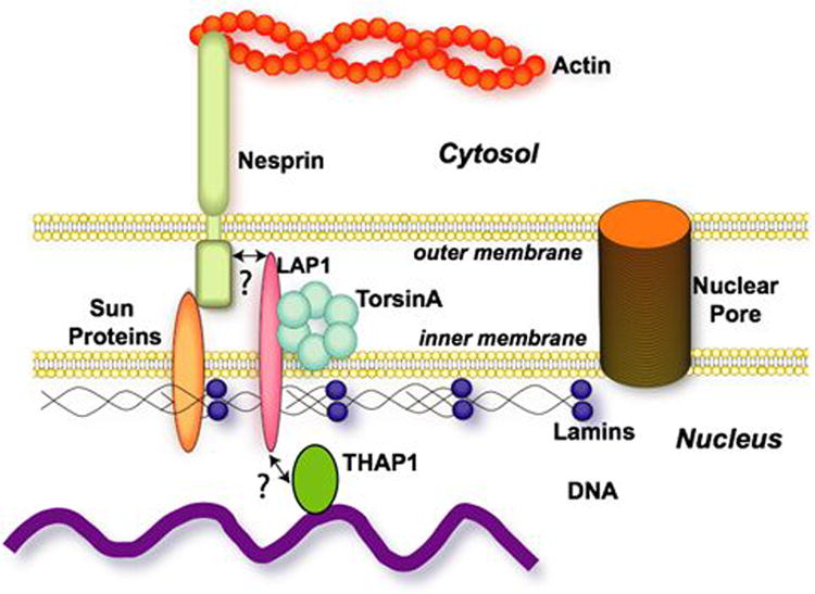
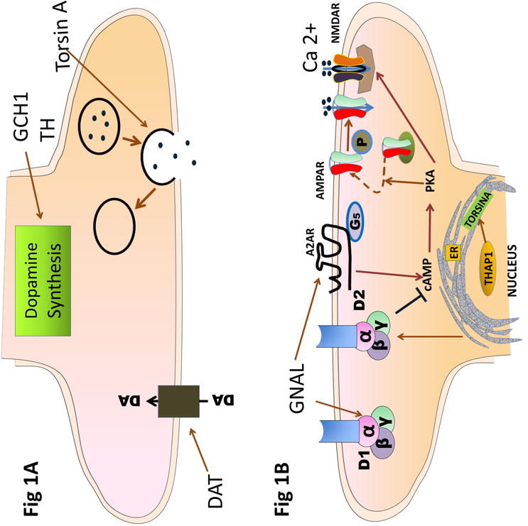
The dystonias are a group of hyperkinetic movement disorders whose principal cause is neuron dysfunction at 1 or more interconnected nodes of the motor system. The study of genes and proteins that cause familial dystonia provides critical information about the cellular pathways involved in this dysfunction, which disrupts the motor pathways at the systems level. In recent years study of the increasing number of DYT genes has implicated a number of cell functions that appear to be involved in the pathogenesis of dystonia. A review of the literature published in English-language publications available on PubMed relating to the genetics and cellular pathology of dystonia was performed. Numerous potential pathogenetic mechanisms have been identified. We describe those that fall into 3 emerging thematic groups: cell-cycle and transcriptional regulation in the nucleus, endoplasmic reticulum and nuclear envelope function, and control of synaptic function. © 2013 Movement Disorder Society.
Keywords: DYT genes; cell cycle; endoplasmic reticulum; nuclear envelope; synaptic function.
© 2013 Movement Disorder Society.
Conflict of interest statement
Relevant conflicts of interest/financial disclosures: Nothing to report.
Figures
References
-
- Phukan J, Albanese A, Gasser T, Warner T. Primary dystonia and dystonia-plus syndromes: clinical characteristics, diagnosis, and pathogenesis. Lancet Neurol. 2011;10:1074–1085. - PubMed
-
- Ozelius L, Kramer P, Page CE, et al. The early-onset torsion dystonia gene (DYT1) encodes an ATP-binding protein. Nature Genet. 1997;17:40–48. - PubMed
-
- Hanson PI, Whiteheart SW. AAA+proteins: have engine will work. Nat Rev Mol Cell Biol. 2005;6:519–529. - PubMed
-
- Kock N, Naismith TV, Boston HE, et al. Effects of genetic variations in the dystonia protein torsinA: identification of polymorphism at residue 216 as protein modifier. Hum Mol Genet. 2006;15:1355–1364. - PubMed
-
- Augood SJ, Penney JB, Friberg IK, et al. Expression of the early onset torsion dystonia gene (DYT1) in human brain. Ann Neurol. 1998;43:669–673. - PubMed
Publication types
MeSH terms
Substances
Grants and funding
LinkOut - more resources
Full Text Sources
Other Literature Sources
