. 2013 Sep 13;288(37):26397-409.
doi: 10.1074/jbc.M113.500223. Epub 2013 Jul 27.

Characterization of the ATPase activity of RecG and RuvAB proteins on model fork structures reveals insight into stalled DNA replication fork repair

Affiliations

Characterization of the ATPase activity of RecG and RuvAB proteins on model fork structures reveals insight into stalled DNA replication fork repair

Syafiq Abd Wahab et al. J Biol Chem. .

Abstract

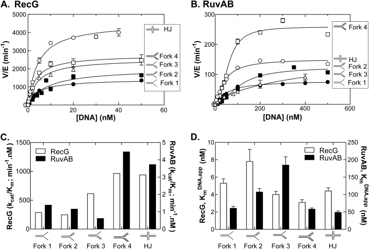
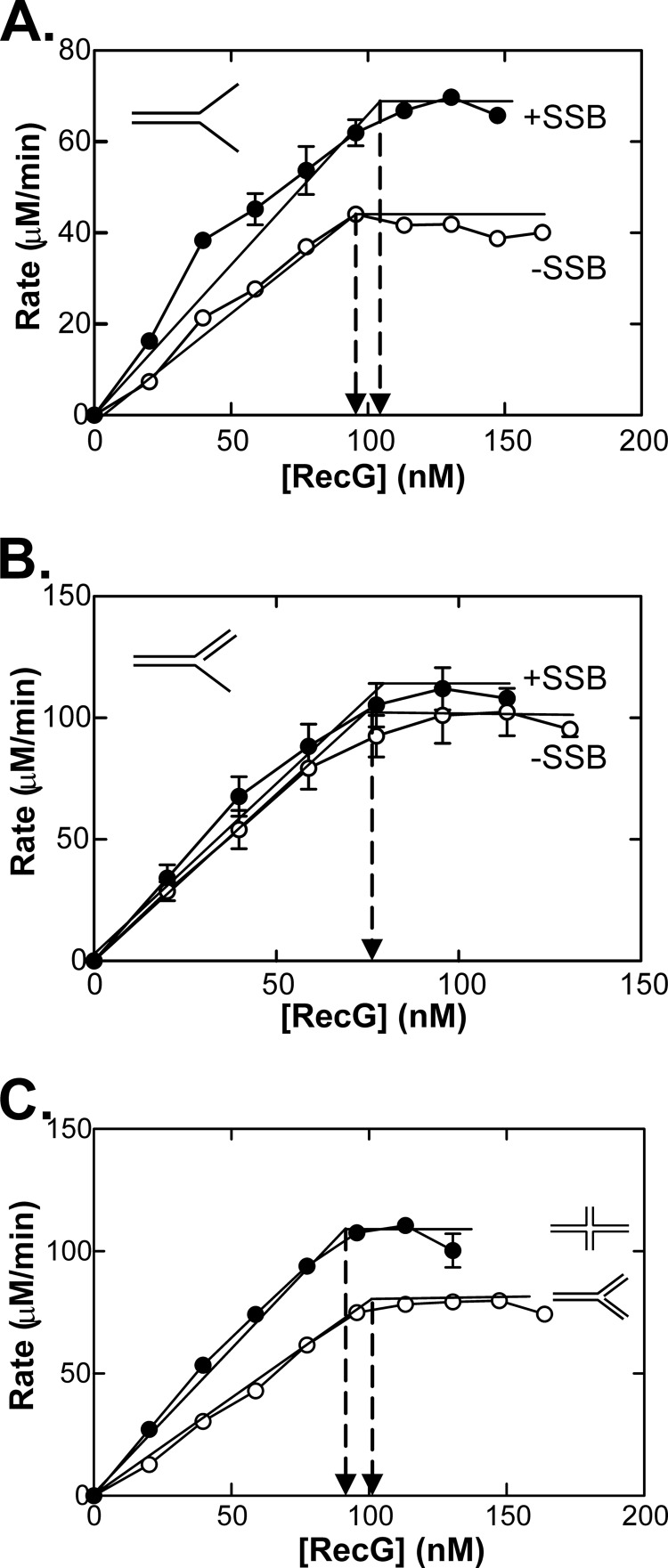
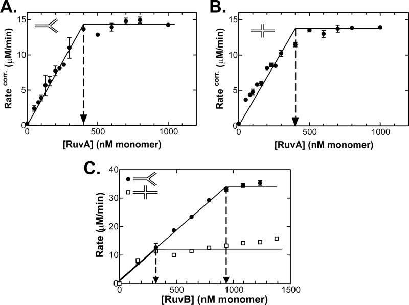
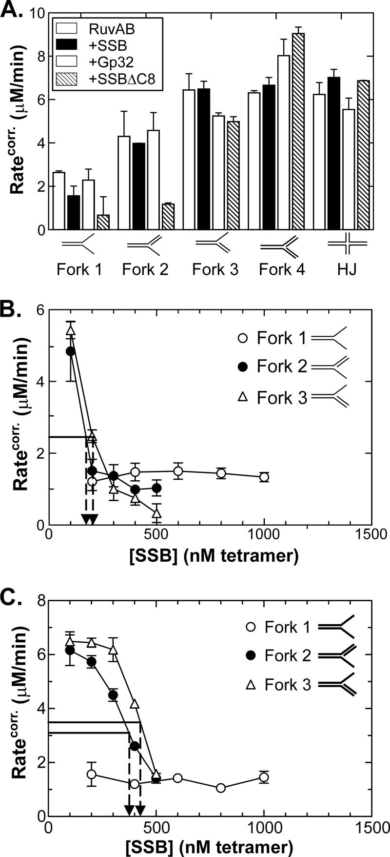
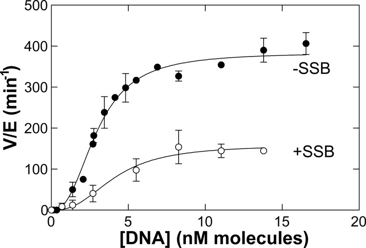
RecG and RuvAB are proposed to act at stalled DNA replication forks to facilitate replication restart. To clarify the roles of these proteins in fork regression, we used a coupled spectrophotometric ATPase assay to determine how these helicases act on two groups of model fork substrates: the first group mimics nascent stalled forks, whereas the second mimics regressed fork structures. The results show that RecG is active on the substrates in group 1, whereas these are poor substrates for RuvAB. In addition, in the presence of group 1 forks, the single-stranded DNA-binding protein (SSB) enhances the activity of RecG and enables it to compete with excess RuvA. In contrast, SSB inhibits the activity of RuvAB on these substrates. Results also show that the preferred regressed fork substrate for RuvAB is a Holliday junction, not a forked DNA. The active form of the enzyme on the Holliday junction contains a single RuvA tetramer. In contrast, although the enzyme is active on a regressed fork structure, RuvB loading by a single RuvA tetramer is impaired, and full activity requires the cooperative binding of two forked DNA substrate molecules. Collectively, the data support a model where RecG is responsible for stalled DNA replication fork regression. SSB ensures that if the nascent fork has single-stranded DNA character RuvAB is inhibited, whereas the activity of RecG is preferentially enhanced. Only once the fork has been regressed and the DNA is relaxed can RuvAB bind to a RecG-extruded Holliday junction.

Keywords: ATPases; DNA Recombination; DNA-Protein Interaction; DNA-binding Protein; Enzyme Kinetics; Fork Regression; Holliday Junction; RecG; Replication Fork; RuvAB.


Figures

FIGURE 1.
FIGURE 1.
The preferred fork substrates for RecG and RuvAB have two duplex arms. A, RecG is more active in the presence of a Holliday junction. B, RuvAB exhibits higher activity on a forked DNA substrate with duplex arms. C, comparison of the catalytic efficiency of each enzyme reveals similar substrate preferences for each DNA helicase. D, RecG binds to model fork substrates more tightly than RuvAB. ATPase assays were performed as described under “Experimental Procedures” and were initiated by the addition of protein. Time courses were analyzed by linear regression to determine reaction rate, and the resulting rates are graphed as a function of DNA concentration. RecG was present at 1.5 nm, and RuvAB was present at 650 nm RuvA and 500 nm RuvB. The data presented are from two to seven experiments per cofactor per enzyme with assays conducted on separate days. For RuvAB, the rates shown have been corrected for the DNA-independent ATPase rate of the enzyme. The nomenclature of the five substrates is indicated with details provided in Table 1. The error bars represent the mean and error.
FIGURE 2.
FIGURE 2.
SSB stabilizes RecG on model fork substrates. ATPase assays were performed as described under “Experimental Procedures” and were initiated by the addition of 1.92 nm RecG. DNA was present at 100 nm molecules, and reactions contained 10 mm MgOAc and 1 mm ATP. In assays with SSB, it was added to the DNA to 250 nm tetramer final concentration prior to reaction initiation by RecG. For each substrate, assays were done in duplicate. The resulting rates were calculated as described under “Experimental Procedures” with the relative activity expressed as a percentage of the hydrolysis rate of RecG in the absence of added NaCl. Arrows indicate the salt titration midpoint for fork 2 in the absence (24 mm) and presence of SSB (103 mm). Only fork 2 is shown for clarity with the STMPs for the remaining substrates shown in Table 4. Open symbols, reactions with RecG only; closed symbols, reactions done in the presence of SSB. The error bars represent the mean and error.
FIGURE 3.
FIGURE 3.
The RecG-DNA stoichiometry is unaffected by the presence of SSB. A, fork 1; B, fork 2; C, Fork 4 and the Holliday junction. Reactions contained 100 nm DNA, 10 mm MgOAc, 1 mm ATP, and stoichiometric ratios of SSB relative to DNA. This was determined to be 200 nm tetramer for fork 1, which has two ssDNA arms, and 100 nm tetramer for fork 2, which has only one ssDNA arm. Reactions were initiated by the addition of RecG. Arrows in each panel indicate the concentration of RecG at which maximum ATPase activity was observed. The error bars represent the mean and error.
FIGURE 4.
FIGURE 4.
A single RuvA tetramer is required for full activity on fork 4 and a Holliday junction. A, a titration of RuvA in the presence of fork 4. The concentration of RuvB was 500 nm monomer. B, a titration of RuvA in the presence of the Holliday junction substrate. The concentration of RuvB was 500 nm monomer. C, separate RuvB titrations in the presence of fork 4 and the Holliday junction. The concentration of RuvA was 400 nm monomer. Reactions contained 100 nm DNA, 10 mm MgOAc, and 1 mm ATP. The error bars represent the mean and error. corr., corrected.
FIGURE 5.
FIGURE 5.
An excess of SSB is required to inhibit the ATPase activity of RuvAB on forks with one ssDNA tail. A, the effects of stoichiometric amounts of single-stranded DNA-binding proteins on the ATPase activity of RuvAB. The concentration of single strand-binding proteins used was 100 nm tetramer for SSB and SSBΔC8 and 100 nm monomer for gp32. B, stoichiometric RuvAB relative to DNA (400 nm RuvA and 800 nm RuvB). C, excess of RuvAB relative to DNA (650 nm RuvA and 500 nm RuvB). In these assays, DNA was present at 100 nm molecules, ATP was present at 1 mm, and MgOAc was present at 10 mm. In all assays, the single-stranded DNA-binding protein was added first followed by a 2-min incubation, then RuvA was added followed by a second 2-min incubation, and finally RuvB was added to initiate reactions. The corrected reaction rates reflect the final rate following subtraction of the RuvAB DNA-independent rate in the presence of each concentration of the single-stranded DNA-binding protein present. The error bars represent the mean and error. corr., corrected.
FIGURE 6.
FIGURE 6.
SSB reduces the catalytic efficiency of RuvAB on ssDNA. ATPase assays were performed as described under “Experimental Procedures” and were initiated by the addition of a preformed complex of RuvAB. The DNA cofactor is M13 ssDNA (10 μm nucleotides), and reactions were done in buffer containing 10 mm MgOAc and 1 mm ATP. In assays with SSB, it was present at 1 μm and was added before RuvAB. Data were fit to the Hill equation (57): V = (Vmax·[DNA]n)/([S0.5]n + [DNA]n). The data presented are from two to four assays per DNA concentration. The error bars represent the mean and error.
FIGURE 7.
FIGURE 7.
SSB helps RecG to compete with RuvA(B) on model fork substrates. A, RecG is modestly inhibited by RuvA. A single concentration of RuvA was used (100 nm tetramer), RecG was present at 2 nm, DNA was present at 100 nm molecules, and reactions contained 1 mm ATP and 10 mm MgOAc. When added last, RuvA was added to an ongoing assay after a steady-state rate of ATP hydrolysis by RecG was observed (typically after 5 min). When RuvA was added first, this was followed by a 5-min incubation prior to initiation by RecG. B, effective inhibition of RecG requires excess RuvA. Reaction conditions were the same as those used in A except increasing amounts of RuvA were added 5 min after a steady-state rate of ATP hydrolysis by RecG was observed. C, RuvA inhibition of RecG is rescued by SSB. Reactions were initiated by addition of 2 nm RecG. Once a steady-state rate was achieved, RuvA was added (455 nm tetramer), and once the lower rate of ATP hydrolysis was established, 250 nm SSB (in tetramers) was added. In reactions where RuvA was added first, it was present at 455 nm tetramer. In reactions initiated by a RecG + SSB mixture, the two proteins were incubated on ice for 30 min prior to addition. D, RuvAB does not inhibit RecG on model fork substrates. In these assays, 100 nm RuvA (in tetramer) was added first to the reaction mixture containing 100 nm junction DNA; 2 min later, 1,200 nm RuvB K68A (monomer) was added followed 2 min later by 2 nm RecG. In reactions where only RecG was present, it was added to reactions following a 2-min preincubation of all components. These experiments were done on the same day using the same tubes of substrates and proteins to eliminate potential complications from variations between preparations. The error bars represent the mean and error.

Similar articles

Cited by

References

    1. Kogoma T. (1997) Stable DNA replication: interplay between DNA replication, homologous recombination, and transcription. Microbiol. Mol. Biol. Rev. 61, 212–238 - PMC - PubMed
    1. Kuzminov A. (1999) Recombinational repair of DNA damage in Escherichia coli and bacteriophage λ. Microbiol. Mol. Biol. Rev. 63, 751–813 - PMC - PubMed
    1. Kowalczykowski S. C. (2000) Initiation of genetic recombination and recombination-dependent replication. Trends Biochem. Sci. 25, 156–165 - PubMed
    1. Seigneur M., Bidnenko V., Ehrlich S. D., Michel B. (1998) RuvAB acts at arrested replication forks. Cell 95, 419–430 - PubMed
    1. Cox M. M., Goodman M. F., Kreuzer K. N., Sherratt D. J., Sandler S. J., Marians K. J. (2000) The importance of repairing stalled replication forks. Nature 404, 37–41 - PubMed

Publication types

LinkOut - more resources