

The effects of methamphetamine self-administration on cortical monoaminergic deficits induced by subsequent high-dose methamphetamine administrations
- PMID: 23893609
- PMCID: PMC3962656
- DOI: 10.1002/syn.21696
The effects of methamphetamine self-administration on cortical monoaminergic deficits induced by subsequent high-dose methamphetamine administrations
Abstract
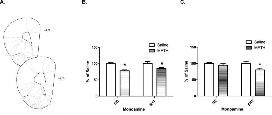
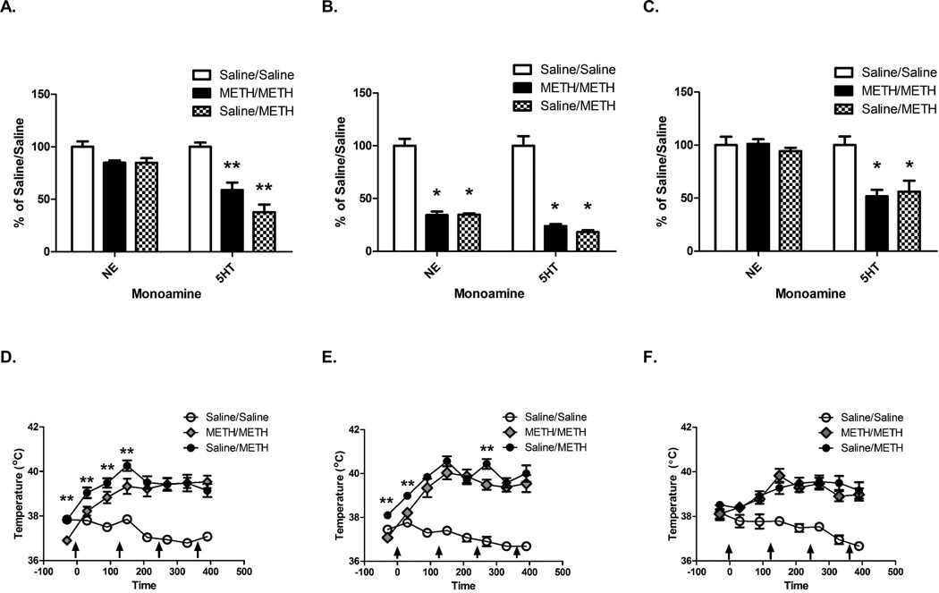
Preclinical models suggest that repeated high-dose methamphetamine (METH) exposures, administered in a "binge-like" pattern, acutely decrease norepinephrine (NE), and acutely and persistently decrease serotonin (5-hydroxytryptamine; 5HT) content in the frontal cortex. However, the impact of METH self-administration on this region is unknown. Because of the importance of the monoaminergic neurons in the frontal cortex to a variety of cognitive and addictive processes, effects of METH self-administration on cortical NE and 5HT content were assessed. Results revealed several novel findings. First, METH self-administration decreased cortical NE content as assessed 24 h after last exposure. Consistent with previous preclinical reports after a binge METH regimen, this decrease was reversed 8 days after the final METH exposure. Second, and in contrast to our previous reports involving the hippocampus or striatum, METH self-administration caused persistent decreases in 5HT content as assessed 8 days after the final METH exposure. Of note, the magnitude of this decrease (≈ 20%) was less than that observed typically after a binge METH treatment. Third, prior METH self-administration attenuated METH-induced serotonergic deficits as assessed 7 days, but not 1 h, following a neurotoxic METH regimen. No protection was observed when the binge exposure occurred 15 days after the last self-administration session. Taken together, these data demonstrate important and selective alterations in cortical serotonergic neuronal function subsequent to METH self-administration. These data provide a foundation to investigate complex questions involving "resistance" to the persistent deficits caused by neurotoxic METH exposure and frontal cortical function.
Keywords: cortex; methamphetamine; norepinephrine; self-administration; serotonin.
Copyright © 2013 Wiley Periodicals, Inc.
Figures
Similar articles
-
Prior methamphetamine self-administration attenuates serotonergic deficits induced by subsequent high-dose methamphetamine administrations.Drug Alcohol Depend. 2012 Nov 1;126(1-2):87-94. doi: 10.1016/j.drugalcdep.2012.04.020. Epub 2012 May 28. Drug Alcohol Depend. 2012. PMID: 22647900 Free PMC article.
-
Prior methamphetamine self-administration attenuates the dopaminergic deficits caused by a subsequent methamphetamine exposure.Neuropharmacology. 2015 Jun;93:146-54. doi: 10.1016/j.neuropharm.2015.01.013. Epub 2015 Jan 31. Neuropharmacology. 2015. PMID: 25645392 Free PMC article.
-
Methamphetamine self-administration causes persistent striatal dopaminergic alterations and mitigates the deficits caused by a subsequent methamphetamine exposure.J Pharmacol Exp Ther. 2012 Feb;340(2):295-303. doi: 10.1124/jpet.111.188433. Epub 2011 Oct 27. J Pharmacol Exp Ther. 2012. PMID: 22034657 Free PMC article.
-
Methamphetamine self-administration attenuates hippocampal serotonergic deficits: role of brain-derived neurotrophic factor.Int J Neuropsychopharmacol. 2014 Aug;17(8):1315-20. doi: 10.1017/S1461145714000327. Epub 2014 Mar 20. Int J Neuropsychopharmacol. 2014. PMID: 24650575 Free PMC article.
-
Methamphetamine self-administration is associated with persistent biochemical alterations in striatal and cortical dopaminergic terminals in the rat.PLoS One. 2010 Jan 20;5(1):e8790. doi: 10.1371/journal.pone.0008790. PLoS One. 2010. PMID: 20098750 Free PMC article.
Cited by
-
Chronic methamphetamine self-administration dysregulates 5-HT2A and mGlu2 receptor expression in the rat prefrontal and perirhinal cortex: Comparison to chronic phencyclidine and MK-801.Pharmacol Biochem Behav. 2018 Dec;175:89-100. doi: 10.1016/j.pbb.2018.09.007. Epub 2018 Sep 18. Pharmacol Biochem Behav. 2018. PMID: 30240581 Free PMC article.
-
Behavioral and Serotonergic Changes in the Frontal Cortex Following Methamphetamine Self-Administration.Int J Neuropsychopharmacol. 2018 Aug 1;21(8):758-763. doi: 10.1093/ijnp/pyy044. Int J Neuropsychopharmacol. 2018. PMID: 29762664 Free PMC article.
References
-
- Bowyer JF, Ali S. High doses of methamphetamine that cause disruption of the blood-brain barrier in limbic regions produce extensive neuronal degeneration in mouse hippocampus. Synapse. 2006;60:521–532. - PubMed
-
- Bowyer JF, Robinson B, Ali S, Schmued LC. Neurotoxic-related changes in tyrosine hydroxylase, microglia, myelin, and the blood-brain barrier in the caudate-putamen from acute methamphetamine exposure. Synapse. 2008;62:193–204. - PubMed
-
- Brennan KA, Colussi-Mas J, Carati C, Lea RA, Fitzmaurice PS, Schenk S. Methamphetamine self-administration and the effect of contingency on monoamine and metabolite tissue levels in the rat. Brain Res. 2010;1317:137–146. - PubMed
Publication types
MeSH terms
Substances
Grants and funding
LinkOut - more resources
Full Text Sources
Other Literature Sources
Medical