. 2013 Jul 24;8(7):e68649.
doi: 10.1371/journal.pone.0068649. Print 2013.

New Australovenator hind limb elements pertaining to the holotype reveal the most complete Neovenatorid leg

Affiliations

New Australovenator hind limb elements pertaining to the holotype reveal the most complete Neovenatorid leg

Matt A White et al. PLoS One. .

Abstract

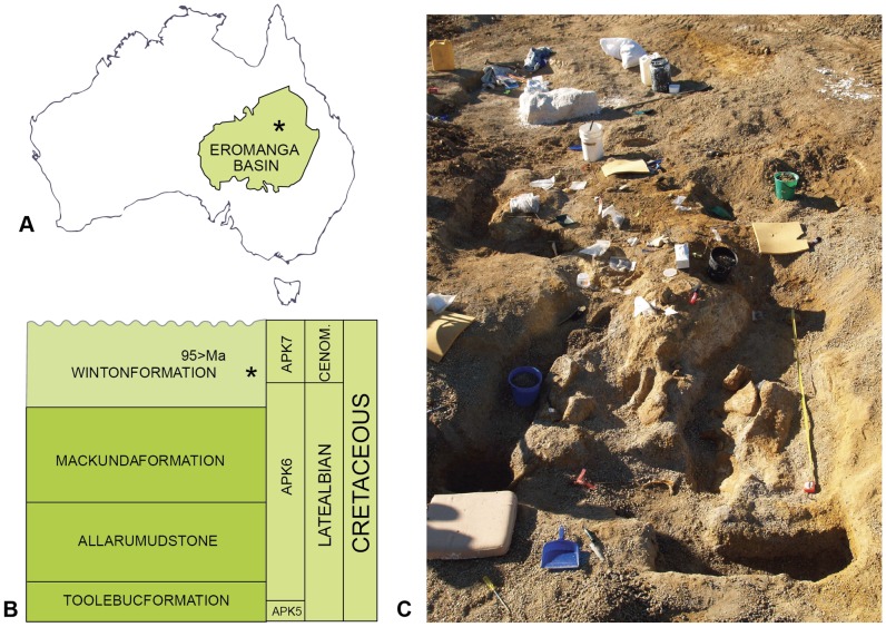
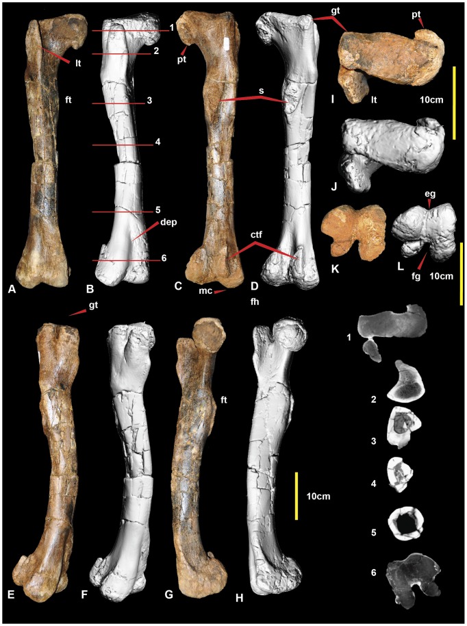
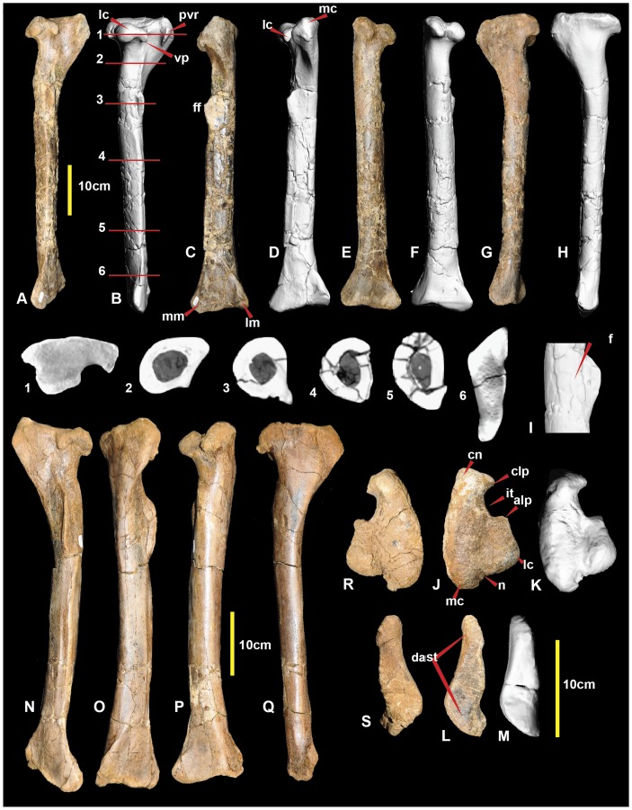
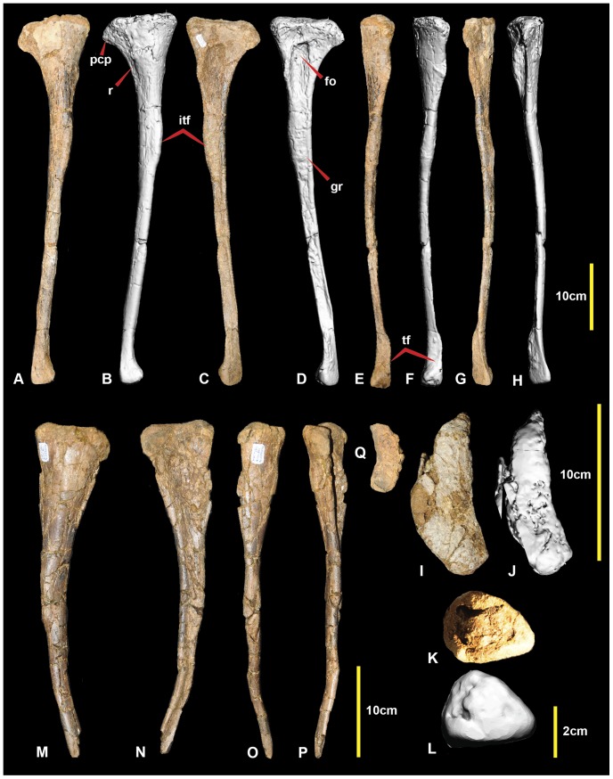
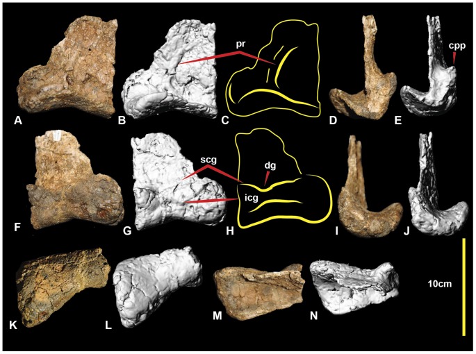
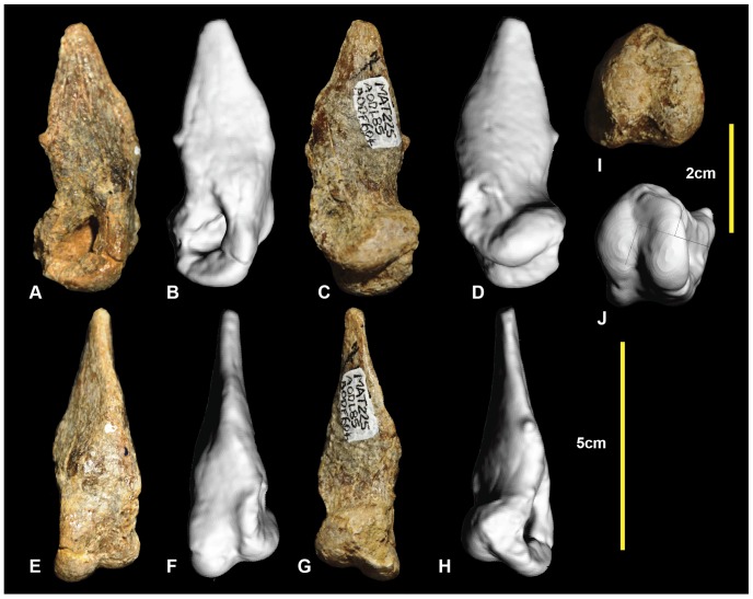
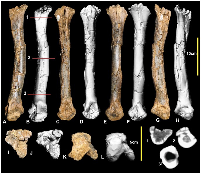
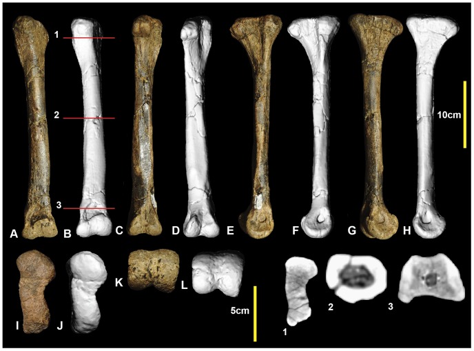
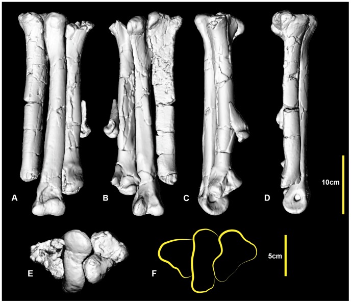
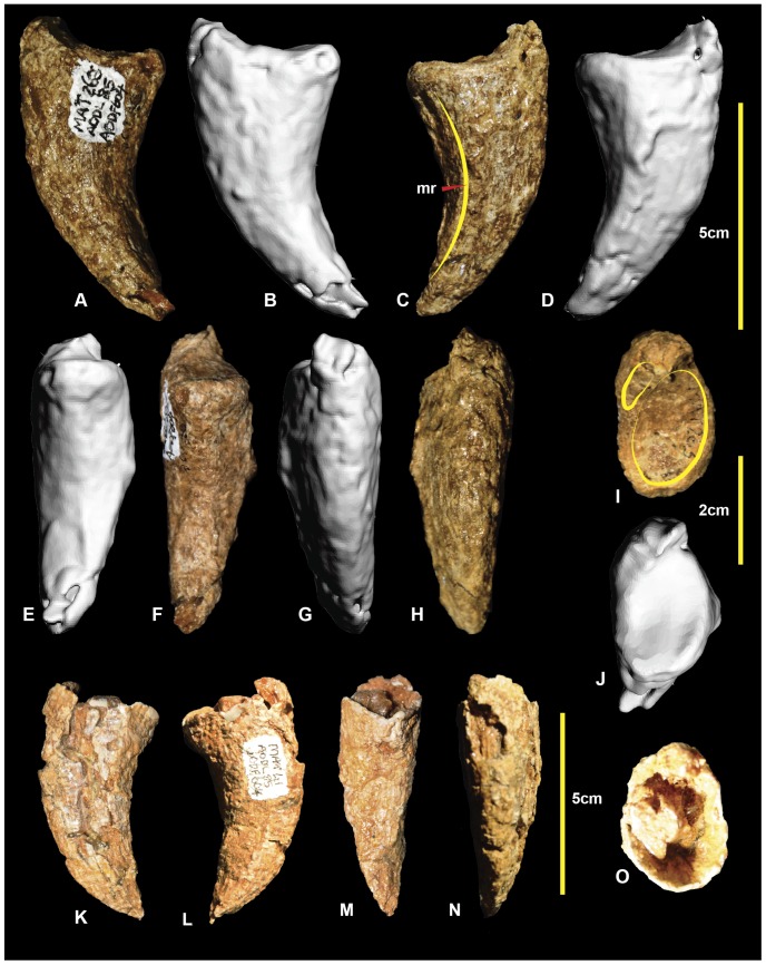
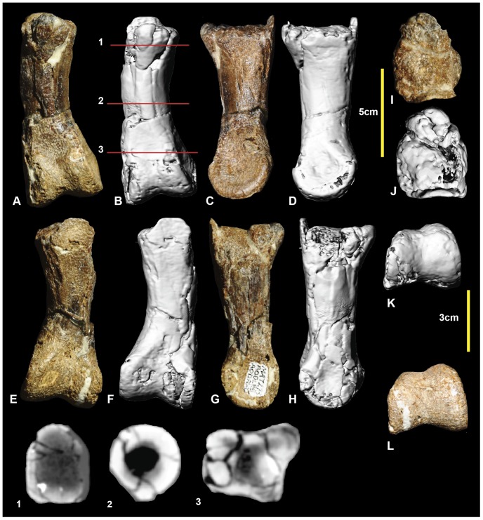
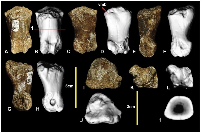
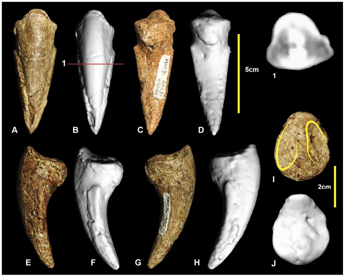
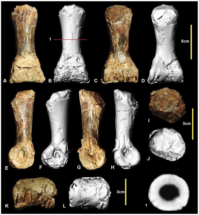
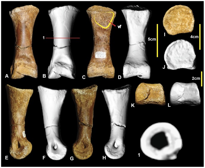
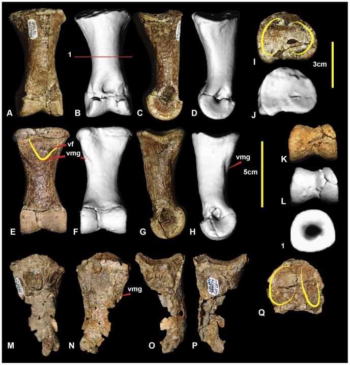
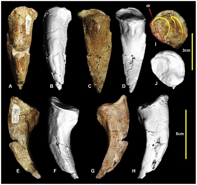
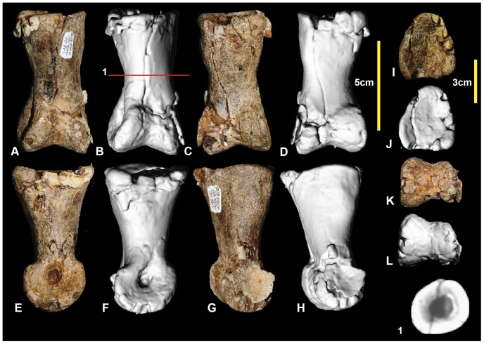
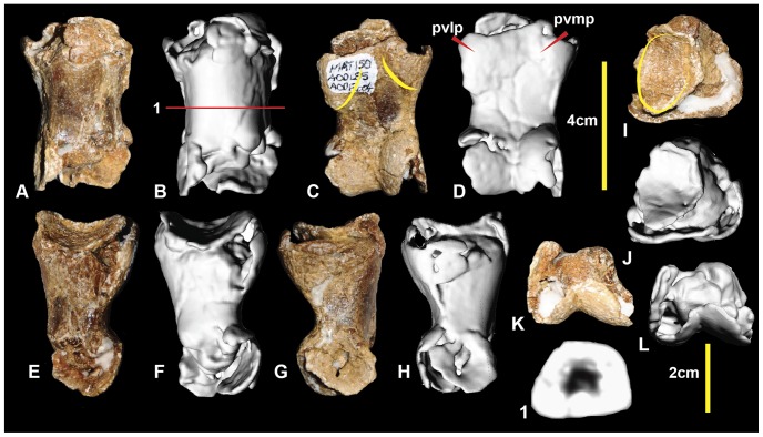
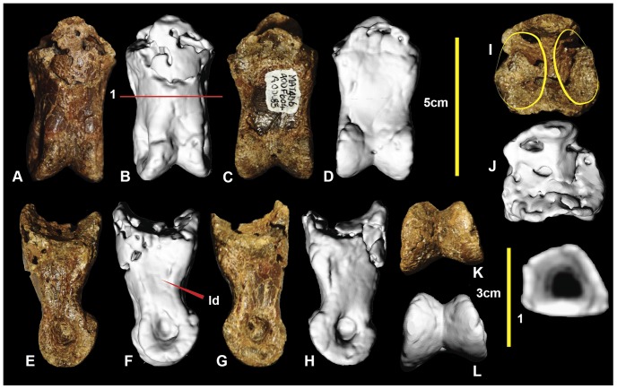
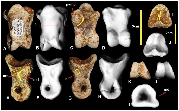
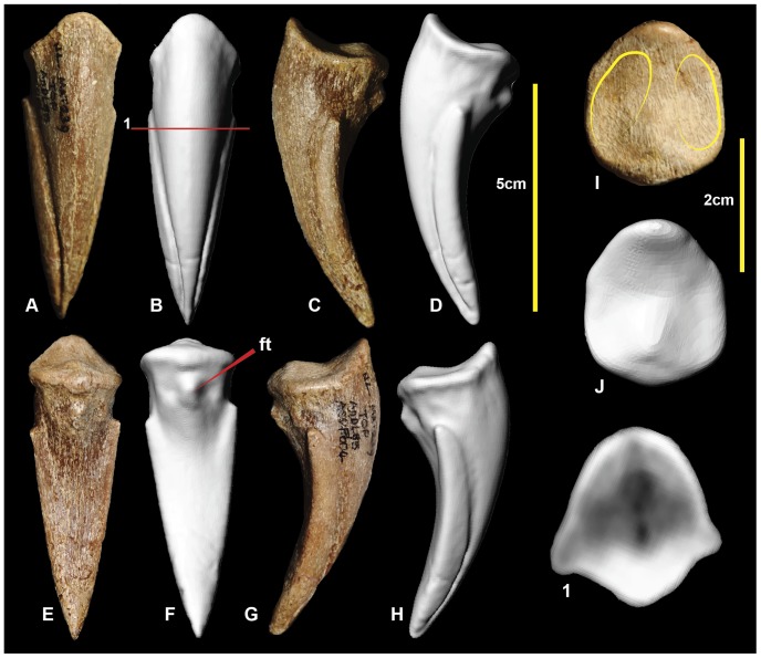
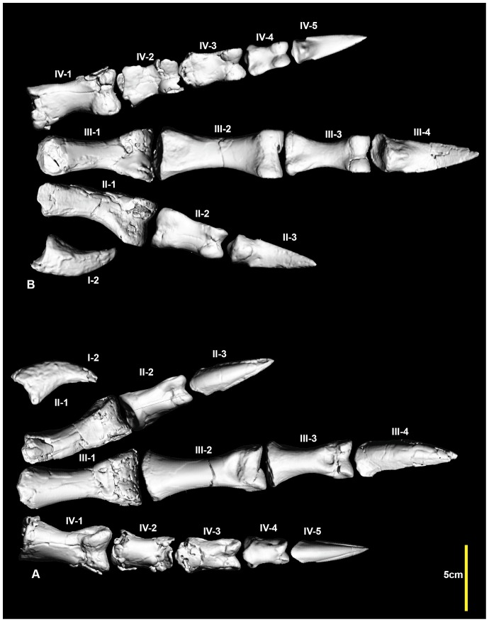
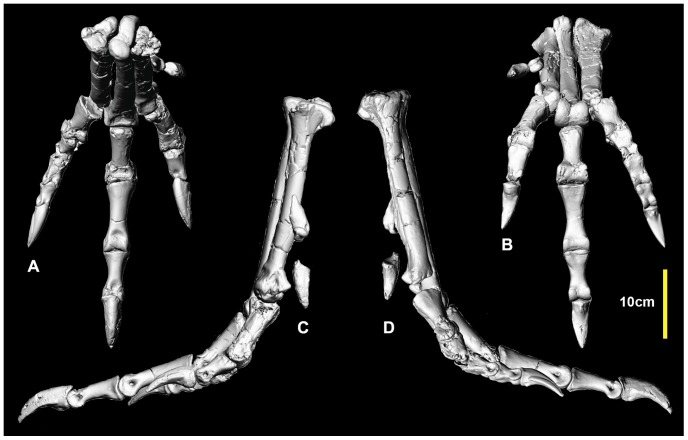
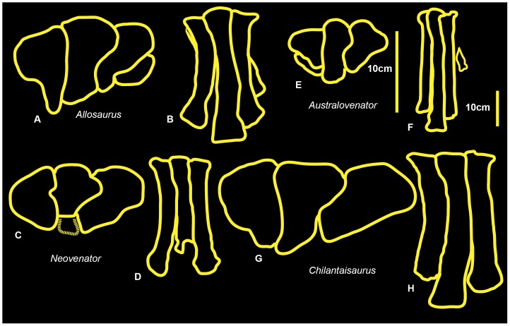
We report new skeletal elements pertaining to the same individual which represents the holotype of Australovenator wintonensis, from the 'Matilda Site' in the Winton Formation (Upper Cretaceous) of western Queensland. The discovery of these new elements means that the hind limb of Australovenator is now the most completely understood hind limb among Neovenatoridae. The new hind limb elements include: the left fibula; left metatarsal IV; left pedal phalanges I-2, II-1, III-4, IV-2, IV-3; and right pedal phalanges, II-2 and III-1. The detailed descriptions are supported with three dimensional figures. These coupled with the completeness of the hind limb will increase the utility of Australovenator in comparisons with less complete neovenatorid genera. These specimens and the previously described hind limb elements of Australovenator are compared with other theropods classified as neovenatorids (including Neovenator, Chilantaisaurus, Fukuiraptor, Orkoraptor and Megaraptor). Hind limb length proportion comparisons indicate that the smaller neovenatorids Australovenator and Fukuiraptor possess more elongate and gracile hind limb elements than the larger Neovenator and Chilantaisaurus. Greater stride lengths to body size exist in both Fukuiraptor and Australovenator with the femur discovered to be proportionally shorter the rest of the hind limb length. Additionally Australovenator is identified as possessing the most elongate metatarsus. The metatarsus morphology varies with body size. The larger neoventorids possess a metatarsus with greater width but shorter length compared to smaller forms.


Conflict of interest statement

Competing Interests: SJW is an employee of Queensland X-Ray. There are no patents, products in development or marketed products to declare. This does not alter the authors' adherence to all the PLOS ONE policies on sharing data and materials.

Figures

Figure 1
Figure 1. Locality and dig site.
Australian Age of Dinosaur locality ‘Matilda site’. The Eromanga basin and site locality (A), formation and age (B), dig site (C).
Figure 2
Figure 2. Right femur.
Right femur in: cranial (A & B); caudal (C & D); lateral (E & F); medial (G & H); proximal (I & J); and distal (K & L) views. Abbreviations: ctf, crista tibiofibularis; dep, depression; eg, extensor groove; fg, flexor groove; ft, fourth trochanter; gt, greater trochanter; lt, lesser trochanter; mc, medial condyle; cf, caudal flange on caput; s, scar; 1-6 CT sections.
Figure 3
Figure 3. Right and left tibia.
Right tibia in: lateral (A & B); cranial (C & D); caudal (E & F); and medial (G & H) views; close up of fibular flange with fossa (I), right tibia in: proximal (J & K); and distal (L & M) views. Left tibia in: lateral (N); cranial (O); caudal (P); medial (Q); proximal (R); and distal (S) views. Abbreviations: alp, anterolateral process; clp, cranio-lateral process; cn, cnemial crest; dast, distal astragular facet; f, fossa; ff, fibular flange; it, incisura tibialis; lc, lateral condyle; lm, lateral malleolus; mc, medial condyle; mm, medial malleolus; n, notch; pvr, postero-ventral ridge; vp, ventral spine-like process.
Figure 4
Figure 4. Right and left fibula.
Right fibula in: medial (A & B); lateral (C & D); caudal (E & F); cranial (G & H); proximal (I & J); and distal (K & L) views. Left fibula in: medial (M); lateral (N); caudal (O); cranial (P); and proximal (Q) views. Abbreviations: fo, fossa; gr, groove; itf, attachment area for interosseum tibiofibulare ligaments; pcp, proximo-cranial process; r, ridge; tf, tibial facet.
Figure 5
Figure 5. Right astragalus.
Right astragalus in: caudal (A, B & C); lateral (D & E); cranial (F, G, H); medial (I & J); distal (K & L); and proximal (M & N) views. Abbreviations: cpp, cranio-proximal process; dg, distal groove; icg, inferior cranial groove; scg, superior cranial groove; pr, caudal ridge.
Figure 6
Figure 6. Left metatarsal I.
Left metatarsal I in: lateral (A & B); medial (C & D); ventral (E & F); dorsal (G & H); and distal (I & J) views.
Figure 7
Figure 7. Right metatarsal II.
Right metatarsal II in: dorsal (A & B); lateral (C & D); medial (E & F); ventral (G & H); proximal (I & J); and distal (K & L) views; and metatarsal II cross-sections (1–3).
Figure 8
Figure 8. Right metatarsal III.
Right metatarsal III in: dorsal (A & B); ventral (C & D); lateral (E & F); medial (G & H); proximal (I & J); and distal (K & L) views; and and metatarsal III cross-sections (1–5).
Figure 9
Figure 9. Left metatarsal IV.
Left metatarsal IV in: ventral (A & B); dorsal (C & D); lateral (E & F); medial (G & H); proximal (I & J); and distal (K) views; and metatarsal IV cross-sections (1–3).
Figure 10
Figure 10. Reconstructed right metatarsus.
Reconstructed right metatarsus in: dorsal (A); ventral (B); medial (C); lateral (D); and proximal (E, F) views.
Figure 11
Figure 11. Left and right pedal phalanges I-2.
Left pedal phalanx I-2 in: lateral (A & B); medial (C & D); ventral (E & F); dorsal (G & H); and proximal (I & J) views. Right pedal phalanx I-2 in; medial (K); lateral (L); ventral (M); dorsal (N); and proximal (O) views.
Figure 12
Figure 12. Left pedal phalanx II-1.
Left pedal phalanx II-1 in: dorsal (A & B); medial (C & D); ventral (E & F); lateral (G & H); proximal (I & J); and distal (K & L) views; and left pedal phalanx II-1 cross-sections (1–3).
Figure 13
Figure 13. Left pedal phalanx II-2.
Left pedal phalanx II-2 in: dorsal (A, B); ventral (C, D); lateral (E, F); medial (G, H); proximal (I, J); and distal (K, L) views; and left pedal phalanx II-2 cross-section (1).
Figure 14
Figure 14. Right pedal phalanx II-3.
Right pedal phalanx II-3 in: dorsal (A & B); ventral (C & D); medial (E & F); lateral (G & H); and proximal (I & J) views; and right pedal phalanx II-3 ungual cross-section (1).
Figure 15
Figure 15. Right pedal phalanx III-1.
Right pedal phalanx III-1 in: dorsal (A & B); ventral (C & D); lateral (E & F); medial (G & H); proximal (I & J); and distal (K & L) views; and right pedal phalanx III-1 mid-shaft cross-section (1).
Figure 16
Figure 16. Right pedal phalanx III-2.
Right pedal phalanx III-2 in: dorsal (A & B); ventral (C & D); medial (E & F); lateral (G & H); proximal (I & J); and distal (K & L) views; and right pedal phalanx III-2 mid-shaft cross-section (1). Abbreviation: vf, ventral facet.
Figure 17
Figure 17. Right and left pedal phalanges III-3.
Right pedal phalanx III-3 in: dorsal (A & B); lateral (C & D); ventral (E & F); medial (G & H); proximal (I & J); and distal (K & L) views; and right pedal phalanx III-3 cross-section (1). Left pedal phalanx in: dorsal (M); ventral (N); lateral (O); medial (P); and proximal (Q) views.
Figure 18
Figure 18. Left pedal phalanx III-4.
Left pedal phalanx III-4 in: dorsal (A & B); ventral (C & D); lateral (E & F); medial (G & H); and proximal (I & J) views; and pedal phalanx III-4 cross-section (1).
Figure 19
Figure 19. Left pedal phalanx IV-1.
Left pedal phalanx IV-1 in: dorsal (A & B); ventral (C & D); medial (E & F); lateral (G & H); proximal (I & J); and distal (K & L) views; and left pedal phalanx IV-1 cross-section (1).
Figure 20
Figure 20. Left pedal phalanx IV-2.
Left pedal phalanx IV-2 in: dorsal (A & B); ventral (C & D); lateral (E & F); medial (G & H); proximal (I & J); and distal (K & L) views; and left pedal phalanx IV-2 cross-section (1). Abbreviations: pvlp, proximal ventral lateral process; pvmp, proximal ventral medial process.
Figure 21
Figure 21. Left pedal phalanx IV-3.
Left pedal phalanx IV-3 in: dorsal (A & B); ventral (C & D); lateral (E & F); medial (G & H); proximal (I & J); and distal (K & L) views; and left pedal phalanx IV-3 cross-section (1). Abbreviation: ld, lateral depression.
Figure 22
Figure 22. Right pedal phalanx IV-4.
Right pedal phalanx IV-4 in: dorsal (A & B); ventral (C & D); medial (E & F); lateral (G & H); proximal (I & J); and distal (K & L) views; and right pedal phalanx IV-4 cross-section (1). Abbreviations: ld, lateral depression; md, medial depression; mr, medial ridge; pvmp, proximal ventral medial process.
Figure 23
Figure 23. Right pedal phalanx IV-5.
Right pedal phalanx IV-5 in: dorsal (A & B); medial (C & D); ventral (E & F); lateral (G & H); and proximal (I & J) views; and right pedal phalanx IV-5 cross-section (1). Abbreviation: ft, flexor tubercle.
Figure 24
Figure 24. Reconstructed right pes.
Reconstructed right pes in: dorsal (A); and ventral (B) views.
Figure 25
Figure 25. Reconstructed right metatarsus and pes.
Reconstructed right metatarsus and pes in: dorsal (A); ventral (B); medial (C) and lateral (D) views.
Figure 26
Figure 26. Right metatarsus comparisons.
Allosaurus in: proximal (A); and dorsal (B) views. Neovenator in: proximal (C) and dorsal (D) views. Australovenator in: proximal (E) and dorsal (F). Chilantaisaurus in: proximal (G) and dorsal (H).

References

    1. Hocknull SA, White MA, Tischler TR, Cook AG, Calleja ND et al... (2009) New Mid Cretaceous (Latest Albian) Dinosaurs from Winton, Queensland, Australia. PLoS ONE 4(7) e6190. doi:10.1371/journal.pone.0006190. - PMC - PubMed
    1. Bryan SE, Cook AG, Allen CM, Siegel C, Purdy DJ, et al. (2012) Early – mid Cretaceous tetonic evolution of eastern Gondwana: From silicic LIP to continental rupture. Episodes Special Issue, Oceania Geology 35(1): 142–152.
    1. White MA, Falkingham PL, Cook AG, Hocknull SA, Elliott DA (2013) Morphological comparisons of metacarpal I for Australovenator wintonensis and Rapator ornitholestoides: implications for their taxonomic relationships. Alcheringa 37: 1–7.
    1. Dettmann ME, Clifford HT (2000) Gemmae of the Marchantiales from the Winton Formation (mid-Cretaceous), Eromanga Basin, Queensland. Memoirs of the Queensland Museum 45: 285–292.
    1. McLoughlin S, Drinnan AN, Rozefelds AC (1995) A Cenomanian flora from the Winton Formation, Eromanga Basin, Queensland, Australia. Memoirs of the Queensland Museum 38: 273–313.

Publication types

MeSH terms

LinkOut - more resources