

Delivery of therapeutic AGT shRNA by PEG-Bu for hypertension therapy
- PMID: 23894329
- PMCID: PMC3716693
- DOI: 10.1371/journal.pone.0068651
Delivery of therapeutic AGT shRNA by PEG-Bu for hypertension therapy
Abstract
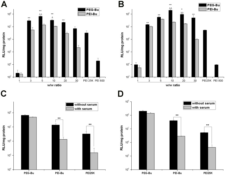
Gene silencing by RNA interference (RNAi) is a promising approach for gene therapy. However, up to today, it is still a major challenge to find safe and efficient non-viral vectors. Previously, we reported PEI-Bu, a small molecular weight PEI derivative, as an efficient non-viral carrier. However, like many PEI-based polymers, PEI-Bu was too toxic. In order to reduce cytotoxicity while maintain or even enhance transfecion efficiency, we modified PEI-Bu with poly(ethylene glycol) (PEG) to obtain PEG-Bu, and used it to delivery a theraputic short hairpin RNA (shRNA) targeting angiotensinogen (AGT) to normal rat liver cells (BRL-3A), which was a key target for the treatment of hypertension. The structure of PEG-Bu was confirmed by proton nuclear magnetic resonance ((1)H-NMR). Gel permeation chromatography (GPC) showed that the weight average molecular weight (Mw) of PEG-Bu was 5880 Da, with a polydispersity of 1.58. PEG-Bu could condense gene cargo into spherical and uniform nanoparticles with particle size (65-88 nm) and zeta potential (7.3-9.6 mV). Interestingly and importantly, PEG-Bu displayed lower cytotoxicity and enhanced tranfection efficiency than PEI-Bu after PEGylation in both normal cells BRL-3A and tumor cells HeLa. Moreover, PEG-Bu could efficiently delivery AGT shRNA to knockdown the AGT expression. To sum up, PEG-Bu would be a promising non-viral vector for delivering AGT shRNA to BRL-3A cells for hypertension therapy.
Conflict of interest statement
Figures


Similar articles
-
Hepatocyte-targeting gene transfer mediated by galactosylated poly(ethylene glycol)-graft-polyethylenimine derivative.Drug Des Devel Ther. 2013;7:211-21. doi: 10.2147/DDDT.S42582. Epub 2013 Mar 26. Drug Des Devel Ther. 2013. PMID: 23576866 Free PMC article.
-
Synthesis of poly(ethylene glycol)-g-chitosan-g-poly(ethylene imine) co-polymer and in vitro study of its suitability as a gene-delivery vector.J Biomater Sci Polym Ed. 2010;21(6-7):741-58. doi: 10.1163/156856209X437941. J Biomater Sci Polym Ed. 2010. PMID: 20482982
-
Poly(ethylene glycol) analogs grafted with low molecular weight poly(ethylene imine) as non-viral gene vectors.Acta Biomater. 2010 Jul;6(7):2650-7. doi: 10.1016/j.actbio.2010.01.022. Epub 2010 Jan 28. Acta Biomater. 2010. PMID: 20114089
-
Polyethylenimine as a promising vector for targeted siRNA delivery.Curr Clin Pharmacol. 2012 May;7(2):121-30. doi: 10.2174/157488412800228857. Curr Clin Pharmacol. 2012. PMID: 22432843 Review.
-
Exploratory Studies on RNAi-Based Therapies Targeting Angiotensinogen in Hypertension: Scoping Review.J Pers Med. 2024 Dec 25;15(1):3. doi: 10.3390/jpm15010003. J Pers Med. 2024. PMID: 39852196 Free PMC article. Review.
Cited by
-
Progresses towards safe and efficient gene therapy vectors.Oncotarget. 2015 Oct 13;6(31):30675-703. doi: 10.18632/oncotarget.5169. Oncotarget. 2015. PMID: 26362400 Free PMC article. Review.
-
Nanoparticles in the diagnosis and treatment of vascular aging and related diseases.Signal Transduct Target Ther. 2022 Jul 11;7(1):231. doi: 10.1038/s41392-022-01082-z. Signal Transduct Target Ther. 2022. PMID: 35817770 Free PMC article. Review.
-
Polymeric Nanostructures for Imaging and Therapy.Chem Rev. 2015 Oct 14;115(19):10967-1011. doi: 10.1021/acs.chemrev.5b00135. Epub 2015 Aug 4. Chem Rev. 2015. PMID: 26463640 Free PMC article. Review. No abstract available.
-
Niclosamide suppresses migration of hepatocellular carcinoma cells and downregulates matrix metalloproteinase-9 expression.Oncol Lett. 2015 Dec;10(6):3515-3518. doi: 10.3892/ol.2015.3789. Epub 2015 Oct 9. Oncol Lett. 2015. PMID: 26788160 Free PMC article.
References
-
- Xi B, Shen Y, Yan Y, Mi J (2012) Association of polymorphisms in the AGT gene with essential hypertension in the Chinese population. J Renin Angiotensin Aldosterone Syst 13: 282–288. - PubMed
-
- Ishigami T, Umemura S, Tamura K, Hibi K, Nyui N, et al. (1997) Essential hypertension and 5' upstream core promoter region of human angiotensinogen gene. Hypertension 30: 1325–30. - PubMed
-
- Tanimoto K, Sugiyama F, Goto Y, Ishida J, Takimoto E, et al. (1994) Angiotensinogen-deficient mice with hypotension. J Biol Chem 269: 31334–31337. - PubMed
-
- Ashihara E, Kawata E, Maekawa T (2010) Future prospect of RNA interference for cancer therapies. Curr Drug Targets 11: 345–360. - PubMed
Publication types
MeSH terms
Substances
LinkOut - more resources
Full Text Sources
Other Literature Sources
Medical
Miscellaneous
