. 2013 Jul 23;8(7):e68879.
doi: 10.1371/journal.pone.0068879. Print 2013.

Magnetic nanoparticles as mediators of ligand-free activation of EGFR signaling

Affiliations

Magnetic nanoparticles as mediators of ligand-free activation of EGFR signaling

Atul A Bharde et al. PLoS One. .

Abstract

Background: Magnetic nanoparticles (NPs) are of particular interest in biomedical research, and have been exploited for molecular separation, gene/drug delivery, magnetic resonance imaging, and hyperthermic cancer therapy. In the case of cultured cells, magnetic manipulation of NPs provides the means for studying processes induced by mechanotransduction or by local clustering of targeted macromolecules, e.g. cell surface receptors. The latter are normally activated by binding of their natural ligands mediating key signaling pathways such as those associated with the epidermal growth factor (EGFR). However, it has been reported that EGFR may be dimerized and activated even in the absence of ligands. The present study assessed whether receptor clustering induced by physical means alone suffices for activating EGFR in quiescent cells.

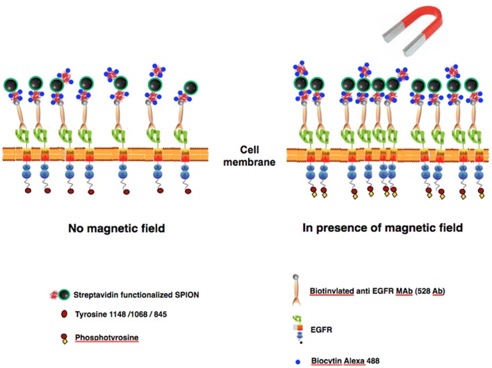
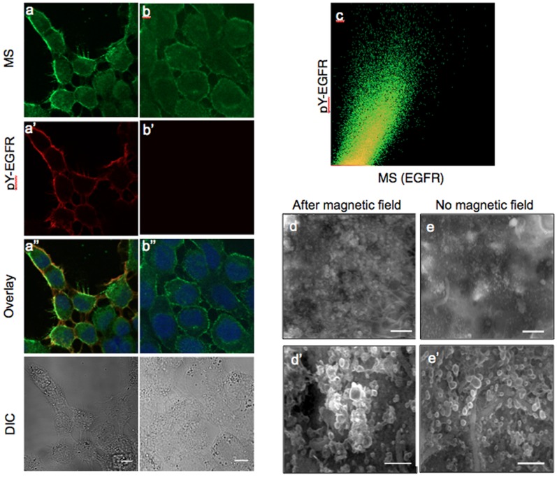
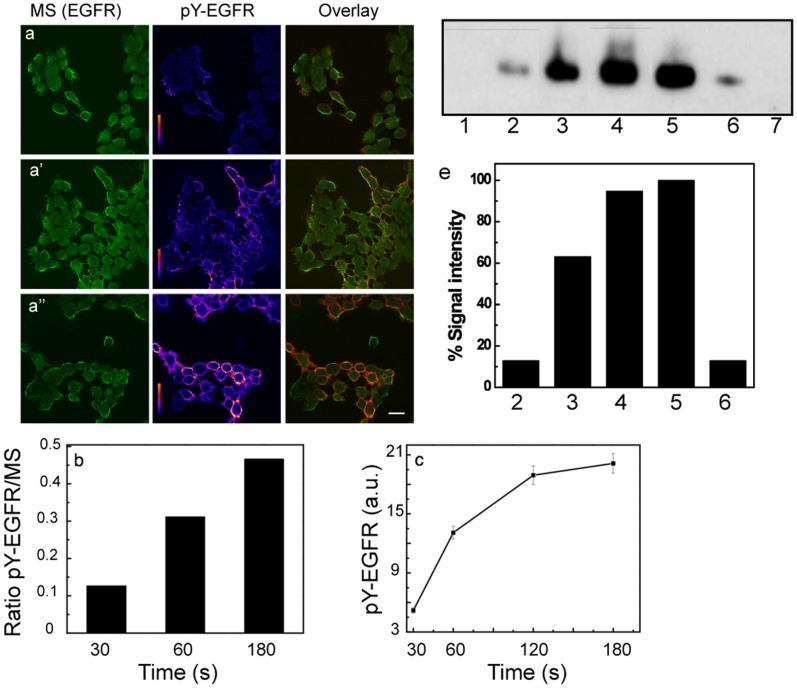
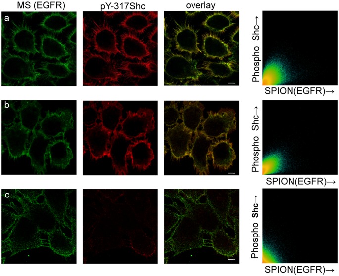
Methodology/principal findings: The EGFR on A431 cells was specifically targeted by superparamagnetic iron oxide NPs (SPIONs) carrying either a ligand-blocking monoclonal anti-EGFR antibody or a streptavidin molecule for targeting a chimeric EGFR incorporating a biotinylated amino-terminal acyl carrier peptide moiety. Application of a magnetic field led to SPION magnetization and clustering, resulting in activation of the EGFR, a process manifested by auto and transphosphorylation and downstream signaling. The magnetically-induced early signaling events were similar to those inherent to the ligand dependent EGFR pathways. Magnetization studies indicated that the NPs exerted magnetic dipolar forces in the sub-piconewton range with clustering dependent on Brownian motion of the receptor-SPION complex and magnetic field strength.

Conclusions/significance: We demonstrate that EGFR on the cell surface that have their ligand binding-pocket blocked by an antibody are still capable of transphosphorylation and initiation of signaling cascades if they are clustered by SPIONs either attached locally or targeted to another site of the receptor ectodomain. The results suggest that activation of growth factor receptors may be triggered by ligand-independent molecular crowding resulting from overexpression and/or sequestration in membrane microdomains.


Conflict of interest statement

Competing Interests: The authors have declared that no competing interests exist.

Figures

Figure 1
Figure 1. Schematic depiction of the composition and use targeted SPION magnetic switches for ligand-free activation of EGFR in a cell membrane.
Magnetic switches (MS) consisting of SPION covalently coupled to streptavidin further reacted with biotinylated anti-EGFR MAb, in most cases, and biocytin-fluorophore.
Figure 2
Figure 2. Magnetic field induced activation of EGFR.
Confocal image analysis-left column after 60 sec magnetization or right column without magnetiztion. (a,b) MS- Alexa488-biocytin, green; (a′, b′) EGFR activation (pY-EGFR1068) red; (a′′,b′′) overlay of red, green and DAPI DNA staining, blue, images; and DIC image (bottom). Scale bar is 10 µm. (c) Two-dimensional colocalization histogram for the same confocal sections a and a′ after background subtraction. SEM images of A431 cells reacted with MS after (d,d′) and without (e,e′) application of magnetic field. Scale bars, 1 µm for (d & e) and 500 nm for (d′ & e′), respectively.
Figure 3
Figure 3. Effect of magnetization time on the level of EGFR phosphorylation.
(a, a′, a′′) The time axis shown vertically. Confocal images of MS 488Alexa biocytin signal (green, left panels) and of anti-pY-EGFR 1068 and GARIG-Cy5 (rainbow intensity scale, middle panels) on A431 cells as a function of the applied magnetic field for time intervals of 30, 60 and 180 s. Overlay images are depicted with green/red LUTs, Alexa488-biocytin/GARIG-Cy5 respectively (right panels). Scale bar, 10 µm. (b) Fluorescence intensity ratio of pY-EGFR to MS signals as a function of magnetization. (c) Mean pixel intensity of the pY-EGFR signal from 5 images for each time point as a function of MS magnetization time. (e) Western blot analysis of A431 cell extracts for pY-EGFR 1148. Lane 1, sample obtained from A431 cells incubated with MS in the absence of a magnetic field. Lanes 2–4, pY-EGFR signals for 30, 60 or 180 s of MS magnetization. Lane 5 and 6, pY-EGFR signals after treatment with 30 nM (saturating) or 100 pM EGF. Lane 7, extract of untreated cells. (e) Signal intensities of the positive pY-EGFR lanes in (d) relative to the lane from 30 nM EGF treated cells.
Figure 4
Figure 4. Shc activation as a result of magnetic activation of EGFR.
Confocal immunofluorescence images of cells incubated for 15 min at 37°C after 0 sec (a) or after 30 sec (b) or 180 sec (c) magnetic field activation. Image columns left to right: MS Alexa-488 biocytin (green); indirect immunofluorescence of MAb against pY-317 Shc protein and GARIG-CY5 (red); overlay of the first 2 columns; two-dimensional colocalization histograms of MS and pY317-Shc fluorescence signals after deconvolution of 50 optical sections using SVI Huygens software.
Figure 5
Figure 5. Localization of MS in endocytic vesicles after magnetic field pulse and 37°C incubation.
(a) Confocal microscopy images showing the endocytosis of MS after 3 min magnetization and subsequent incubation for 20 min at 37°C in the absence of magnetic field. Green, MS signal; red, immunofluorescence staining of early endosomes by Mab EEA1 and GAMIG-CY3. Scale bar is 10 µm. (b) Two-dimensional colocalization histogram of MS and EEA1 from a z stack (0.7 µm sections) of 20 images.
Figure 6
Figure 6. Magnetic activation is not dependent upon the targeting moiety.
Stably transfected HeLa cells expressing chimeric ACP-EGFR, enzymatically modified by biotin CoA and carrying bound Strv-SPIONs. (a) 30 seconds, (b) 180 seconds of magnetization, (c) no magnetization. Green channel, Strv-SPION-biocytin-Alexa546; red channel, phosphorylated-EGFR (pY-EGFR 1068 and GARIG-CY5), overlay of green and red channels. Scale bar, 10 µm.

References

    1. Alivisatos AP, Gu W, Larabell C (2005) Quantum Dots as cellular probes. Annu Rev Biomed Eng 7: 55–76. - PubMed
    1. Kim J, Lee JE, Lee SH, Lee JH, Park TG, et al. (2008) Designed fabrication of a multifunctional polymer nanomedical platform for simultaneous cancer- targeted imaging and magnetically guided drug delivery. Adv Mater 20: 478–483.
    1. Jun YW, Seo JW, Cheon J (2008) Nanoscaling Laws of magnetic nanoparticles and their applicabilities in biomedical sciences. Acc Chem Res 41: 179–189. - PubMed
    1. Parak WJ, Pellegrino T, Plank C (2005) Labelling of cells with Quantum Dots. Nanotechnology 16: R9–R25. - PubMed
    1. Colombo M, Carregal-Romero S, Casula MF, Gutierrez L, Morales MP, et al. (2012) Biological applications of magnetic nanoparticles. Chem Soc Rev 41: 4306–4334 10.1039/c2cs15337h. - PubMed

Publication types