. 2013 Jul 19;8(7):e68893.
doi: 10.1371/journal.pone.0068893. Print 2013.

Interleukin-16 promotes cardiac fibrosis and myocardial stiffening in heart failure with preserved ejection fraction

Affiliations

Interleukin-16 promotes cardiac fibrosis and myocardial stiffening in heart failure with preserved ejection fraction

Shunsuke Tamaki et al. PLoS One. .

Abstract

Background: Chronic heart failure (CHF) with preserved left ventricular (LV) ejection fraction (HFpEF) is observed in half of all patients with CHF and carries the same poor prognosis as CHF with reduced LV ejection fraction (HFrEF). In contrast to HFrEF, there is no established therapy for HFpEF. Chronic inflammation contributes to cardiac fibrosis, a crucial factor in HFpEF; however, inflammatory mechanisms and mediators involved in the development of HFpEF remain unclear. Therefore, we sought to identify novel inflammatory mediators involved in this process.

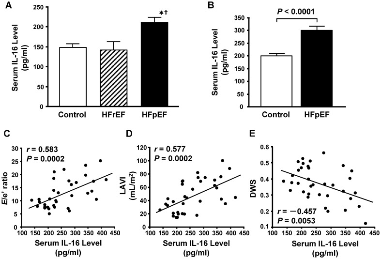
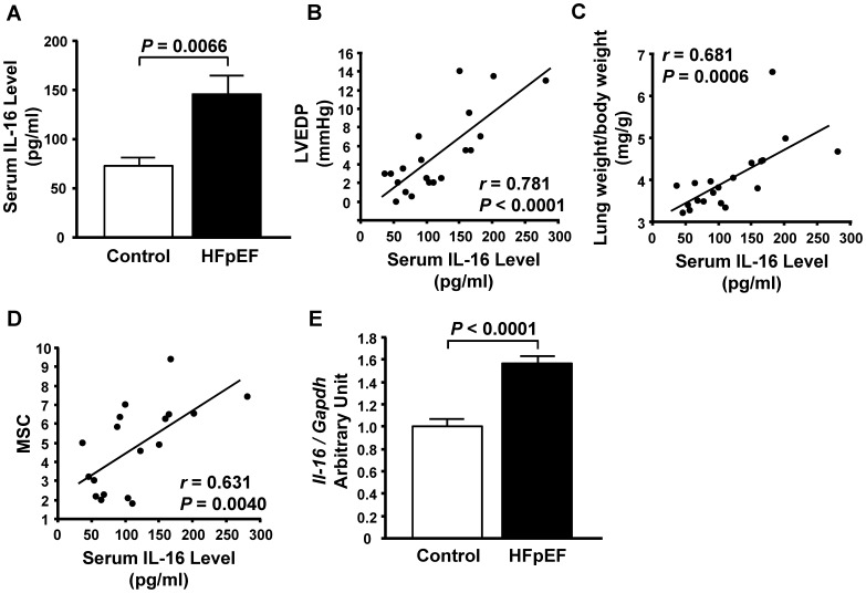
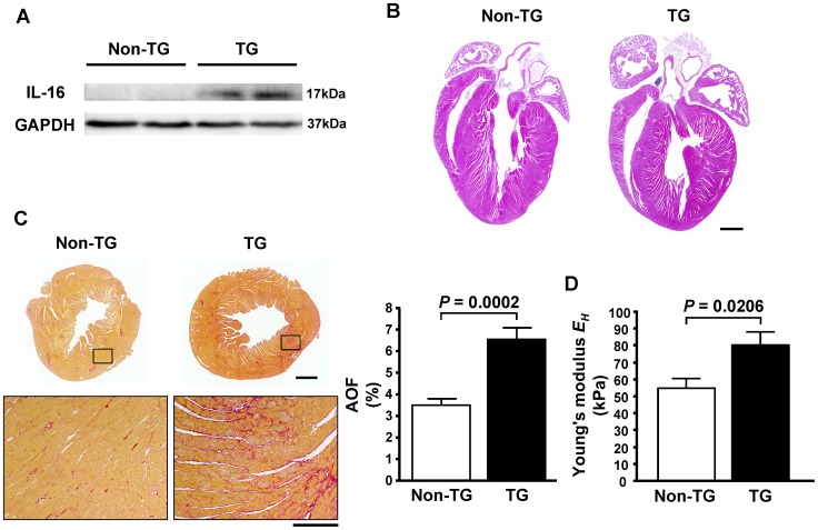
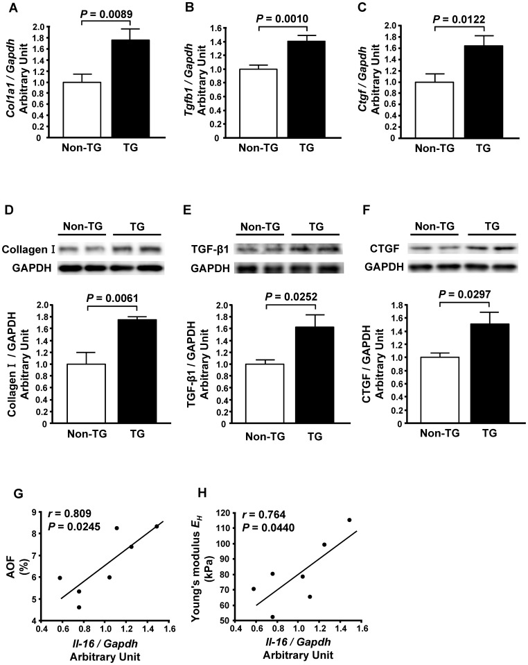
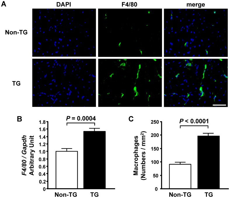
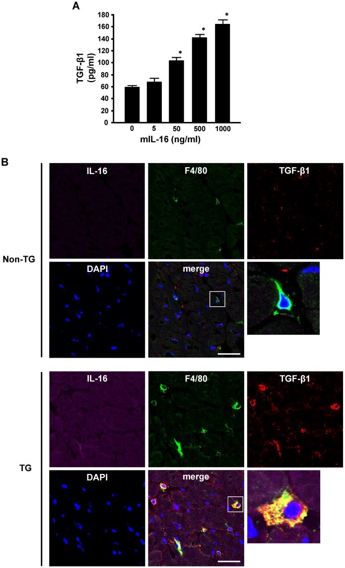
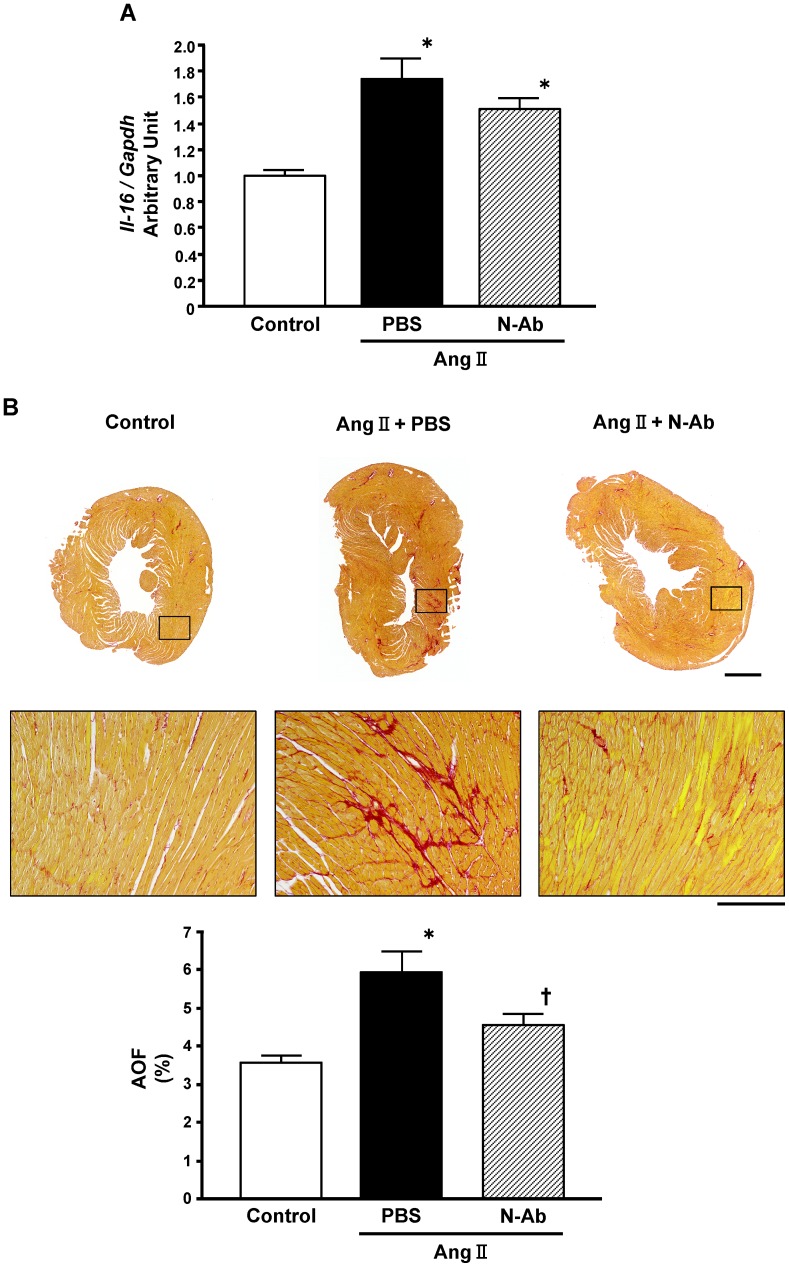
Methods and results: An analysis by multiplex-bead array assay revealed that serum interleukin-16 (IL-16) levels were specifically elevated in patients with HFpEF compared with HFrEF and controls. This was confirmed by enzyme-linked immunosorbent assay in HFpEF patients and controls, and serum IL-16 levels showed a significant association with indices of LV diastolic dysfunction. Serum IL-16 levels were also elevated in a rat model of HFpEF and positively correlated with LV end-diastolic pressure, lung weight and LV myocardial stiffness constant. The cardiac expression of IL-16 was upregulated in the HFpEF rat model. Enhanced cardiac expression of IL-16 in transgenic mice induced cardiac fibrosis and LV myocardial stiffening accompanied by increased macrophage infiltration. Treatment with anti-IL-16 neutralizing antibody ameliorated cardiac fibrosis in the mouse model of angiotensin II-induced hypertension.

Conclusion: Our data indicate that IL-16 is a mediator of LV myocardial fibrosis and stiffening in HFpEF, and that the blockade of IL-16 could be a possible therapeutic option for HFpEF.


Conflict of interest statement

Competing Interests: The authors have declared that no competing interests exist.

Figures

Figure 1
Figure 1. Elevated serum interleukin-16 (IL-16) levels in patients with heart failure with preserved ejection fraction.
A, Serum IL-16 levels in controls and patients with heart failure with reduced (HFrEF) or preserved ejection fraction (HFpEF) measured by a multiplex-bead array assay. *P<0.05 vs. control group. P<0.05 vs. HFrEF group. B, Serum IL-16 levels in controls and patients with HFpEF measured by enzyme-linked immunosorbent assay. C through E, Correlations of serum IL-16 level and the ratio of early transmitral flow velocity to septal mitral annular early diastolic velocity (E/e′ ratio) (C), left atrial volume index (LAVI) (D) and diastolic wall strain (DWS) (E) in controls and HFpEF patients combined.
Figure 2
Figure 2. Elevated serum interleukin-16 (IL-16) levels in rats with heart failure with preserved ejection fraction (HFpEF).
A, Serum IL-16 levels in control rats and the rats with HFpEF. B through D, Correlations of serum IL-16 level and left ventricular end-diastolic pressure (LVEDP) (B), lung weight to body weight ratio (lung weight/body weight) (C) and myocardial stiffness constant (MSC) (D) in control and HFpEF rats combined. E, mRNA level of IL-16 in the left ventricle of control and HFpEF rats.
Figure 3
Figure 3. Enhanced cardiac expression of interleukin-16 (IL-16) causes increased myocardial fibrosis and stiffness in mice.
A, Cardiac expression of IL-16 protein in non-transgenic (Non-TG) and transgenic (TG) mice. B, Four-chamber view of the hearts from Non-TG and TG mice stained with hematoxylin and eosin. Bar = 1 mm. C, Representative photomicrograph of Sirius Red-stained heart sections and the percent area of fibrosis (AOF) of Non-TG and TG mice. Bar: Upper panel = 1 mm; Lower panel = 200 µm. D, Young’s modulus EH in Non-TG and TG mice.
Figure 4
Figure 4. Effect of enhanced cardiac expression of interleukin-16 (IL-16) on markers of cardiac fibrosis in mice.
A through C, Left ventricular mRNA levels of Collagen I (A), transforming growth factor-beta 1 (TGF-β1) (B) and connective tissue growth factor (CTGF) (C) in non-transgenic (Non-TG) and transgenic (TG) mice. D through F, Left ventricular protein levels of Collagen I (D), TGF-β1 (E) and CTGF (F) in Non-TG and TG mice. Top panels in each figure show a representative Western blot. n = 5 per group. G and H, Correlations of IL-16 mRNA levels with AOF (G) and Young’s modulus EH (H) in TG mice.
Figure 5
Figure 5. Enhanced cardiac expression of interleukin-16 induces cardiac macrophage infiltration in mice.
A, Representative photomicrographs of immunofluorescence staining of the left ventricle for F4/80 in non-transgenic (Non-TG) and transgenic (TG) mice. Bar = 50 µm. B, Left ventricular mRNA levels of F4/80 in Non-TG and TG mice. C, Quantitative analysis of macrophage infiltration into left ventricular myocardium in Non-TG and TG mice. n = 6 per group.
Figure 6
Figure 6. Effect of interleukin-16 (IL-16) on transforming growth factor-beta 1 (TGF-β1) production by macrophages.
A, Production of TGF-β1 by macrophages treated with murine recombinant IL-16 (mIL-16). Mouse peritoneal macrophages (2×10⁶ cells/well) were treated with the indicated concentration of mIL-16 for 24 hours. The supernatants of macrophage cultures were collected and TGF-β1 was measured as described in the Methods. n = 5 for each group. *P<0.05 vs. 0 ng/ml group. B, Representative photomicrographs of confocal immunofluorescence staining of the left ventricle in non-transgenic (Non-TG) and transgenic (TG) mice for IL16 (violet), F4/80 (green), and TGF-β1 (red).
Figure 7
Figure 7. Neutralization of interleukin-16 (IL-16) ameliorates the development of cardiac fibrosis.
A, mRNA level of IL-16 in the left ventricle of control mice and mice injected intraperitoneally with phosphate buffered saline (PBS) or anti-IL-16 neutralizing antibody (N-Ab) during angiotensin II (Ang II) infusion for 14 days. *P<0.05 vs. control group. B, Representative photomicrograph of Sirius Red-stained heart sections and the percent area of fibrosis (AOF) in control mice and mice injected intraperitoneally with PBS or N-Ab during Ang II infusion for 28 days. Bar: Upper panel = 1 mm; Lower panel = 200 µm. *P<0.05 vs. control group. P<0.05 vs. mice with PBS treatment during Ang II infusion.

References

    1. Roger VL, Weston SA, Redfield MM, Hellermann-Homan JP, Killian J, et al. (2004) Trends in heart failure incidence and survival in a community-based population. JAMA 292: 344–350. - PubMed
    1. Bhatia RS, Tu JV, Lee DS, Austin PC, Fang J, et al. (2006) Outcome of heart failure with preserved ejection fraction in a population-based study. N Engl J Med 355: 260–269. - PubMed
    1. Owan TE, Hodge DO, Herges RM, Jacobsen SJ, Roger VL, et al. (2006) Trends in prevalence and outcome of heart failure with preserved ejection fraction. N Engl J Med 355: 251–259. - PubMed
    1. Yusuf S, Pfeffer MA, Swedberg K, Granger CB, Held P, et al. (2003) Effects of candesartan in patients with chronic heart failure and preserved left-ventricular ejection fraction: the CHARM-Preserved Trial. Lancet 362: 777–781. - PubMed
    1. Zile MR, Baicu CF, Gaasch WH (2004) Diastolic heart failure–abnormalities in active relaxation and passive stiffness of the left ventricle. N Engl J Med 350: 1953–1959. - PubMed

Publication types

MeSH terms