. 2013 Jul 24;8(7):e69194.
doi: 10.1371/journal.pone.0069194. Print 2013.

Mutation types and aging differently affect revertant fiber expansion in dystrophic mdx and mdx52 mice

Affiliations

Mutation types and aging differently affect revertant fiber expansion in dystrophic mdx and mdx52 mice

Yusuke Echigoya et al. PLoS One. .

Abstract

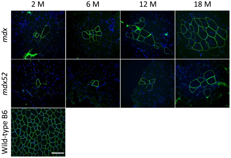
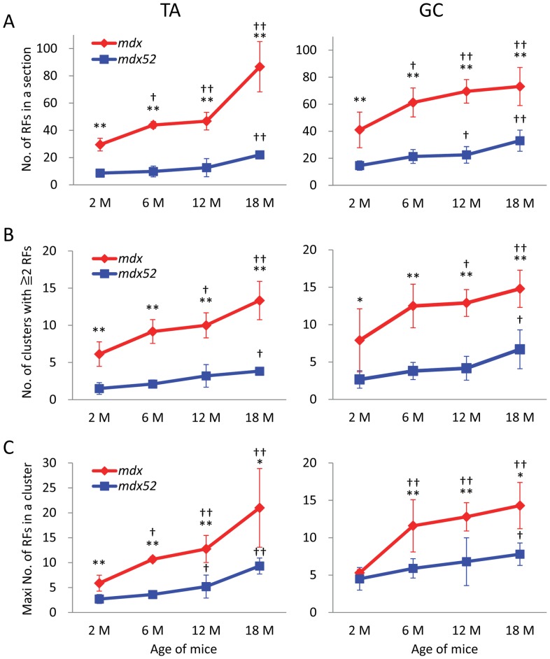
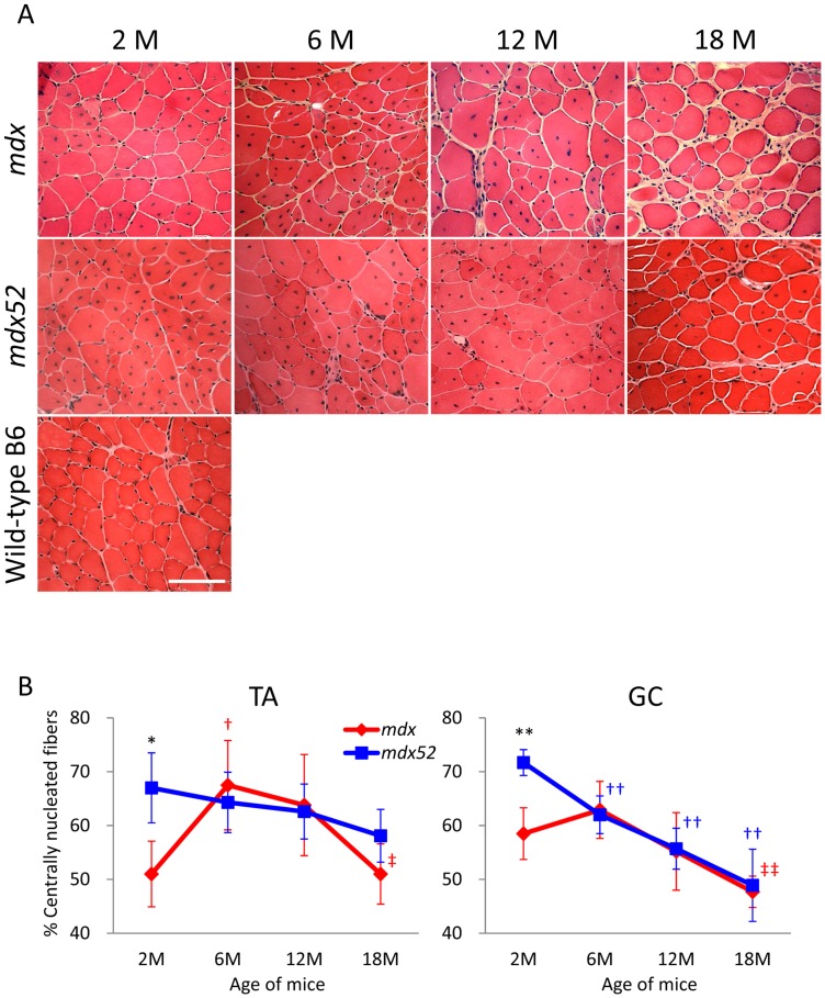
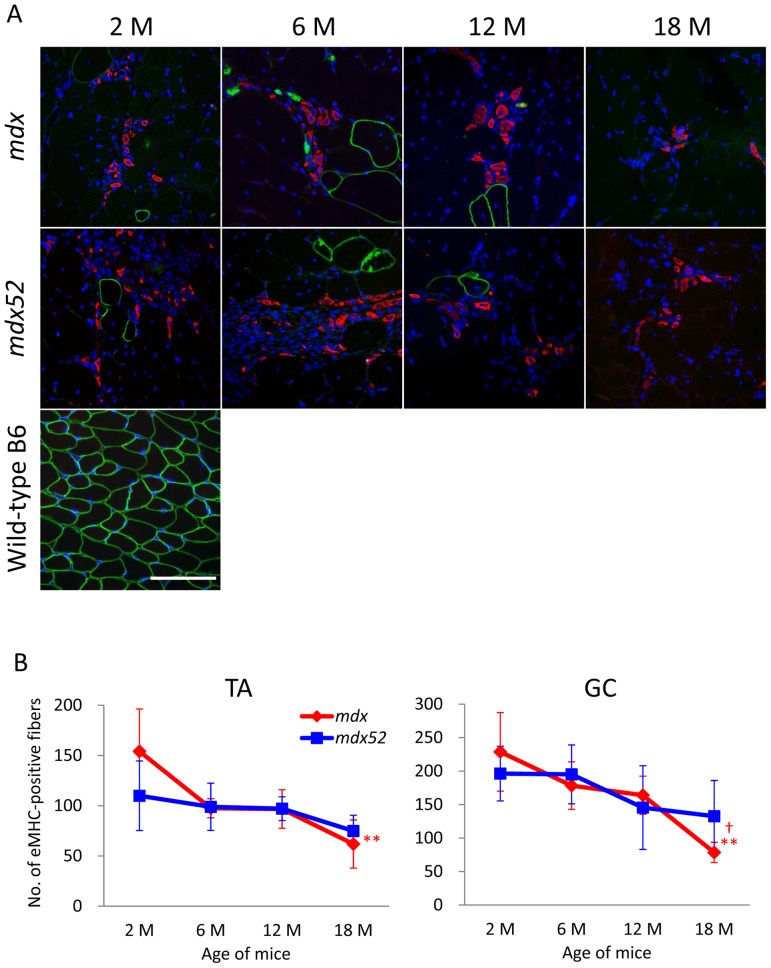
Duchenne muscular dystrophy (DMD), one of the most common and lethal genetic disorders, and the mdx mouse myopathies are caused by a lack of dystrophin protein. These dystrophic muscles contain sporadic clusters of dystrophin-expressing revertant fibers (RFs), as detected by immunohistochemistry. RFs are known to arise from muscle precursor cells with spontaneous exon skipping (alternative splicing) and clonally expand in size with increasing age through the process of muscle degeneration/regeneration. The expansion of revertant clusters is thought to represent the cumulative history of muscle regeneration and proliferation of such precursor cells. However, the precise mechanisms by which RFs arise and expand are poorly understood. Here, to test the effects of mutation types and aging on RF expansion and muscle regeneration, we examined the number of RFs in mdx mice (containing a nonsense mutation in exon 23) and mdx52 mice (containing deletion mutation of exon 52) with the same C57BL/6 background at 2, 6, 12, and 18months of age. Mdx mice displayed a significantly higher number of RFs compared to mdx52 mice in all age groups, suggesting that revertant fiber expansion largely depends on the type of mutation and/or location in the gene. A significant increase in the expression and clustering levels of RFs was found beginning at 6months of age in mdx mice compared with mdx52 mice. In contrast to the significant expansion of RFs with increasing age, the number of centrally nucleated fibers and embryonic myosin heavy chain-positive fibers (indicative of cumulative and current muscle regeneration, respectively) decreased with age in both mouse strains. These results suggest that mutation types and aging differently affect revertant fiber expansion in mdx and mdx52 mice.


Conflict of interest statement

Competing Interests: The authors have declared that no competing interests exist.

Figures

Figure 1
Figure 1. Dystrophin-positive revertant fibers with central nuclei at ages of 2, 6, 12, and 18 months in mdx and mdx52 mice.
Representative immunohistochemical images of maximum clusters of RFs in TA muscles are shown in each group. Mdx shows a higher maximum number of RFs than mdx52 in all age groups. Wild-type C57BL/6 muscle at 2months old is displayed as a control. An anti-dystrophin C-terminal antibody (green) and DAPI staining (blue) were used. M: months. 20x objective lens, scale bar  = 100μm.
Figure 2
Figure 2. Mutation- and age-related expression of dystrophin-positive revertant fibers in TA and GC muscles from mdx and mdx52 mice.
(A) The number of RFs in one TA or GC section. (B) The number of RF clusters containing 2 or more positive fibers. (C) The maximum number of RFs in a single cluster. Mdx mice have a significantly higher number of RFs in all criteria than mdx52 mice except for 2months of age in maximum number of RFs per cluster. The number of RFs in all criteria increases with age. Values are mean ± S.D. (n = 3–6 mice per each group). *P<0.05, **P<0.01 between mdx and mdx52 mice; †P<0.05, ††P<0.05 compared to 2months old. M: months.
Figure 3
Figure 3. Distinct changes in the percentage of centrally nucleated fibers by mutations and age in mdx and mdx52 mice.
(A) Representative images of TA muscles from mdx and mdx52 mice at ages 2, 6, 12 and 18months with hematoxylin and eosin staining. Wild-type C57BL/6 muscle at 2months of age is displayed as a control. M: months. Scale bar  = 100μm. (B) The percentage of centrally nucleated fibers in TA and GC muscles from mdx and mdx52 mice. Three hundred to one thousand myofibers were counted in left and right muscles and the percentage of CNFs was averaged between the two muscles per mouse. Values are mean ± S.D. (n = 3–6 mice per group). *P<0.05, **P<0.01 between mdx and mdx52 mice; †P<0.05, ††P<0.01 compared to 2months old; ‡P<0.05, ‡‡P<0.01 compared to 6months old. Symbol colors are accordant with the color of mice (red; mdx, blue; mdx52).
Figure 4
Figure 4. No expression of eMHC in RFs and attenuation of ongoing muscle regeneration in aged mdx and mdx52 mice.
(A) Triple staining of mdx and mdx52 mice for RF (green), eMHC (red), and nucleus (blue). Revertant dystrophin is not co-localized with newly regenerated eMHC-positive fibers in TA and GC muscles from mdx and mdx52 mice at any age. The pictures are representative GC muscles from mdx and mdx52 mice at each age. 20x objective lens, scale bar  = 100μm. (B) The number of eMHC-positive fibers. Values are mean ± S.D. (n = 3–6 mice per group). A significant decrease in the number of eMHC-positive fibers is found only at 18months old in mdx mice (**P<0.01 compared to 2months old, †P<0.05 compared to 6 and 12months old). Symbol colors are accordant with the color of mice (red; mdx, blue; mdx52).

Similar articles

Cited by

References

    1. Lovering RM, Porter NC, Bloch RJ (2005) The muscular dystrophies: from genes to therapies. Phys Ther 85: 1372–1388. - PMC - PubMed
    1. Biggar WD, Klamut HJ, Demacio PC, Stevens DJ, Ray PN (2002) Duchenne muscular dystrophy: current knowledge, treatment, and future prospects. Clin Orthop Relat Res: 88–106. - PubMed
    1. Duchenne (1867) The pathology of paralysis with muscular degeneration (paralysie myosclerotique), or paralysis with apparent hypertrophy. Br Med J 2: 541–542. - PMC - PubMed
    1. McNally EM, Pytel P (2007) Muscle diseases: the muscular dystrophies. Annu Rev Pathol 2: 87–109. - PubMed
    1. Hyser CL, Mendell JR (1988) Recent advances in Duchenne and Becker muscular dystrophy. Neurol Clin 6: 429–453. - PubMed

Publication types

Substances