. 2013 Jul 24;8(7):e69222.
doi: 10.1371/journal.pone.0069222. Print 2013.

Lymphatic territories (lymphosomes) in a canine: an animal model for investigation of postoperative lymphatic alterations

Affiliations

Lymphatic territories (lymphosomes) in a canine: an animal model for investigation of postoperative lymphatic alterations

Hiroo Suami et al. PLoS One. .

Abstract

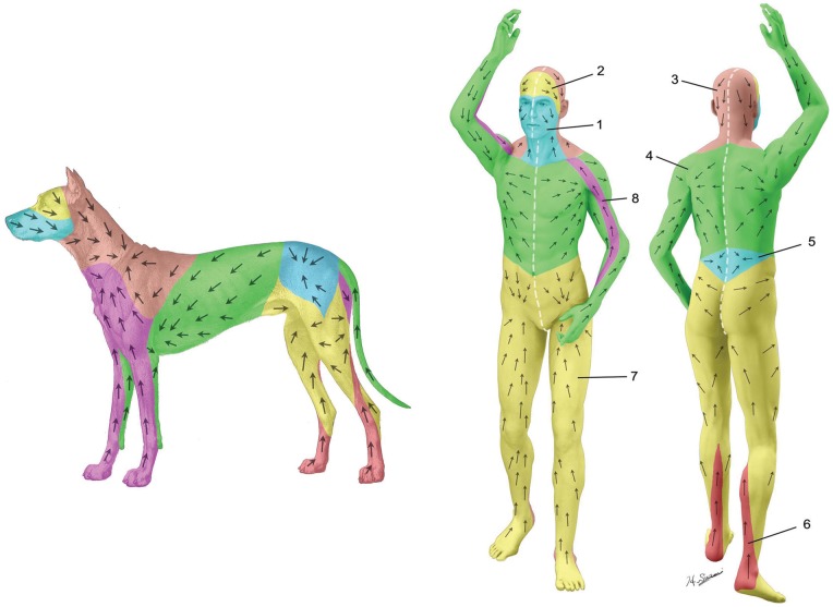
Background: Lymph node dissection is often performed as a part of surgical treatment for breast cancer and malignant melanoma to prevent malignant cells from traveling via the lymphatic system. Currently little is known about postoperative lymphatic drainage pattern alterations. This knowledge may be useful for management of recurrent cancer and prevention of breast cancer related lymphedema. We mapped the complete superficial lymphatic system of a dog and used this canine model to perform preliminary studies of lymphatic architectural changes in postoperative condition.

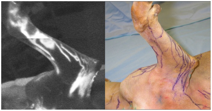
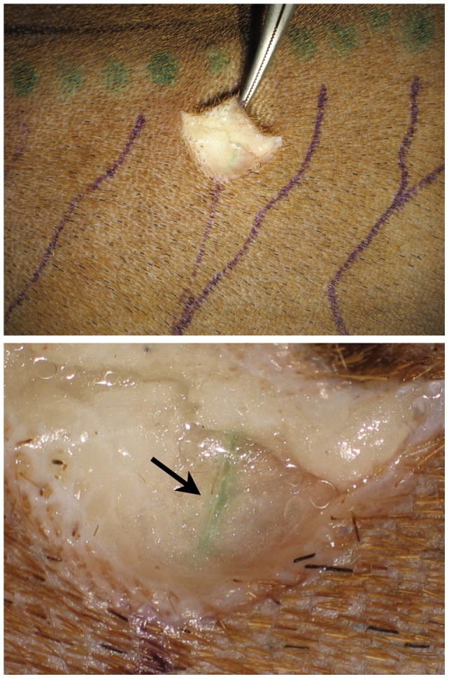
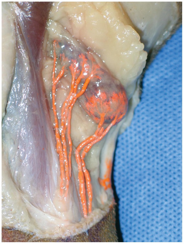
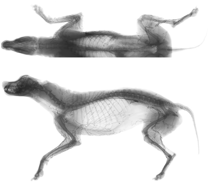
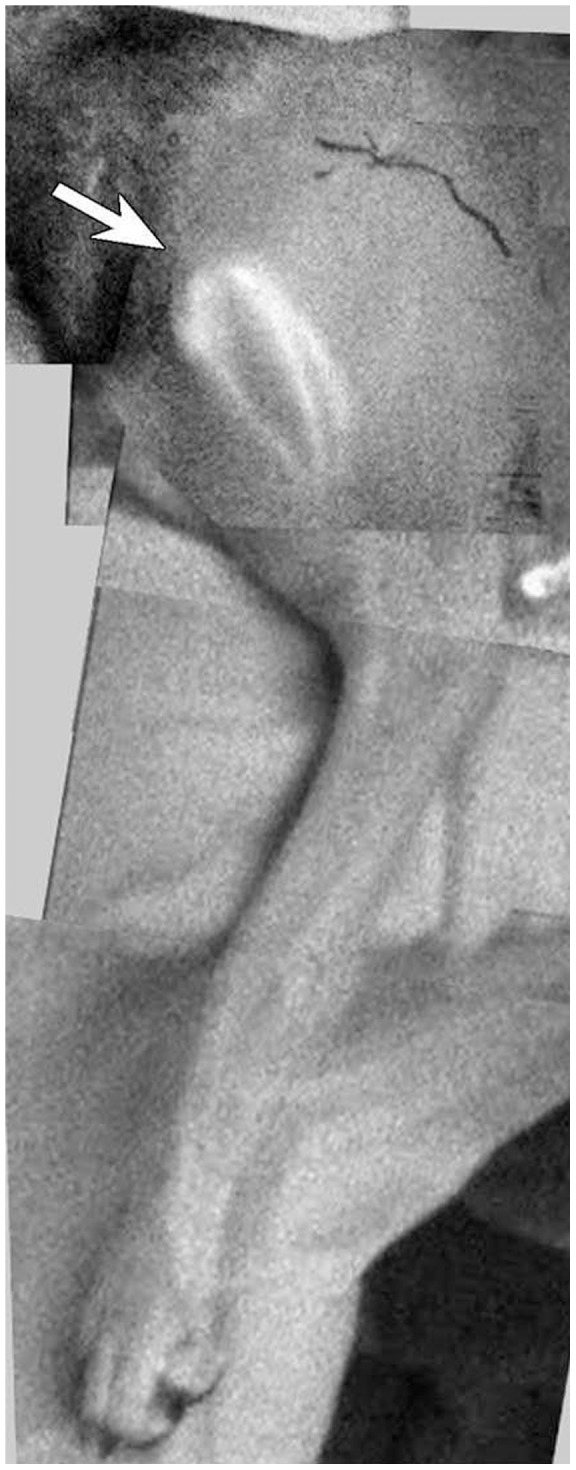
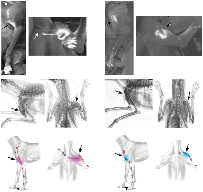
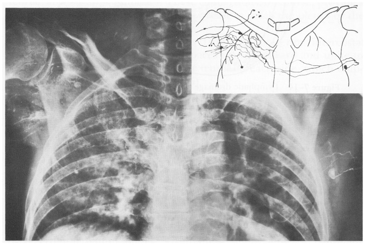
Methods: Lymphatic territories (lymphosomes) were mapped with 4 female mongrel carcasses using an indocyanine green (ICG) fluorescent lymphography and a radiographic microinjection technique. Two live dogs were then subjected to unilateral lymph node dissection of lymph basins of the forelimb, and ICG lymphography and lymphangiogram were performed 6 months after the surgery to investigate lymphatic changes. Lymphatic patterns in the carcass were then compared with postoperative lymphatic patterns in the live dogs.

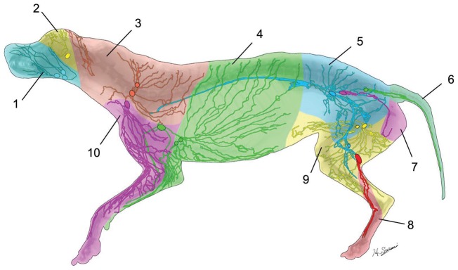
Results: Ten lymphosomes were identified, corresponding with ten lymphatic basins. Postoperative fluorescent lymphographic images and lymphangiograms in the live dogs revealed small caliber lymphatic network fulfilling gaps in the surgical area and collateral lymphatic vessels arising from the network connecting to lymph nodes in the contralateral and ipsilateral neck in one dog and the ipsilateral subclavicular vein in another dog.

Conclusion: Our canine lymphosome map allowed us to observe lymphatic collateral formations after lymph node dissection in live dogs. This canine model may help clarify our understanding of postoperative lymphatic changes in humans in future studies.


Conflict of interest statement

Competing Interests: The authors have declared that no competing interests exist.

Figures

Figure 1
Figure 1. Indocyanine green (ICG) fluorescent lymphographic image of the medial side of the left forelimb in a dog carcass (left).
Tracing of the lymphatic vessels visualized using ICG lymphography (right).
Figure 2
Figure 2. Marked lymphatic vessels near the dorsal midline in the torso (indocyanine green injection sites are shown as green dots) (top).
A magnified photo shows that ICG was specifically taken into the lymph vessel (arrow) (bottom).
Figure 3
Figure 3. The popliteal lymph node with afferent lymphatic vessels (stained orange by the lead tetroxide radiocontrast mixture).
Figure 4
Figure 4. Anteroposterior (top) and lateral (bottom) radiographs of the whole body carcass after lymphatic injection.
Figure 5
Figure 5. Montage of indocyanine green (ICG) lymphographic images of the left forelimb of the live dog prior to lymph node dissection.
The ventral superficial lymph node (arrow) was identified using injections of ICG at interdigital webspaces.
Figure 6
Figure 6. Color-coded diagram of the lymphatic territories (lymphosomes) with lymphatic vessels shown distally from their corresponding lymph nodes: 1, submandibular; 2, parotid; 3, dorsal superficial cervical; 4, axillary; 5, medial iliac; 6, lateral sacral; 7, hypogastric; 8, popliteal; 9, superficial inguinal; 10, ventral superficial cervical.
Figure 7
Figure 7. A montage of indocyanine green lymphographic images of the left forelimbs of 2 live dogs 6 months after lymph node dissection (top).
Bright spots were seen in the area in which the surgery took place (black arrow). Locations of lymph nodes are marked (white arrows). Lymphangiograms from the same dogs from lateral (left) and antero-posterior (right) views showing capillary-like network (black arrows) and bypassed lymph nodes (white arrows) (middle). Diagrams show changes of lymphatic pathways (bottom).
Figure 8
Figure 8. Histological image with hematoxylin and eosin staining of a cross section of a skin in the capillary-like network (scale bar: 5 mm) (left).
Small lymphatics lined with pigment is found in the entire fibrous scar tissue (scale bar: 2 mm) (right: white square field of the left).
Figure 9
Figure 9. Human lymphosomes, similar to the canine lymphosomes.
1, Frontal cervical; 2, parotid; 3, posterior cervical; 4, axillary; 5, retroperitoneal; 6, popliteal; 7, superficial inguinal; 8, subclavicular.
Figure 10
Figure 10. Postoperative lymphangiogram of a patient who underwent right breast mastectomy and axillary node dissection.
Injected radiocontrast from the affected hand reached the contralateral axillary and ipsilateral supraclavicular nodes. Compare with the lymphangiogram of the live dog in Fig. 7. (Reprinted with permission from Bobbio P, Peracchia G, Pellegrino F. (1962) Connessioni linfatiche presternali fra le regioni mammarie dei due lati. Ateneo Parmensa, 33(supp.): 95–109).

References

    1. Moore CH (1867) On the influence of inadequate operations on the theory of cancer. Med Chir Trans 50: 245–280. - PMC - PubMed
    1. Sappey MPC (1874) Anatomie, physiologie, pathologie des vaisseaux lymphatiques consideres chez l'homme at les vertebres. Paris: A. Delahaye and E. Lecrosnier.
    1. Sugarbaker EV, McBride CM (1976) Melanoma of the trunk: the results of surgical excision and anatomic guidelines for predicting nodal metastasis. Surgery 80: 22–30. - PubMed
    1. Herd-Smith A, Russo A, Muraca MG, Del Turco MR, Cardona G (2001) Prognostic factors for lymphedema after primary treatment of breast carcinoma. Cancer 92: 1783–1787. - PubMed
    1. Lee TS, Kilbreath SL, Refshauge KM, Herbert RD, Beith JM (2008) Prognosis of the upper limb following surgery and radiation for breast cancer. Breast Cancer Res Treat 110: 19–37. - PubMed

Publication types

LinkOut - more resources