. 2013 Jul 22;8(7):e69362.
doi: 10.1371/journal.pone.0069362. Print 2013.

Coenzyme Q10 ameliorates pain and cartilage degradation in a rat model of osteoarthritis by regulating nitric oxide and inflammatory cytokines

Affiliations

Coenzyme Q10 ameliorates pain and cartilage degradation in a rat model of osteoarthritis by regulating nitric oxide and inflammatory cytokines

Jennifer Lee et al. PLoS One. .

Abstract

Objective: To investigate the effect of CoenzymeQ10 (CoQ10) on pain severity and cartilage degeneration in an experimental model of rat osteoarthritis (OA).

Materials and methods: OA was induced in rats by intra-articular injection of monosodium iodoacetate (MIA) to the knee. Oral administration of CoQ10 was initiated on day 4 after MIA injection. Pain severity was assessed by measuring secondary tactile allodynia using the von Frey assessment test. The degree of cartilage degradation was determined by measuring cartilage thickness and the amount of proteoglycan. The mankin scoring system was also used. Expressions of matrix metalloproteinase-13 (MMP-13), interleukin-1β (IL-1β), IL-6, IL-15, inducible nitric oxide synthase (iNOS), nitrotyrosine and receptor for advanced glycation end products (RAGE) were analyzed using immunohistochemistry.

Results: Treatment with CoQ10 demonstrated an antinociceptive effect in the OA animal model. The reduction in secondary tactile allodynia was shown by an increased pain withdrawal latency and pain withdrawal threshold. CoQ10 also attenuated cartilage degeneration in the osteoarthritic joints. MMP-13, IL-1β, IL-6, IL-15, iNOS, nitrotyrosine and RAGE expressions were upregulated in OA joints and significantly reduced with CoQ10 treatment.

Conclusion: CoQ10 exerts a therapeutic effect on OA via pain suppression and cartilage degeneration by inhibiting inflammatory mediators, which play a vital role in OA pathogenesis.


Conflict of interest statement

Competing Interests: The authors have declared that no competing interests exist.

Figures

Figure 1
Figure 1. Therapeutic effect of Coenzyme Q10 in an early phase of MIA-induced OA in rats.
Rats were injected with 3 mg of monosodium iodoacetate (MIA) in the right knee. Coenzyme Q10 (CoQ10) was administered orally every day from day 4 after MIA injection. Behavioral tests of secondary tactile allodynia in MIA-injected rats treated with vehicle or CoQ10 were evaluated using a dynamic plantar esthesiometer (n = 10 on each day for each group). Compared with vehicle-treated OA rats, OA animals treated with CoQ10 at a dose of 100 mg/kg showed a significant increase in PWL and PWT. The data are expressed as mean and error bars for three animals per group. PWL and PWT were conducted right before the administration of CoQ10. Significant differences between vehicle- and CoQ10-treated groups: *P<0.05, **P<0.01 and ***P<0.001 compared with the vehicle-treated OA group.
Figure 2
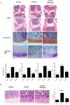
Figure 2. Histological evaluation of joints and osteoclastic activity after treatment with CoenzymeQ10 in MIA-induced OA.
Rats were injected with 3 mg of monosodium iodoacetate (MIA) in the right knee. Coenzyme Q10 (CoQ10) was administered orally every day from the day 4 after MIA injection. The knee joints were resected on day 7 after MIA injection. (A) The knee joints from the OA rats treated with CoQ10 or vehicle were stained with HE, Safranin O-fast green, and toluidine blue. Extensive cartilage degradation, bone destruction, and fibrosis are seen in the vehicle-treated group, whereas the treatment with CoQ10 preserved the articular space and prevented the depletion of proteoglycan. CoQ10 treatment preserved the cartilage structure and decreased the depth and the extent of cartilage damage. The joint lesions were graded on a scale of 0–13 using the modified Mankin scoring system. Total mankin score is a sum of the scores for cartilage structure, cellular abnormalities, and matrix staining. CoQ10 prevented damage to the cartilage structure, reduced cellular abnormalities, and preserved matrix staining. The data are expressed as mean and error bars for six animals per group. (B) The number of osteoclasts were measured in the knee joint sections. Osteoclasts were more frequently found in vehicle-treated group than CoQ10-treated group. *P<0.05, **P<0.01, and ***P<0.001 compared with the MIA-injected vehicle-treated group.
Figure 3
Figure 3. Effect of Coenzyme Q10 on the expression of MMP-13, IL-1β, IL-6 and IL-15 in OA joints.
Rats were injected with 3 mg of MIA in the right knee. Coenzyme Q10 was administered orally every day from the day 4 after MIA injection. Immunohistochemical staining was used to identify the expression of MMP-13, IL-1b, IL-6, and IL-15 in the articular cartilage. In the articular cartilage of MIA-injected rats, the expression of MMP-13, IL-1β, IL-6 and IL-15 increased, whereas treatment with CoQ10 inhibited the expression of these molecules. The data are expressed as mean and error bars for six animals per group. Original magnification 200x. *P<0.05, **P<0.01, and ***P<0.001 compared with the MIA-injected vehicle-treated group.
Figure 4
Figure 4. Attenuated iNOS, nitrotyrosine and RAGE expression in Coenzyme Q10-treated OA rats.
Rats were injected with 3 mg of monosodium iodoacetate (MIA) in the right knee. Coenzyme Q10 (CoQ10) was administered orally every day from the day 4 after MIA injection. The knees were resected on day 7 after MIA injection. Immunohistochemical staining was used to identify the expression of induced nitric oxide synthase (iNOS), nitrotyrosine and receptor for advanced glycation end products (RAGE) in the joint tissue. Expression of iNOS, nitrotyrosine and RAGE in chondrocytes was increased after MIA-injection, and was reduced in CoQ10-treated OA animals. The data are expressed as mean and error bar for three animals per group. Original magnification 200x. *P<0.05, **P<0.01, and ***P<0.001 compared with the MIA-injected vehicle-treated group.

References

    1. Berenbaum F (2012) Osteoarthritis as an inflammatory disease (osteoarthritis is not osteoarthrosis!). Osteoarthritis Cartilage 21: 16–21. - PubMed
    1. Kapoor M, Martel-Pelletier J, Lajeunesse D, Pelletier JP, Fahmi H (2011) Role of proinflammatory cytokines in the pathophysiology of osteoarthritis. Nat Rev Rheumatol 7: 33–42. - PubMed
    1. Green DE, Wharton DC (1963) STOICHIOMETRY OF THE FIXED OXIDATION-REDUCTION COMPONENTS OF THE ELECTRON TRANSFER CHAIN OF BEEF HEART MITOCHONDRIA. Biochem Z 338: 335–348. - PubMed
    1. Stocker R, Bowry VW, Frei B (1991) Ubiquinol-10 protects human low density lipoprotein more efficiently against lipid peroxidation than does alpha-tocopherol. Proc Natl Acad Sci U S A 88: 1646–1650. - PMC - PubMed
    1. Fuller B, Smith D, Howerton A, Kern D (2006) Anti-inflammatory effects of CoQ10 and colorless carotenoids. J Cosmet Dermatol 5: 30–38. - PubMed

Publication types

MeSH terms