. 2013 Jul 24;8(7):e69389.
doi: 10.1371/journal.pone.0069389. Print 2013.

p16 Stimulates CDC42-dependent migration of hepatocellular carcinoma cells

Affiliations

p16 Stimulates CDC42-dependent migration of hepatocellular carcinoma cells

Ya-Wen Chen et al. PLoS One. .

Abstract

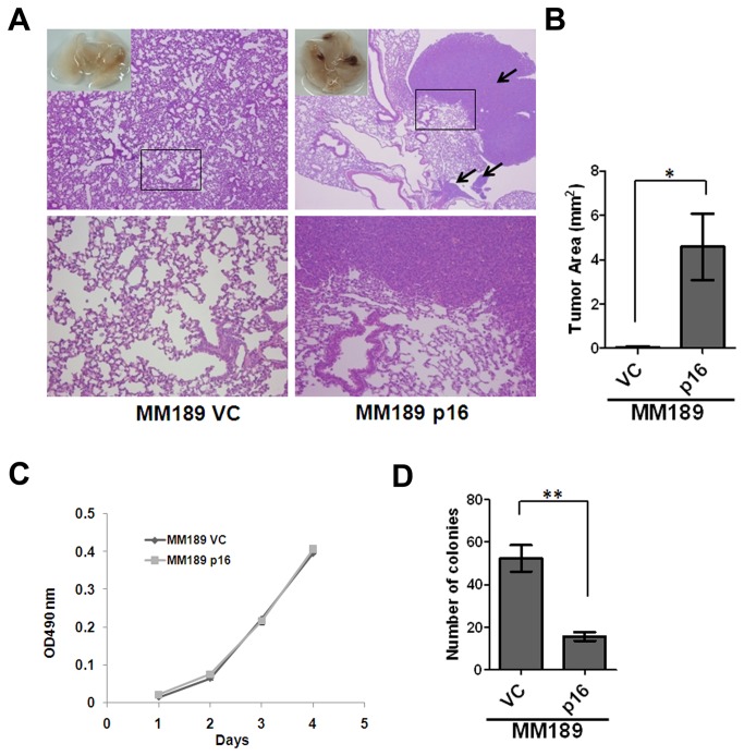
Hepatocellular carcinoma (HCC) is a leading cause of cancer-related deaths worldwide. Tumor dissemination to the extra-hepatic region of the portal vein, lymph nodes, lungs or bones contributes to the high mortality seen in HCC; yet, the molecular mechanisms responsible for HCC metastasis remain unclear. Prior studies have suggested a potential link between accumulated cytoplasm-localized p16 and tumor progression. Here we report that p16 enhances metastasis-associated phenotypes in HCC cells - ectopic p16 expression increased cell migration in vitro, and lung colonization after intravenous injection, whereas knockdown of endogenous p16 reduced cell migration. Interestingly, analysis of p16 mutants indicated that the Cdk4 interaction domain is required for stimulation of HCC cell migration; however, knockdown of Cdk4 and Cdk6 showed that these proteins are dispensable for this phenomenon. Intriguingly, we found that in p16-positive HCC samples, p16 protein is predominantly localized in the cytoplasm. In addition, we identified a potential role for nuclear-cytoplasmic shuttling in p16-stimulated migration, consistent with the predominantly cytoplasmic localization of p16 in IHC-positive HCC samples. Finally, we determined that p16-stimulated cell migration requires the Cdc42 GTPase. Our results demonstrate for the first time a pro-migratory role for p16, and suggest a potential mechanism for the observed association between cytoplasmic p16 and tumor progression in diverse tumor types.


Conflict of interest statement

Competing Interests: The authors have declared that no competing interests exist.

Figures

Figure 1
Figure 1. p16 enhances HCC cell migration.
(A) Upper panel: Immunoblot confirming ectopic p16 expression in MM189 and BL322 mouse HCC cells infected with either pBabe-puro retrovirus (Vector control, VC) or pBabe-puro retrovirus encoding wild type p16 (p16). β-actin serves as a loading control. Lower panel: Migration activity of MM189 and BL322 cells expressing p16, and their vector controls. Data are from a representative experiment performed in duplicate. Bar, SEM. (B) Upper panel: Immunoblot detection of ectopic p16 protein in BL185 and HepG2 HCC cells. Lower panel: Migration activity of BL185 and HepG2 cells expressing ectopic p16, and their vector controls. Data are from a representative experiment performed in duplicate. Bar, SEM. (C) Upper panel: Immunoblot detection of specific knockdown of p16 and p19 in BL185 HCC cells. pLKO-GFP is the non-silencing control for the p16-targeting shRNA. MLP-Luci is the non-silencing control for the p19-specific shRNA. Lower panel: Migration activity of BL185 cells with p16- or p19-specific knockdown relative to their non-silencing controls. Data are from a representative experiment performed in duplicate. Bar, SEM. (D) Left panel: Immunoblot detection of p16-specific knockdown in HepG2 cells infected with lentiviral vectors encoding p16-targeting shRNA, or a non-silencing control pLKO-GFP. Right panel: Migration activity of HepG2 cells with p16 knockdown relative to cells expressing the non-silencing control (pLKO-GFP). Data are from a representative experiment performed in duplicate. Bar, SEM. *, p<0.05; **, p<0.01; ***, p<0.001.
Figure 2
Figure 2. p16 enhances lung colonization by HCC cells.
(A) Representative lung fields of nude mice after the delivery, via tail vein injection, of MM189 cells with ectopic p16 expression (MM189 p16) or vector controls (MM189 VC). The boxed area in the upper panel is shown at higher magnification in the lower panel. (B) Quantification of the total area of the lung lesions in mice injected with MM189 p16 or MM189 VC cells. Bar, SEM. (C) Representative cell proliferation assay for MM189 p16 and MM189 VC cells. (D) Representative anchorage-independent growth assay for MM189 VC and MM189 p16 cells. Bar, SEM. *, p<0.05; **, p<0.01; ***, p<0.001.
Figure 3
Figure 3. Cdk4 and Cdk6 are dispensable for p16-enhanced migration.
(A) Immunoblot detection of p16 mutants in MM189 cells. (B) IP-Western Blot detection of binding of p16 mutants to Cdk4 and Cdk6. (C) Migration activity of MM189 cells expressing listed p16 mutants. Bar, SEM. (D) Immunoblot for Cdk4 or Cdk6 knockdown in MM189 p16 cells. (E) Migration activity of MM189 p16 cells with Cdk4 or Cdk6 knockdown relative to that of cells infected with a non-silencing control, pLKO-GFP. Bar, SEM. *, p<0.05; **, p<0.01; ***, p<0.001.
Figure 4
Figure 4. Human HCC samples display cytoplasmic localization of p16.
Control gastric (A) and endometrial (B) carcinoma samples displaying nuclear and cytoplasmic staining for p16. (CF) Representative images of HCC samples displaying different levels and localization of p16 staining. Note the absence of nuclear staining in panels (D and E). All images are at 200X magnification.
Figure 5
Figure 5. Nuclear-cytoplasmic shuttling is involved in p16-induced cell migration.
(A) Immunofluorescent detection of sub-cellular localization of p16 proteins. p16-NES, p16 tagged with NES at the C-terminus; p16-2NES, p16 tagged with NES at both N- and C-termini; p16-NLS, p16 tagged with NLS at the C-terminus; p16-2NLS, p16 tagged with NLS at both N- and C-termini. (B) Immunoblot detection of p16 proteins tagged with NES or NLS sequences. β-actin serves as a loading control. (C) IP-Western blot confirming the ability of NES- or NLS-tagged p16 to bind to Cdk4 and Cdk6. (D) Migration activity of MM189 cells expressing NES-tagged p16 proteins. Bar, SEM. (E) Migration activity of MM189 cells expressing NLS-tagged p16 proteins. Bar, SEM. *, p<0.05; **, p<0.01; ***, p<0.001.
Figure 6
Figure 6. Enhanced Cdc42 and Rac1 activity in p16-expressing HCC cells.
(A) Immunoblot analysis of epithelial and mesenchymal proteins and EMT-associated transcription factors in MM189 p16 and MM189 VC cells. BL185 cells and 3T3L1 cells serve as positive controls for the expression of E-cadherin and vimentin, respectively. α-tubulin serves as a loading control. The lower panel shows the quantification of 3 independent immunoblots. (B) Immunoblot detection of active Cdc42, Rac1, and Rho proteins bound by GST-Pak1 and GST-Rhotekin. GTPγS serves as a positive control to maintain the active form of GTPase in the reaction. Immunoblotting of whole cell lysate serves as an input control, and detection of GST serves as a loading control. (C) The relative density of the bands corresponding to Cdc42, Rac1 and Rho in MM189 p16 cells and control MM189 VC cells.
Figure 7
Figure 7. Cdc42 is required for p16-mediated migration.
Immunoblot analysis for knockdown of (A) Cdc42, (C) Rac1, and (E) RhoA in MM189 p16 cells. Cells infected with a vector encoding an shRNA targeting GFP serve as a non-silencing control (pLKO-GFP). α-tubulin serves as a loading control. Migration activity of cells with knockdown of (B) Cdc42, (D) Rac1, and (F) RhoA relative to MM189 cells infected with a non-silencing control. Data are from a representative experiment performed in duplicate. Bar, SEM. *, p<0.05; **, p<0.01; ***, p<0.001.

References

    1. Jemal A, Bray F, Center MM, Ferlay J, Ward E et al. (2011) Global cancer statistics. CA Cancer J Clin 61: 69-90. doi:10.3322/caac.20107. PubMed: 21296855. - DOI - PubMed
    1. Farazi PA, DePinho RA (2006) Hepatocellular carcinoma pathogenesis: from genes to environment. Nat Rev Cancer 6: 674-687. doi:10.1038/nrc1934. PubMed: 16929323. - DOI - PubMed
    1. Robinson WS (1994) Molecular events in the pathogenesis of hepadnavirus-associated hepatocellular carcinoma. Annu Rev Med 45: 297-323. doi:10.1146/annurev.med.45.1.297. PubMed: 8198385. - DOI - PubMed
    1. Buendia MA (2000) Genetics of hepatocellular carcinoma. Semin Cancer Biol 10: 185-200. doi:10.1006/scbi.2000.0319. PubMed: 10936068. - DOI - PubMed
    1. Liew CT, Li HM, Lo KW, Leow CK, Chan JY et al. (1999) High frequency of p16INK4A gene alterations in hepatocellular carcinoma. Oncogene 18: 789-795. doi:10.1038/sj.onc.1202359. PubMed: 9989830. - DOI - PubMed

Publication types

MeSH terms

Substances