. 2013 Jul 24;8(7):e69407.
doi: 10.1371/journal.pone.0069407. Print 2013.

Analysis of transcription factor mRNAs in identified oxytocin and vasopressin magnocellular neurons isolated by laser capture microdissection

Affiliations

Analysis of transcription factor mRNAs in identified oxytocin and vasopressin magnocellular neurons isolated by laser capture microdissection

Madison Humerick et al. PLoS One. .

Abstract

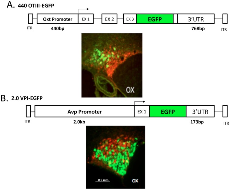
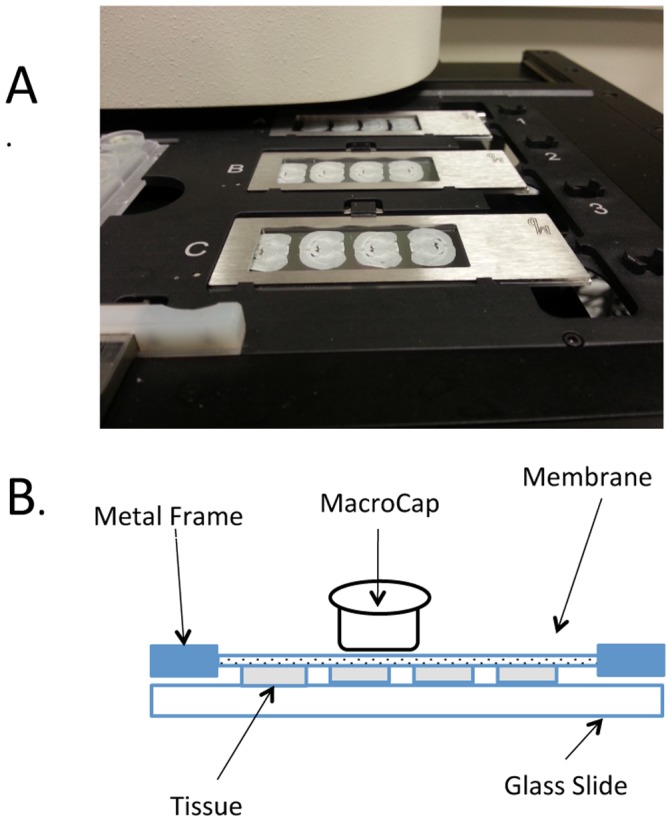
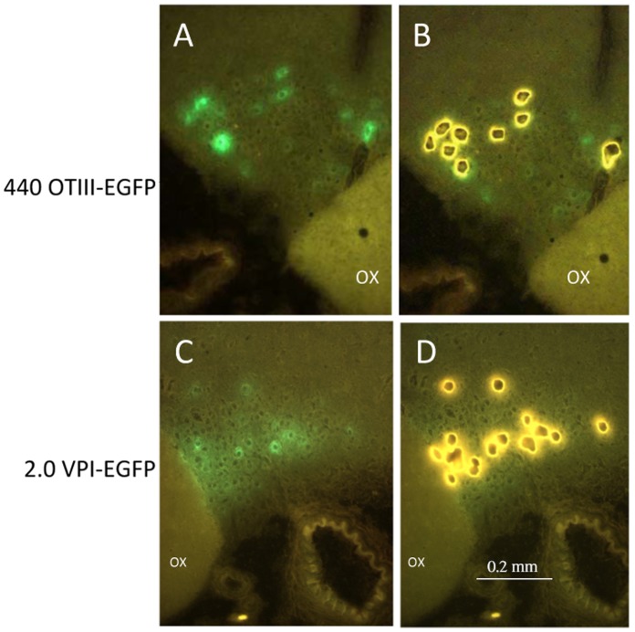
The oxytocin (Oxt) and vasopressin (Avp) magnocellular neurons (MCNs) in the hypothalamus are the only neuronal phenotypes that are present in the supraoptic nucleus (SON), and are characterized by their robust and selective expression of either the Oxt or Avp genes. In this paper, we take advantage of the differential expression of these neuropeptide genes to identify and isolate these two individual phenotypes from the rat SON by laser capture microdissection (LCM), and to analyze the differential expression of several of their transcription factor mRNAs by qRT-PCR. We identify these neuronal phenotypes by stereotaxically injecting recombinant Adeno-Associated Viral (rAAV) vectors which contain cell-type specific Oxt or Avp promoters that drive expression of EGFP selectively in either the Oxt or Avp MCNs into the SON. The fluorescent MCNs are then dissected by LCM using a novel Cap Road Map protocol described in this paper, and the purified MCNs are extracted for their RNAs. qRT-PCR of these RNAs show that some transcription factors (RORA and c-jun) are differentially expressed in the Oxt and Avp MCNs.


Conflict of interest statement

Competing Interests: The authors have declared that no competing interests exist.

Figures

Figure 1
Figure 1. Specificity of Expression of the Oxytocin and vasopressin rAAV constructs in the SON.
A. The 440OTIII-EGFP rAAV that was injected into the SON contains 440 bp of the promoter sequence upstream of the transcription start site (TSS) of the oxytocin gene, all three exons, fused to the EGFP sequence within the 3rd exon (EX 3), followed by 768 bp of the 3′UTR of the oxytocin gene . The immunohistochemical (IHC) image shown below the construct shows that the EGFP from the 440 OTIII-EGFP virus is specifically expressed in Oxt MCNs in the dorsal SON. The green color represents the fluorescence of the EGFP immunoreactivity in Oxt MCNs and the red fluorescence depicts the PS 41 antibody immunoreactivity in the Avp MCNs. Scale line for both panels A and B is shown in lower panel B. B. The 2.0VPI-EGFP rAAV that was injected contains 2.0 bp of the promoter sequence found upstream of the vasopressin gene TSS, exon 1 of the vasopressin gene, fused to the EGFP sequence, and 173 bp of the 3′UTR downstream of the Avp gene . The immunohistochemical (IHC) image shown below the construct shows that the EGFP from the 2.0VPI-EGFP virus is specifically expressed in Avp MCNs in the ventral SON. The green color represents the fluorescence of the EGFP immunoreactivity in Avp MCNs and the red fluorescence depicts the PS 38 antibody immunoreactivity in the Oxt MCNs. Abbreviation: OX, optic chiasm.
Figure 2
Figure 2. View of brain sections on the Frame metal slide LCM slides, and the LCM Cap position.
A. Illustration of three metal frame membrane slides each containing four hypothalamic sections mounted on the stage of the Arcturus XT instrument ready for LCM. B. Schematic view of the Metal Frame Membrane slide and LCM cap. With these slides the LCM cap sits directly on the membrane without touching the tissue. This allows the cap to sit evenly without being affected by any folds in the tissue and adjustments to the laser’s power and alignment usually do not need to be made. Use of the Macro LCM caps also reduces the danger of contaminating the sample with surrounding tissue.
Figure 3
Figure 3. LCM sample dissection as viewed through the Arcturus XT fluorescent LCM microscope.
A. View of 440OTIII-EGFP fluorescent expression in oxytocin MCNs in the SON on PEN Membrane Metal Frame slides. 15× magnification. Abbreviation: OX, optic chiasm. B. View of fluorescent Oxt MCN ghosts that have been captured and cut out by LCM. 15× magnification. C. View of 2.0VPI-EGFP fluorescent expression in Avp MCNs in the SON on PEN Membrane Metal Frame slides. 15× magnification. D. View of fluorescent Avp MCN ghosts that have been captured and cut out by LCM. OX represents the optic chiasm. 15× magnification. Scale line shown in panel D is representative for all panels.

Similar articles

Cited by

References

    1. Stevens CF (1998) Neuronal diversity: too many cell types for comfort? Curr Biol 8: R708–710. - PubMed
    1. Brownstein MJ, Russell JT, Gainer H (1980) Synthesis, transport, and release of posterior pituitary hormones. Science 207: 373–378. - PubMed
    1. Soltesz I (2006) Diversity in the neuronal machine : order and variability in interneuronal microcircuits. Oxford; New York: Oxford University Press. xvii, 238 p. p.
    1. Armstrong WE (1995) Morphological and electrophysiological classification of hypothalamic supraoptic neurons. Prog Neurobiol 47: 291–339. - PubMed
    1. Rhodes CH, Morrell JI, Pfaff DW (1981) Immunohistochemical analysis of magnocellular elements in rat hypothalamus: distribution and numbers of cells containing neurophysin, oxytocin, and vasopressin. J Comp Neurol 198: 45–64. - PubMed

Publication types

LinkOut - more resources