. 2013 Jul 22;8(7):e69410.
doi: 10.1371/journal.pone.0069410. Print 2013.

Alignment of synaptic vesicle macromolecules with the macromolecules in active zone material that direct vesicle docking

Affiliations

Alignment of synaptic vesicle macromolecules with the macromolecules in active zone material that direct vesicle docking

Mark L Harlow et al. PLoS One. .

Abstract

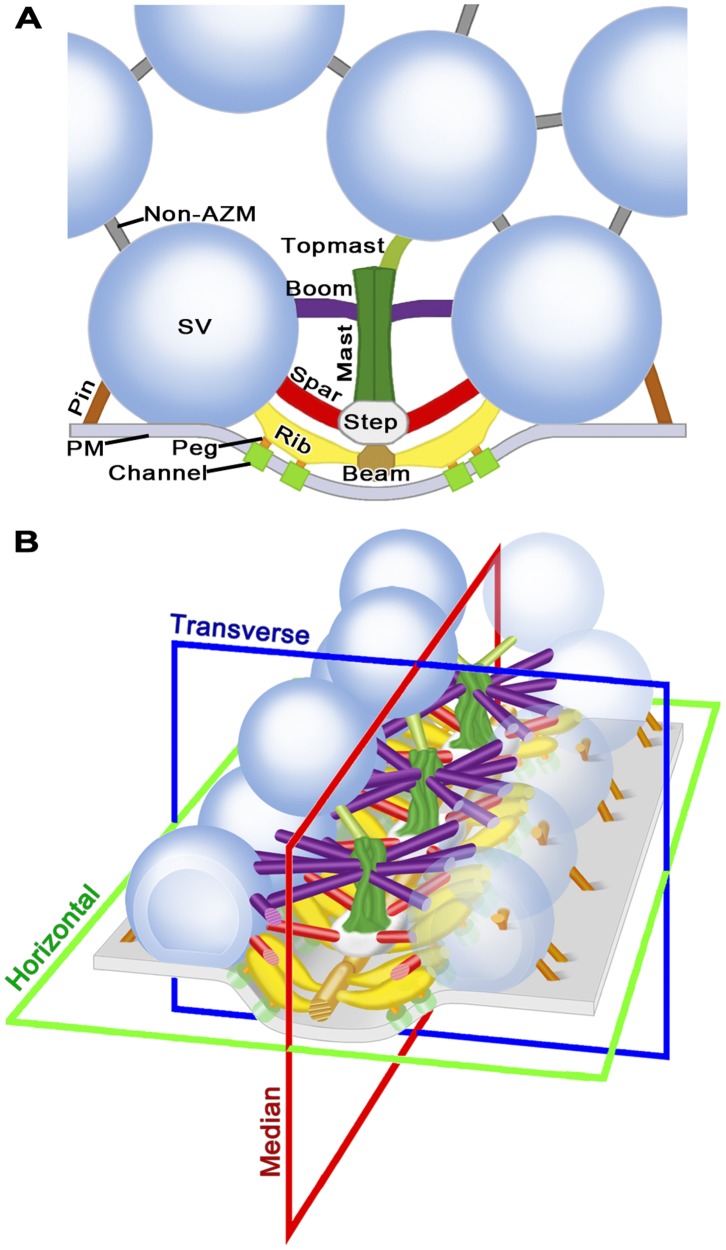
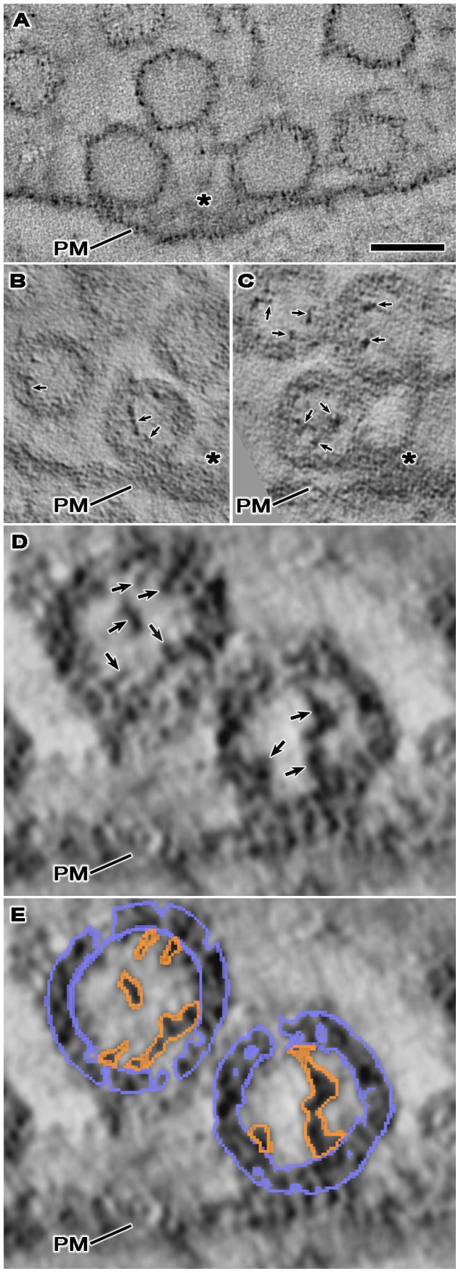
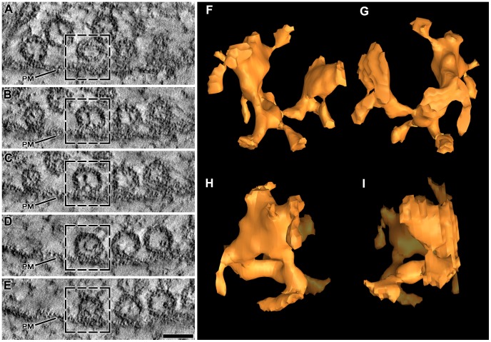
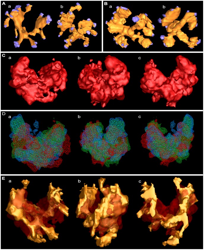
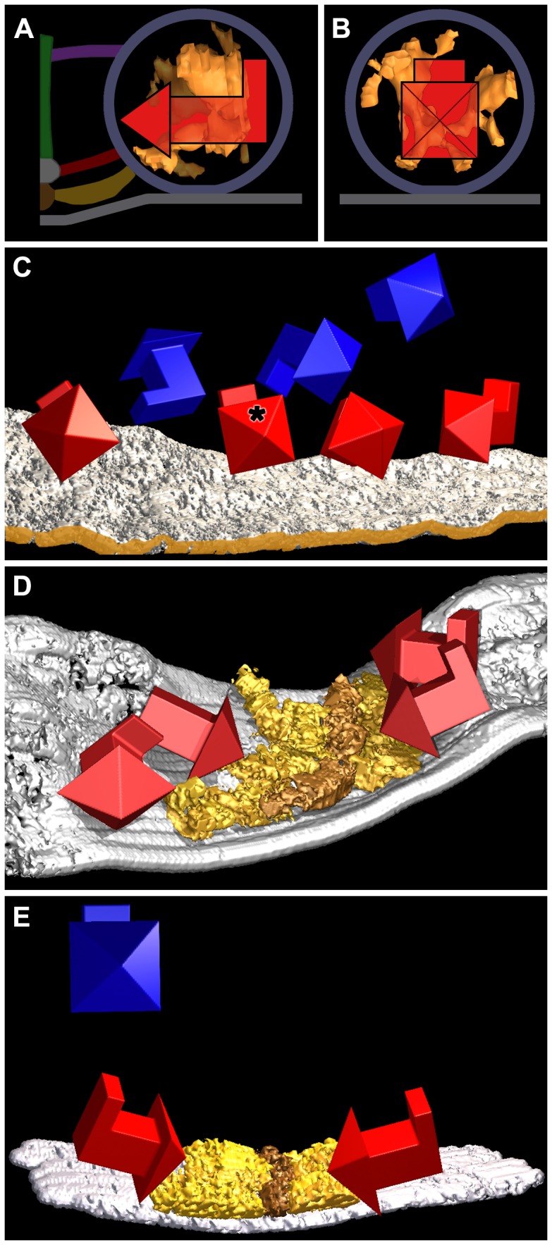
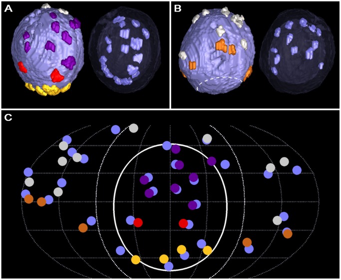
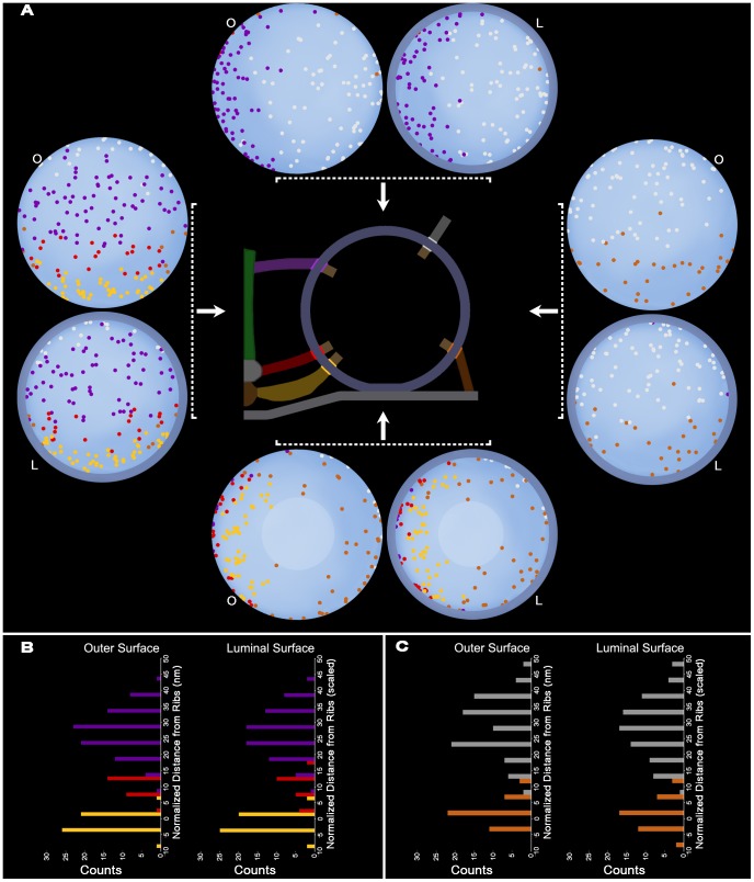
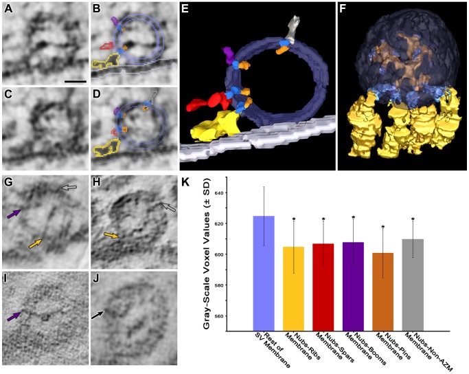
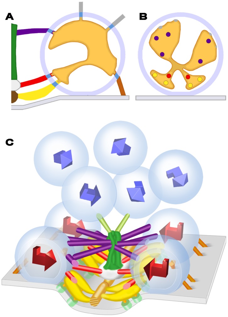
Synaptic vesicles dock at active zones on the presynaptic plasma membrane of a neuron's axon terminals as a precondition for fusing with the membrane and releasing their neurotransmitter to mediate synaptic impulse transmission. Typically, docked vesicles are next to aggregates of plasma membrane-bound macromolecules called active zone material (AZM). Electron tomography on tissue sections from fixed and stained axon terminals of active and resting frog neuromuscular junctions has led to the conclusion that undocked vesicles are directed to and held at the docking sites by the successive formation of stable connections between vesicle membrane proteins and proteins in different classes of AZM macromolecules. Using the same nanometer scale 3D imaging technology on appropriately stained frog neuromuscular junctions, we found that ∼10% of a vesicle's luminal volume is occupied by a radial assembly of elongate macromolecules attached by narrow projections, nubs, to the vesicle membrane at ∼25 sites. The assembly's chiral, bilateral shape is nearly the same vesicle to vesicle, and nubs, at their sites of connection to the vesicle membrane, are linked to macromolecules that span the membrane. For docked vesicles, the orientation of the assembly's shape relative to the AZM and the presynaptic membrane is the same vesicle to vesicle, whereas for undocked vesicles it is not. The connection sites of most nubs on the membrane of docked vesicles are paired with the connection sites of the different classes of AZM macromolecules that regulate docking, and the membrane spanning macromolecules linked to these nubs are also attached to the AZM macromolecules. We conclude that the luminal assembly of macromolecules anchors in a particular arrangement vesicle membrane macromolecules, which contain the proteins that connect the vesicles to AZM macromolecules during docking. Undocked vesicles must move in a way that aligns this arrangement with the AZM macromolecules for docking to proceed.


Conflict of interest statement

Competing Interests: The authors have declared that no competing interests exist.

Figures

Figure 1
Figure 1. Layout of the active zone at the frog’s NMJ.
(A) Composite diagram viewed in the active zone’s transverse plane. The main body of the AZM is between the two synaptic vesicles (SV) docked on the presynaptic membrane (PM). At the core of the main body are the beam, step and mast, each connected to the docked vesicles by the horizontally arranged ribs, spars and booms, respectively. Pegs, which were not included in this study (but see –[10]) connect the ribs to channels in the presynaptic membrane, while pins, which are AZM macromolecules away from the AZM’s main body connect the docked vesicles directly to the presynaptic membrane. A topmast links the mast to an undocked vesicle. Non-AZM macromolecules connect the docked vesicles to nearby undocked vesicles and are similar in appearance to macromolecules linking undocked vesicles to each other. (B) Schematic of a short segment of the active zone showing the 3D relationship of AZM macromolecules to docked vesicles and those undocked vesicles linked to topmasts, with indicators of the active zone’s horizontal, median and transverse planes. The color code for the structures shown here is the same for all Figures in this report. A complete description of the organization of AZM and non-AZM macromolecules can be found in .
Figure 2
Figure 2. The lumen of synaptic vesicles after staining under different conditions.
Each panel shows a virtual slice, 1–2 nm thick, through vesicles at or near an active zone (PM, presynaptic membrane; asterisk, main body of AZM). (A) The NMJ was fixed and stained with glutaraldehyde, osmium tetroxide and uranyl acetate at room temperature. (B) The NMJ was fixed with glutaraldehyde at room temperature and, after rapid freezing, fixed further and stained with osmium tetroxide and uranyl acetate in acetone by freeze-substitution. (C–E) The NMJ’s were fixed by rapid freezing, stained with osmium tetroxide and uranyl acetate in acetone by freeze-substitution. While the lumen of synaptic vesicles at the NMJ fixed and stained with glutaraldehyde, osmium tetroxide and uranyl acetate at room temperature appears empty (A), the lumen of synaptic vesicles at the NMJ’s, stained with osmium tetroxide and uranyl acetate in acetone by freeze-substitution, regardless of whether they were fixed by rapid freezing or with glutaraldehyde at room temperature, contains an assembly of macromolecules (B–D; arrows). The staining of the vesicle membrane and luminal assembly in all cases is particulate. The particles in the luminal assemblies have greater electron density than those in the membrane. In (E), the vesicle membrane and luminal assemblies shown in (D) are overlaid with the portion of the 3D surface models of the entire vesicle membrane (blue) and the luminal assembly (orange) that were generated from this virtual slice, which establishes the limits of the membrane width throughout the membrane’s circumference, the edges of the luminal assemblies, and the sites of connection of the luminal assemblies to the membrane. Scale bar (A–C) = 50 nm, (D–E) = 25 nm.
Figure 3
Figure 3. Shape of the luminal assembly of macromolecules in the principal vesicle.
(A–E) Selected 1 nm thick virtual slices from a series made through one of the primary reconstructions. The section from which the reconstruction was made was cut in the active zone’s median plane; the virtual slices are shown in the same plane. Four vesicles are docked in a row on the presynaptic membrane (PM). Undocked vesicles are nearby. All of the vesicles contain luminal assemblies of macromolecules. The box in each virtual slice outlines the so-called principal vesicle. (F–I) 3D surface model of the principal vesicle’s luminal assembly shown in different degrees of rotation. In (F) the assembly is viewed from the median plane of the AZM with the portion facing the presynaptic membrane downward. In (G) the assembly is rotated 180 degrees around the vertical axis of its orientation in (F). In (H) and (I) the assembly is rotated 90 degrees to the right and left, respectively, around the vertical axis of the orientation in (F). The assembly has a bilateral arrangement of four irregular arms, which radiate from below and behind the center of the vesicle. Nubs of varying lengths arise from the arms. Scale bar (A–E) = 50 nm.
Figure 4
Figure 4. Similarity in the shape of the luminal assemblies and their association with the vesicle membrane.
(Aa) Surface model of the assembly from the principal docked vesicle shown in Figure 3F–I. (Ab) Surface model of an assembly from another docked vesicle. (Ba,b) Surface models of assemblies from undocked vesicles. The assemblies in (Ab,Ba,Bb) were rotated until their shape matched that of the principal vesicle. The NMJ’s used for the assemblies in (A) and (B) were fixed by rapid freezing. In all cases, the assemblies are bilateral with irregular arms radiating from near the center of the vesicle. Nubs arise from the arms to connect at their end to the vesicle membrane; the terminal 3 voxels of each nub were made blue to mark its connection site on the membrane. (Ca–c) Alignment model generated by gray-scale density alignment of the 12 docked and undocked vesicles in the two primary reconstructions, which included those shown in (Aa,Ba). In (Ca) the alignment model is oriented according to the orientation of the surface model of the assembly from the principal vesicle as shown in (Aa). In (Cb) the alignment model is rotated 90 degrees to the right around the vertical axis in (Ca). In (Cc) the alignment model is rotated 180 degrees around the vertical axis in (Ca). The alignment model shows a bilateral arrangement of 4 radiating arms as do the models of individual assemblies in (Aa,b,Ba,b). (Da–c) The alignment model (red) generated from gray-scale density alignment of the 12 luminal assemblies in (Ca–c) aligned together with two alignment models (blue and green) generated from the same 12 assemblies represented according to their surface models. The different orientations of the superimposed alignment models are the same as for that in (Ca–c). The alignment models, which were calculated to have 95% 3D overlap, were used to measure the similarity in the shape of the assemblies from vesicle to vesicle and for establishing the orientation of the shape from vesicle to vesicle. (Ea–c) The surface model in (Aa) inserted into the alignment model in (Ca–c) according to its position in the alignment model. The different orientations of the combined models are the same as in (Ca–c). The portions of the surface model least included in the alignment model are the nubs, probably due to the nubs being smaller and/or having a somewhat greater variability in positioning than the arms among the surface models used for generating the alignment model.
Figure 5
Figure 5. Orientation of the shape of the luminal assemblies.
In (A), the surface model of the luminal assembly of the principal vesicle is viewed in the transverse plane of its active zone, while in (B) it is viewed from the median plane of the active zone. The 3D arrow superimposed on the surface model in both (A) and (B) is oriented so its head points to the median plane of the AZM, its shaft is orthogonal to the median plane and parallel to the presynaptic membrane and its tail is vertical to the presynaptic membrane. The vesicle membrane, presynaptic membrane, and AZM macromolecules are schematized for reference. (C) A surface model of the presynaptic membrane in a primary reconstruction, shown in the virtual slices in Figure 3A–E that include the principal vesicle, three other docked vesicles and three undocked vesicles. The edge of the membrane nearest the median plane of the AZM is brown-gold. The 3D arrows show the orientation of the shape of the luminal assembly in the docked (red) and undocked (blue) vesicles relative to that of the principal docked vesicle (asterisk) with respect to the median plane of the active zone and to the presynaptic membrane as shown in (A) and (B). The shape of the assembly in all of the docked vesicles has the same orientation (±30 degrees) as that of the principal vesicle, while the shape of the assembly in undocked vesicles does not share a common orientation. (D) Surface models of the presynaptic membrane and superficial layer of the AZM from the other primary reconstruction. The superficial layer of the AZM, which contains ribs (gold) and beams (brown-gold), is viewed from near the active zone’s transverse plane. There is a slight angular change midway along the AZM’s long axis . The 3D arrows show the orientation of the shape of the luminal assembly in four docked vesicles relative to that of the primary docked vesicle in (A–C) with respect to the median plane of the active zone and presynaptic membrane. The shape of the assembly for three of the docked vesicles had the same orientation (±30 degrees) with respect to the median plane of the active zone and to the presynaptic membrane as the docked vesicles in (C). While the shape of the assembly for the fourth vesicle (arrowhead and shaft lacking a tail) was similarly oriented with respect to the median plane of the active zone, there was not sufficient information to determine its orientation with respect to the presynaptic membrane. (E) Surface models of the presynaptic membrane and superficial layer of the AZM from another reconstruction. The ribs and beams of the AZM are viewed from near the active zone’s transverse plane. The 3D arrows show the orientation of the shape of the luminal assembly in two docked vesicles (red) and one undocked vesicle (blue) relative to that of the primary docked vesicle in (A–C) with respect to the median plane of its active zone and presynaptic membrane. The shape of the assembly for the two docked vesicles had the same orientation (±30 degrees) with respect to the median plane of the active zone and to the presynaptic membrane as the docked vesicles in (C) and (D), while the shape of the assembly in the undocked vesicle did not.
Figure 6
Figure 6. Pairing of connection sites of nubs with connection sites of AZM and non-AZM macromolecules.
(A,B) Surface model of the membrane of a docked vesicle in one of the primary reconstructions; (A) shows the hemisphere facing the main body of the AZM and (B) shows the hemisphere facing away from the main body. (A-left, B-left) Three-voxel-thick terminal portions of the AZM’s ribs (gold), spars (red), booms (purple) and pins (copper) and of non-AZM macromolecules (pewter), mark the connection site of each on the outer cytosolic surface of the vesicle membrane (pale blue). The vesicle’s fusion domain is indicated by the dashed line (B). (A-right, B-right) The AZM and non-AZM macromolecules are disabled on the surface model and the membrane has been made almost transparent. Three-voxel-thick terminal portions of the luminal assembly’s nubs (blue) mark the connection site of each on the luminal surface of the vesicle membrane. The distribution of the connection sites of the nubs is non-uniform and appears similar to the asymmetric distribution of the connection sites of the AZM and non-AZM macromolecules, although some of the similarity is obscured in these 2D images by the vesicle’s 3D curvature and the difference in the vesicle’s luminal and outer diameters. (C) Using x,y,z coordinates of the connection sites in (A,B), after correcting for the difference between luminal and outer diameters of the vesicle membrane, the relative positions of the centroids of the connection sites of the AZM and non-AZM macromolecules and of the nubs were plotted on a 2D Robinson map. The centroids were overlaid by filled circles slightly smaller in diameter than the connection sites and color-coded as in (A,B). The hemisphere of the vesicle facing the main body of the AZM lies between the bold longitudinal lines. The main AZM binding domain, which covers about 20% of the outer surface area of this vesicle and includes about half of the total nub and AZM/non-AZM macromolecule connection sites on the membrane, is encircled by the solid white line. Each nub is paired with an AZM or non-AZM macromolecule lying opposite to, or slightly offset from, it.
Figure 7
Figure 7. Composite maps of nub, AZM and non-AZM macromolecule connection sites.
(A) The centroids of the connection sites of different classes of AZM macromolecules, non-AZM macromolecules and nubs on the membrane of 11 docked vesicles from 4 active zones are plotted on the outer (O) and luminal (L) surfaces of an idealized vesicle. The hemispheres are viewed from (→) or toward (←) the midline of the main body of the AZM or from (↑) or toward (↓) the presynaptic membrane as indicated in the schematic of the active zone. The connection sites of ribs (gold), spars (red), booms (purple), pins (copper) and non-AZM macromolecules (pewter) were plotted on the idealized vesicle, while maintaining their relative positions per vesicle, using a cross correlation method that maximizes the degree of overlap of the rib connections to the outer surface (see Methods). The connection sites of nubs were plotted according to the same method using the nub connection sites paired with rib connection sites as the reference. Nub connection sites are color-coded according to the class of AZM/non-AZM macromolecules with which they were paired. There is an asymmetric distribution of AZM/non-AZM connection sites between the hemisphere facing the median plane of the AZM and the hemisphere facing away from the AZM, which is mirrored by an asymmetric distribution of nubs. Similarly, the asymmetric distribution of AZM/non-AZM connection sites between the hemisphere facing the presynaptic membrane and away from the presynaptic membrane, also is mirrored by an asymmetric distribution of nubs. The distribution of nub and AZM/non-AZM connection sites on the hemisphere facing the presynaptic membrane is greatly affected by the absence of such connections in the fusion domain (within the pale circular area). (B,C) The frequency of the connection sites of the different classes of main body AZM macromolecules on the outer surface and their paired nubs on the luminal surface (B), and non-AZM macromolecules and pins on the outer surface and their paired nubs on the luminal surface (C), as a function of distance from the average rib position per vesicle (see methods). The plots of the nub connections in (B) and (C) are normalized (scaled) for differences between the luminal and outer diameters of the vesicle membrane. The asymmetric distribution of the connection sites of the different classes of AZM and non-AZM macromolecules on the outer surface of the vesicle membrane reflects the asymmetric distribution of connection sites of their paired nubs on the luminal surface of the membrane.
Figure 8
Figure 8. Transmembrane bands of stain linking the connection sites of nubs to AZM and non-AZM macromolecules.
(A) Virtual slice ∼2–3 nm thick through a docked vesicle. (B) Same slice as in (A) with color-coded outlines of portions of nubs (orange), transmembrane bands of stain (darker blue), and portions of a rib (gold), a spar (red), and a boom (purple). The pale blue outlines marking the luminal and outer surfaces of the vesicle membrane are an overlay of this section’s contribution to a 3D surface model of the entire vesicle. The nubs are linked to the rib and boom by the transmembrane bands of stain. The band of stain and nub to which the spar is linked is evident in the next virtual slice, shown in (C). (C) A virtual slice adjacent to the one shown in (A) and (B). (D) Same slice as in (C) with outlines of portions of nubs, transmembrane bands of stain and portions of AZM macromolecules and the surfaces of the vesicle membrane color-coded as in (B). Portion of a non-AZM macromolecule is outlined in pewter. Nubs are connected by the transmembrane bands of stain to the spar, boom and non-AZM macromolecule. (E) Surface model ∼10 nm thick generated from a series of virtual slices showing in 3D the nubs linked by transmembrane bands of stain to the rib, spar, boom and non-AZM macromolecule outlined in (B) and (D). The membrane has been made partially transparent to enable viewing the extent of the transmembrane bands in the z-axis. (F) The membrane of the principal vesicle has been made transparent to reveal the luminal assembly of macromolecules (orange). Four ribs (gold) are connected by transmembrane bands of stain (dark blue) to nubs of the luminal assembly at their sites of connection to the luminal surface of the vesicle membrane. The connection sites of other nubs on the membrane were marked by coloring the portion of the nubs within three voxels of their connection site dark blue. (G–I) Virtual slices from different docked vesicles showing nubs linked to transmembrane bands of stain. In adjacent slices (not shown) the transmembrane bands of stain were linked to an AZM or non-AZM macromolecule as indicated by the colored arrows (ribs, gold; booms, purple; non-AZM macromolecules, pewter). (J) Virtual slice from an undocked vesicle showing a transmembrane band of stain linked to a nub. In the adjacent section, the transmembrane band of stain was linked to a non-AZM macromolecule as indicated by the black arrow. Scale bar (A–D,G–J) = 25 nm. (K) For a single vesicle, the frequency distribution of the voxel gray-scale values (ranging from 0, black, to 1000, white) of the narrow region of the vesicle membrane between nub connection sites and the connection sites of opposed AZM and non-AZM macromolecules, i.e. the region containing the transmembrane band of stain, was compared with the voxel gray-scale values for the rest of the synaptic vesicle (SV) membrane. The voxel gray-scale values in the regions between 24 nubs and their opposed ribs (4), spars (2), booms (6), pins (4) and non-AZM macromolecules (8) were on average significantly darker than the rest of the vesicle membrane (asterisks) as determined by ANOVA with a Tukey Post Hoc Test (p<0.05). An additional 7 vesicles were similarly tested and the results are displayed in Table 1 (details of results from this vesicle are shown at #8 in Table 1). Altogether, the findings indicate that, on average, the transmembrane regions linking nubs to AZM and non-AZM macromolecules have a greater density of stained material than the remainder of the vesicle membrane.
Figure 9
Figure 9. Schematized relationships of the luminal assembly of macromolecules at frog NMJ’s.
(A) Profile of a vesicle (pale blue) docked on the presynaptic membrane (gray) viewed in the transverse plane of the active zone. AZM macromolecules, including a rib (gold), a spar (red), a boom (purple) and a pin (copper), connect to the outer surface of the vesicle membrane as do non-AZM macromolecules (pewter). The luminal assembly of macromolecules is orange. Nubs arise from the luminal assembly’s arms to connect to the vesicle membrane, where they are linked to macromolecules that span the membrane (dark blue) and connect to the AZM and non-AZM macromolecules. (B) Profile of a docked vesicle viewed from the active zone’s median plane. The arms of the luminal assembly radiate from a point near the center of the vesicle and are larger near the vesicle membrane than at the vesicle’s center. The arms in the hemisphere facing the presynaptic membrane are shorter than those away from the presynaptic membrane. Colored spots on the luminal assembly mark regions connected by nubs and their membrane spanning macromolecules to specific classes of AZM and non-AZM macromolecules, as in (A). C) 3D arrangement of docked and nearby undocked vesicles relative to the AZM and presynaptic membrane. The orientation of the stereotypic shape of the luminal assembly with respect to the median plane of the AZM and presynaptic membrane is indicated by 3D arrows. The orientation is the same for docked vesicles (red arrows) while for undocked vesicles (blue arrows) it is not. Thus, in order for an undocked vesicle to replace a docked vesicle that fuses with the presynaptic membrane during synaptic transmission, it must, typically, rotate so the appropriate vesicle membrane macromolecules linked to the luminal assembly (A,B) can sequentially interact with the different classes of AZM macromolecules that direct it to the docking site on the presynaptic membrane.

References

    1. Katz B (1969) The release of neural transmitter substances. Springfield, Ill.,: Thomas. ix, 60 p.
    1. Peters A, Palay SL, Webster Hd (1991) The fine structure of the nervous system : neurons and their supporting cells. New York: Oxford University Press. xviii, 494 p.
    1. Couteaux R, Pecot-Dechavassine M (1970) [Synaptic vesicles and pouches at the level of “active zones” of the neuromuscular junction]. C R Acad Sci Hebd Seances Acad Sci D 271: 2346–2349. - PubMed
    1. Heuser JE, Reese TS, Dennis MJ, Jan Y, Jan L, et al. (1979) Synaptic vesicle exocytosis captured by quick freezing and correlated with quantal transmitter release. J Cell Biol 81: 275–300. - PMC - PubMed
    1. Südhof TC (2004) The synaptic vesicle cycle. Annu Rev Neurosci 27: 509–547. - PubMed

Publication types

Substances

LinkOut - more resources