. 2013 Jul 23;8(7):e69572.
doi: 10.1371/journal.pone.0069572. Print 2013.

A simple model system enabling human CD34(+) cells to undertake differentiation towards T cells

Affiliations

A simple model system enabling human CD34(+) cells to undertake differentiation towards T cells

Antonio Lapenna et al. PLoS One. .

Abstract

Background: Channelling the development of haematopoietic progenitor cells into T lymphocytes is dependent upon a series of extrinsic prompts whose temporal and spatial sequence is critical for a productive outcome. Simple models of human progenitor cells development depend in the main on the use of xenogeneic systems which may provide some limitations to development.

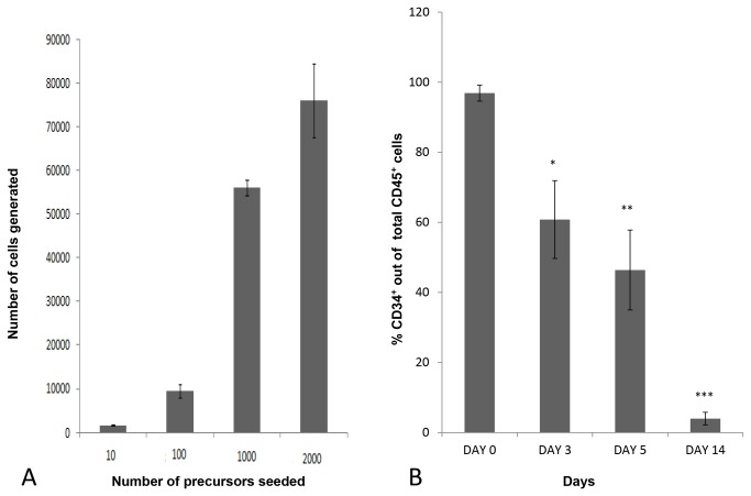
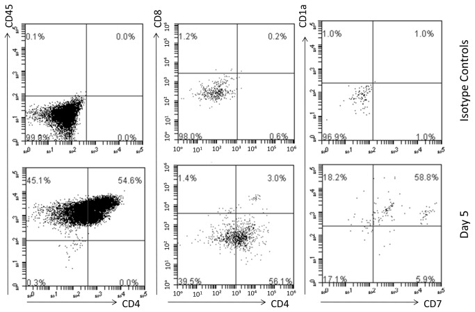
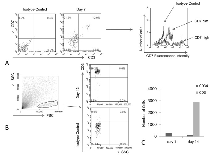
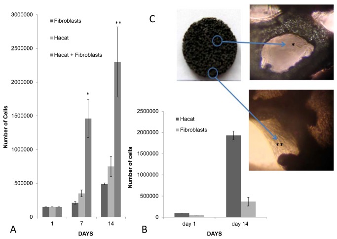
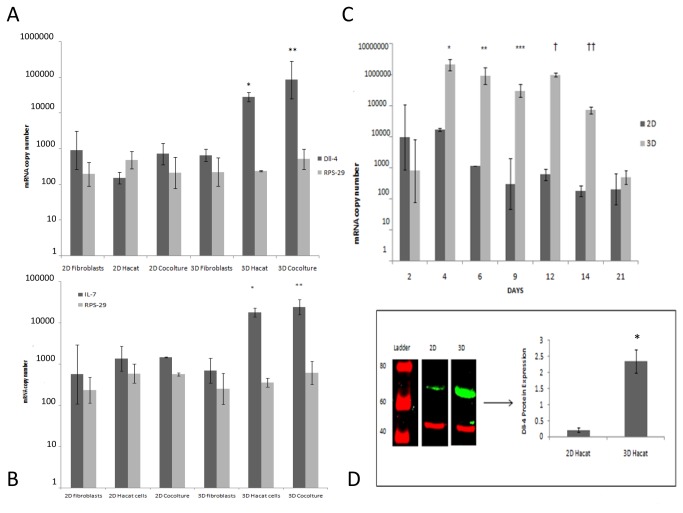
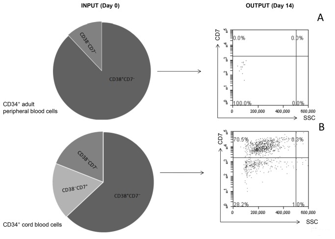
Methods and findings: Here we provide evidence that a simple model system which utilises both human keratinocyte and fibroblast cell lines arrayed on a synthetic tantalum coated matrix provides a permissive environment for the development of human CD34⁺ haematopoietic cells into mature CD4⁺ or CD8⁺ T lymphocytes in the presence of Interleukin 7 (IL-7), Interleukin 15 (IL-15) and the Fms-like tyrosine kinase 3 ligand (Flt-3L). This system was used to compare the ability of CD34(+) cells to produce mature thymocytes and showed that whilst these cells derived from cord blood were able to productively differentiate into thymocytes the system was not permissive for the development of CD34(+) cells from adult peripheral blood.

Conclusions/significance: Our study provides direct evidence for the capacity of human cord blood CD34(+) cells to differentiate along the T lineage in a simple human model system. Productive commitment of the CD34⁺ cells to generate T cells was found to be dependent on a three-dimensional matrix which induced the up-regulation of the Notch delta-like ligand 4 (Dll-4) by epithelial cells.


Conflict of interest statement

Competing Interests: The authors have declared that no competing interests exist.

Figures

Figure 1
Figure 1. Expansion and differentiation of CD34+ cells.
(A) Correlation between the initial number of CD34+ cells seeded and the amount of mature cells generated at day 14th. The results are the average ± standard derivation of three different experiments. (B) Progressive decline with time of CD34 expression among cord blood cellscultured in the matrix. The results are the average of three different experiments ± standard derivation. The differences between the 3rd, 5th and 14th day and the seeded population are all significant (*p< 0.001; **p< 0.001; ***p< 0.001).
Figure 2
Figure 2. Kinetics of thymocytes generation.
(A) By day 5 CD4 dimintermediate single positive and some double positive CD4+CD8+ cells were present. These progenitors all expressed CD45, either high or dim and analysis of cultures also showed the presence of CD1a+CD7+ and CD1a+CD7 cells The images are representative of three different experiments.
Figure 3
Figure 3. Generation of CD3+ thymocytes.
(A) CD7hiCD3hi and CD7 dim CD3⁻ cells were detected at day 7. (B) By day 12 approximately 90% of all the cells generated were CD3+ thymocytes. (C) A matrix seeded with approximately 300 CD34+ cord blood derived progenitors generated about 2900 CD3+ cells after 14 days. At that time about 150 CD34+ progenitors were still present whereas no other cell types were detected. The image A is representative of three different experiments while images B and C show a single experiment.
Figure 4
Figure 4. Most of generated cells are mature thymocytes by day12.
The presence of double positive CD4+CD8+ and either CD4+ or CD8+ single positive CD3+ thymocytes was evident by day 12 when only about 2% of total CD45+ cells still expressed CD34. The images are representative of three different experiments.
Figure 5
Figure 5. Hacat keratinocytes and fibroblasts growth in the matrices.
(A) Growth curves of Hacat keratinocytes and/or fibroblasts cultured in the matrices: The differences between the co-culture and the separated components are all statistically significant (*p < 0.001; **p < 0.001) and the results shown are the average of three different experiments ± standard deviation. (B) At the 14th day of culture the matrices seeded with Hacat keratinocytes and fibroblasts at a 2:1 ratio were predominantly constituted by the former cell type. The cells were distinguished by CD10 expression and the results shown are the average of three different experiments. (C) Images of cells attached either to the matrix borders (*) or inner niches (**). The tantalum skeleton of the matrix appears in black. Light microscope image (100X).
Figure 6
Figure 6. Dll-4 and IL-7 are up-regulated by three-dimensional cultured keratinocytes.
(A) Dll-4 gene expression is strongly up-regulated in 3D cultured Hacat keratinocytes either alone or in the presence of fibroblasts. The differences between 3D and 2D either Hacat (*p<0.01) or cocultures (**p<0.05) are significant. No differences were observed in the housekeeping gene expression in all the conditions tested. The results shown are the average of three different experiments ± standard deviation. (B) IL-7 gene expression is strongly up-regulated in 3D cultured Hacat keratinocytes either alone or in the presence of fibroblasts. The differences between 3D and 2D either Hacat (*p<0.05) or co-cultures (**p<0.05) are significant. No differences were observed in the housekeeping gene expression in all the conditions tested. The results shown are the average of three different experiments ± standard deviation. (C) Time dependent up-regulation of the Dll-4 gene in three-dimensional Hacat keratinocytes/ fibroblasts co-cultures. A strong induction is observed during the first week and this high expression is maintained for about 10 days. The results shown are the average of three different experiments ± standard deviation and the differences between 2D and 3D within days 4-14 (* p < 0.01; ** p < 0.01; *** p <0.05; † p < 0.01; † † p < 0.01), are all statistically significant. (D) Dll-4 protein expression in 2D and 3D cultured Hacat keratinocytes. The western blot image shows different parts of one single gel. The average of Dll-4 level of expression normalized to actin from three different experiments ± standard deviation differs between the 2D and 3D environment and the difference is significant (p< 0.001).
Figure 7
Figure 7. TREC analysis shows that thymocytes were generated de novo.
(A) TREC was amplified from DNA from cells generated in the matrices after 10 days of co-culture but not from DNA from cord blood separated precursors. RPS-29 housekeeping gene was amplified in both cases. TREC and RPS-29 were respectively identified as a band of 192 and 142 base pairs. (B) TREC/CD3+ ratios from cord blood T cells and thymocytes generated in the matrices. The latter cells show higher level of TREC expression per cell compared to T cells which were separated from cord blood. The results shown are the average of three different experiments± standard deviation and the difference is significant (p < 0.001).
Figure 8
Figure 8. Cord and peripheral CD34⁺ precursors are dissimilar.
(A) Thymocytes were never found in the supernatant of matrices seeded with CD34⁺ adult peripheral blood cells and checked up to 2 weeks of co-culture. (B) Cord blood cells gave rise to CD7+thymocytes. The images are representative of three different experiments all performed in parallel and reports initial differences in the CD34⁺ cells subsets composition.

Similar articles

Cited by

References

    1. De la Hera A, Marston W, Aranda C, Toribio ML, Martinez C (1989) Thymic stroma is required for the development of human T cell lineages in vitro. Int Immunol 1(5): 471-478. doi:10.1093/intimm/1.5.471. PubMed: 2489037. - DOI - PubMed
    1. Mitchell WA, Lang PO, Aspinall R (2010) Tracing thymic output in older individuals. Clin Exp Immunol 161(3): 497-503. doi:10.1111/j.1365-2249.2010.04209.x. PubMed: 20646007. - DOI - PMC - PubMed
    1. Law LW (1966) Restoration of thymic function in neonatally thymectomized mice bearing xenogeneic thymic grafts. Nature 210: 1118. doi:10.1038/2101118a0. PubMed: 5336677. - DOI - PubMed
    1. Anderson G, Jenkinson EJ (2008) Bringing the thymus to the bench. J Immunol 181(11): 7435-7436. PubMed: 19017928. - PubMed
    1. de Pooter R, Zúñiga-Pflücker JC (2007) T-cell potential and development in vitro: the OP9-DL1 approach. Curr Opin Immunol 19: 163-168. doi:10.1016/j.coi.2007.02.011. PubMed: 17303399. - DOI - PubMed

Publication types