. 2013 Jul 23;8(7):e69950.
doi: 10.1371/journal.pone.0069950. Print 2013.

Porous chitosan scaffolds with embedded hyaluronic acid/chitosan/plasmid-DNA nanoparticles encoding TGF-β1 induce DNA controlled release, transfected chondrocytes, and promoted cell proliferation

Affiliations

Porous chitosan scaffolds with embedded hyaluronic acid/chitosan/plasmid-DNA nanoparticles encoding TGF-β1 induce DNA controlled release, transfected chondrocytes, and promoted cell proliferation

Huading Lu et al. PLoS One. .

Abstract

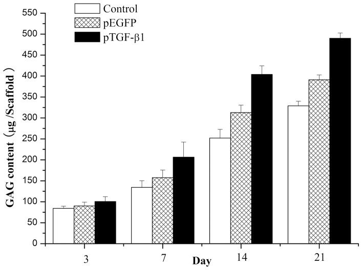
Cartilage defects resulting from traumatic injury or degenerative diseases have very limited spontaneous healing ability. Recent progress in tissue engineering and local therapeutic gene delivery systems has led to promising new strategies for successful regeneration of hyaline cartilage. In the present study, tissue engineering and local therapeutic gene delivery systems are combined with the design of a novel gene-activated matrix (GAM) embedded with hybrid hyaluronic acid(HA)/chitosan(CS)/plasmid-DNA nanoparticles encoding transforming growth factor (TGF)-β1. A chitosan scaffold functioned as the three-dimensional carrier for the nanoparticles. Results demonstrated that scaffold-entrapped plasmid DNA was released in a sustained and steady manner over 120 days, and was effectively protected in the HA/CS/pDNA nanoparticles. Culture results demonstrated that chondrocytes grown in the novel GAM were highly proliferative and capable of filling scaffold micropores with cells and extracellular matrix. Confocal laser scanning microscopy indicated that chondrocytes seeded in the GAM expressed exogenous transgenes labeled with green fluorescent protein. ELISA results demonstrated detectable TGF-β1 expression in the supernatant of GAM cultures, which peaked at the sixth day of culture and afterwards showed a moderate decline. Histological results and biochemical assays confirmed promotion of chondrocyte proliferation. Cell culture indicated no affects on phenotypic expression of ECM molecules, such as GAG. The results of this study indicate the suitability of this novel GAM for enhanced in vitro cartilage tissue engineering.


Conflict of interest statement

Competing Interests: The authors have declared that no competing interests exist.

Figures

Figure 1
Figure 1. Scanning electron microscopy (SEM) micrographs of nanoparticles and scaffolds.
Panel A shows a representative image of lyophilized HA/CS/pDNA nanoparticles prepared with a weight ratio of CS:HA of 4∶1 and 25 µg/mL of pDNA. Panel B shows the porous chitosan scaffolds without nanoparticles. Panel C shows a GAM scaffold embedding HA/CS/pDNA nanoparticles. Panel D–F shows a gradually magnified view of GAM surface morphology featuring entrapped HA/CS/pDNA nanoparticles. Panel F shows many spherical nanoparticles on the surface of the scaffold, ranging in size from 100–300 nm.
Figure 2
Figure 2. Gel electrophoresis of HA/CS/pTGF-β1 nanoparticles, digests, and control samples.
Lane A shows the DNA molecular weight marker. Lane B shows HA/CS/pTGF-β1 nanoparticles. Lane C shows normal naked pTGF-β1. Lane D shows HA/CS/pTGF-β1 nanoparticles digested by DNase I. Lane E shows naked pTGF-β1 digested by DNase I. Lane F shows HA/CS/pTGF-β1 nanoparticles digested by chitosanase (2.78 µg/µL).
Figure 3
Figure 3. Cumulative pDNA release profiles of HA/CS/pDNA nanoparticles and the GAM embedding HA/CS/pDNA nanoparticles as a function of time up to 120 days.
Figure 4
Figure 4. SEM images of chondrocytes cultured for 7 days with plain chitosan scaffolds (A and D), GAM scaffolds embedding empty plasmid scaffolds (B and E), and GAM scaffolds embedding TGF-β1 nanoparticles (C and F). Chondrocytes formed confluent cells on the surface of GAM scaffolds.
Panels A, B, C: ×100; D, E, F: ×400.
Figure 5
Figure 5. Confocal laser scanning microscopic observation of chondrocyte-scaffolds cultures on the 7th and 21st day.
Panels A, B, C show chondrocytes cultured for 7 days while panels D, E, F show chondrocytes cultured for 21 days. A large number of GFP positive chondrocytes clusters are visible for culture with GAM embedding HA/CS/pDNA nanoparticles encoding either EGFP (B and E) or TGF-β1(C and F), indicating transfection by expression of GFP. No GFP-positive cells were detected in chitosan-only scaffolds (A and C). Images come from screenshots of 3-D scanning images of the examined area.
Figure 6
Figure 6. The TGF-β1 expressed in cultures was analyzed by ELISA.
Chondrocytes incubated in the GAM embedding HA/CS/pTGF-β1 nanoparticles produced a higher level of TGF-β1 during the culture period. The maximum concentration of TGF-β1 in the culture media was detected on day 6, after which point levels declined moderately (n = 3).
Figure 7
Figure 7. Histology (toluidine blue staining) of chondrocytes seeded to chitosan-only scaffolds, GAM embedding empty plasmid nanoparticles, and GAM embedding HA/CS/pTGF-β1 nanoparticles for 7 and 21 days of culture.
Toluidine blue stains ECM, therefore cell clusters intensely stained by toluidine blue indicate newly synthesized proteoglycans. Cell clusters were much denser and evenly distributed in GAM embedding HA/CS/pTGF-β1 nanoparticles than the other two control groups. Panels A, B, C: ×100; D, E, F: ×400; G, H, I: ×40; J, K, L: ×200.
Figure 8
Figure 8. Immunohistochemistry staining for type II collagen in chondrocytes cultured on the chitosan-only scaffold, GAM embedding HA/CS/pEGFP nanoparticles, and GAM embedding HA/CS/pTGF-β1 nanoparticles for 7 and 21 days.
Panels A, B, C: ×100; D, E, F: ×200; G, H, I: ×100; J, K, L: ×400.
Figure 9
Figure 9. DNA content per sample for chitosan-only scaffolds encapsulating chondrocytes, chondrocytes/GAM embedding HA/CS/pEGFP nanoparticles, or chondrocytes/GAM embedding HA/CS/pTGF-β1 nanoparticles.
At a given time point, samples marked by (*) exhibited significantly higher cellularity than chitosan scaffolds of DNA-free samples containing only chondrocytes. Samples indicated with (**) had significantly higher DNA amount than the other two groups (p<0.05). Error bars represent mean (n = 4) ± s.d.
Figure 10
Figure 10. GAG content per sample for chitosan-only scaffolds encapsulating chondrocytes, chondrocytes/GAM embedding HA/CS/pEGFP nanoparticles, or chondrocytes/GAM embedding HA/CS/pTGF-β1 nanoparticles.
At a given time point, (*) indicates that samples of chondrocytes/GAM embedding HA/CS/pTGF-β1 nanoparticles exhibit significant higher (p<0.05) GAG than samples of the other two group. Error bars represent mean (n = 4) ± s.d.
Figure 11
Figure 11. GAG content per amount of DNA of chondrocyte in chitosan-only scaffolds, GAM embedding HA/CS/pEGFP nanoparticles, or GAM embedding HA/CS/pTGF-β1 nanoparticles.
No significant differences were observed when values were compared amongst experimental groups at a given time point. Error bars represent mean (n = 4) ± s.d.

Similar articles

Cited by

References

    1. Bachmann G, Basad E, Lommel D, Steinmeyer J (2004) [MRI in the follow-up of matrix-supported autologous chondrocyte transplantation (MACI) and microfracture]. Radiologe 44: 773–782 doi:10.1007/s00117-004-1084-y. PMID: 15278206 - DOI - PubMed
    1. Macchiarini P, Jungebluth P, Go T, Asnaghi MA, Rees LE, et al. (2008) Clinical transplantation of a tissue-engineered airway. Lancet 372: 2023–2030 doi:10.1016/S0140-6736(08)61598-6. PMID: 19022496 - DOI - PubMed
    1. Asnaghi MA, Jungebluth P, Raimondi MT, Dickinson SC, Rees LE, et al. (2009) A double-chamber rotating bioreactor for the development of tissue-engineered hollow organs: from concept to clinical trial. Biomaterials 30: 5260–5269 doi:10.1016/j.biomaterials.2009.07.018. PMID: 19647867 - DOI - PubMed
    1. Chung C, Burdick JA (2008) Engineering cartilage tissue. Adv Drug Deliv Rev 60: 243–262 doi:10.1016/j.addr.2007.08.027. PMID: 17976858 - DOI - PMC - PubMed
    1. Guo T, Zhao J, Chang J, Ding Z, Hong H, et al. (2006) Porous chitosan-gelatin scaffold containing plasmid DNA encoding transforming growth factor-beta1 for chondrocytes proliferation. Biomaterials 27: 1095–1103 doi:10.1016/j.biomaterials.2005.08.015, PMID: 16143394. - DOI - PubMed

Publication types