

Unravelling the functional biomechanics of dental features and tooth wear
- PMID: 23894570
- PMCID: PMC3720920
- DOI: 10.1371/journal.pone.0069990
Unravelling the functional biomechanics of dental features and tooth wear
Abstract
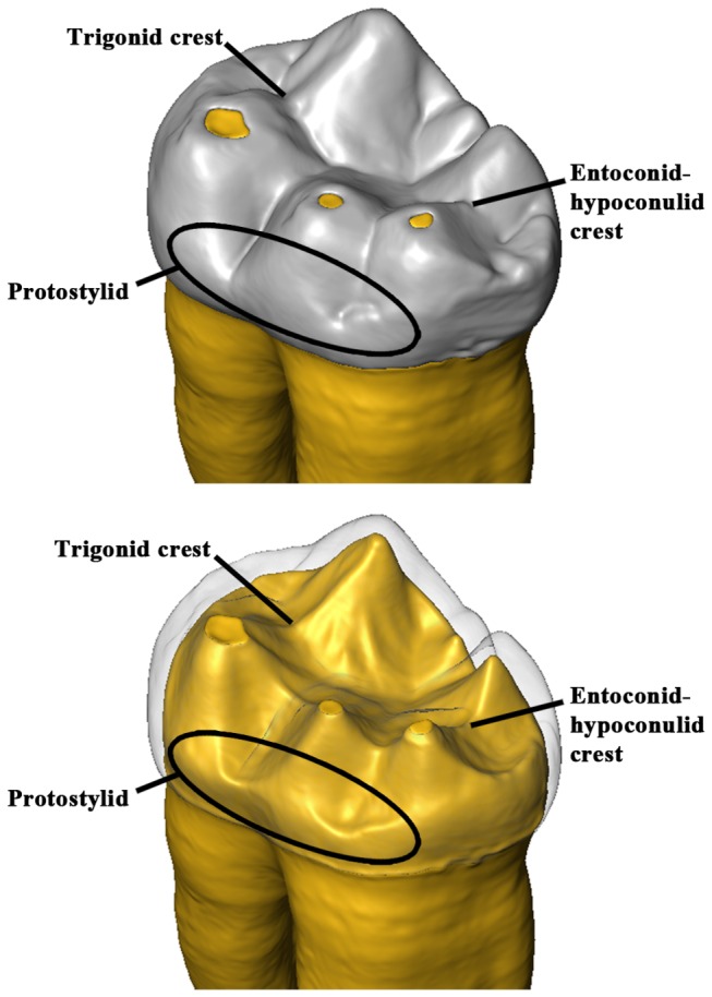
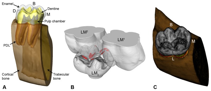
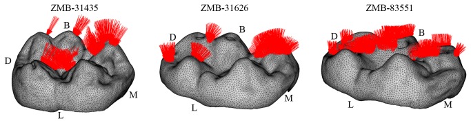
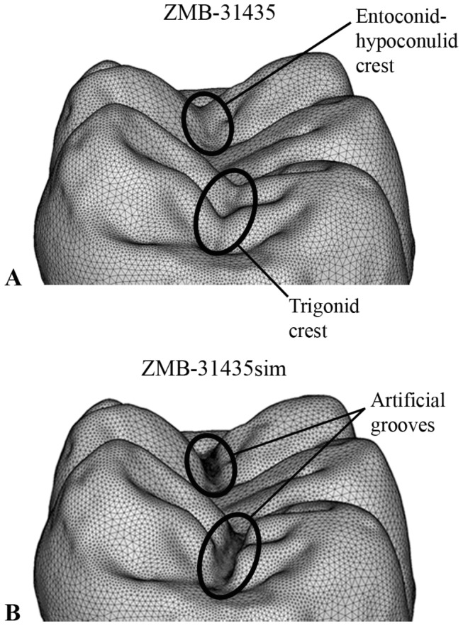
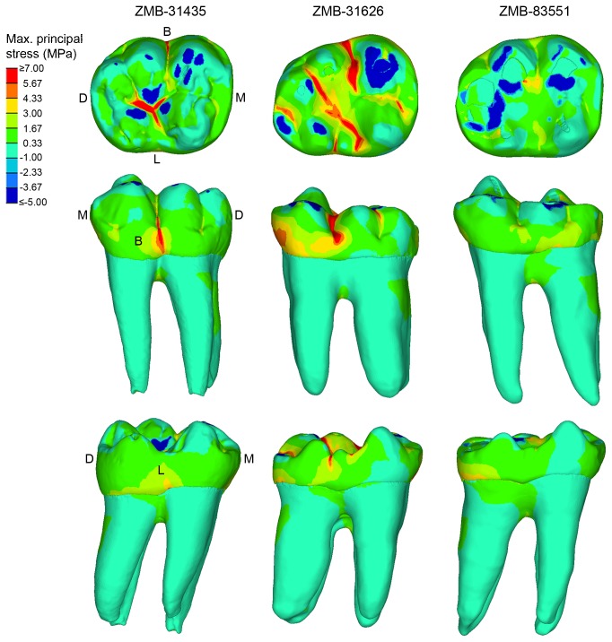
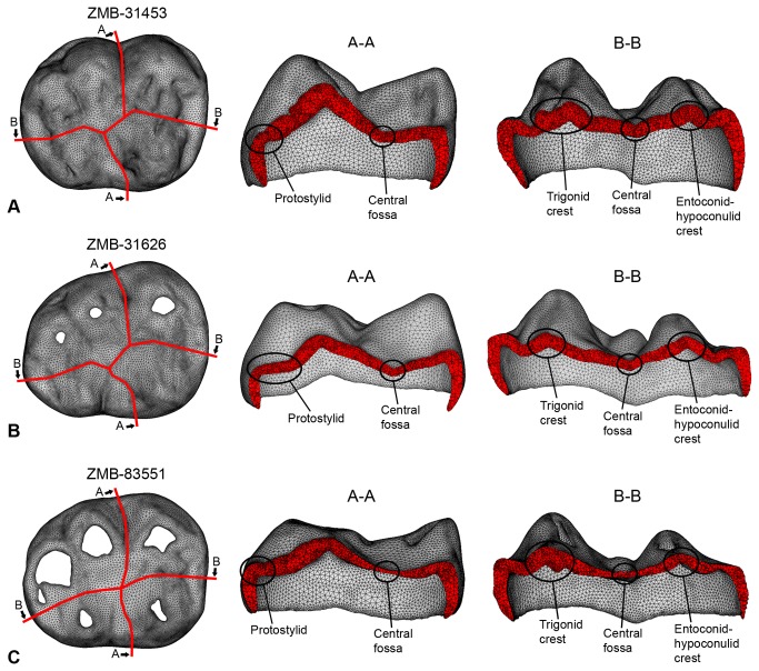
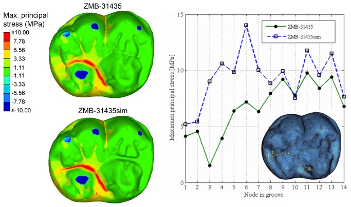
Most of the morphological features recognized in hominin teeth, particularly the topography of the occlusal surface, are generally interpreted as an evolutionary functional adaptation for mechanical food processing. In this respect, we can also expect that the general architecture of a tooth reflects a response to withstand the high stresses produced during masticatory loadings. Here we use an engineering approach, finite element analysis (FEA), with an advanced loading concept derived from individual occlusal wear information to evaluate whether some dental traits usually found in hominin and extant great ape molars, such as the trigonid crest, the entoconid-hypoconulid crest and the protostylid have important biomechanical implications. For this purpose, FEA was applied to 3D digital models of three Gorillagorilla lower second molars (M2) differing in wear stages. Our results show that in unworn and slightly worn M2s tensile stresses concentrate in the grooves of the occlusal surface. In such condition, the trigonid and the entoconid-hypoconulid crests act to reinforce the crown locally against stresses produced along the mesiodistal groove. Similarly, the protostylid is shaped like a buttress to suffer the high tensile stresses concentrated in the deep buccal groove. These dental traits are less functional in the worn M2, because tensile stresses decrease physiologically in the crown with progressing wear due to the enlargement of antagonistic contact areas and changes in loading direction from oblique to nearly parallel direction to the dental axis. This suggests that the wear process might have a crucial influence in the evolution and structural adaptation of molars enabling to endure bite stresses and reduce tooth failure throughout the lifetime of an individual.
Conflict of interest statement
Figures
References
-
- Berthaume M, Grosse IR, Patel ND, Strait DS, Wood S et al. (2010) The effect of early hominin occlusal morphology on the fracturing of hard food items. Anat Rec 293: 594-606. doi:10.1002/ar.21130. - DOI - PubMed
-
- Kay RF (1975) The functional adaptations of primate molar teeth. Am J Phys Anthropol 43: 195-216. doi:10.1002/ajpa.1330430207. PubMed: 810034. - DOI - PubMed
-
- Scott RS, Ungar PS, Bergstrom TS, Brown CA, Grine FE et al. (2005) Dental microwear texture analysis shows within-species diet variability in fossil hominins. Nature 436: 693-695. doi:10.1038/nature03822. PubMed: 16079844. - DOI - PubMed
-
- Ungar P (2004) Dental topography and diets of Australopithecus afarensis and early Homo. J Hum Evol 46: 605-622. doi:10.1016/j.jhevol.2004.03.004. PubMed: 15120268. - DOI - PubMed
-
- Ungar PS, Sponheimer M (2011) The diets of early hominins. Science 334: 190-193. doi:10.1126/science.1207701. PubMed: 21998380. - DOI - PubMed
Publication types
MeSH terms
LinkOut - more resources
Full Text Sources
Other Literature Sources
Research Materials
Miscellaneous
