. 2013 Jul 23;8(7):e70177.
doi: 10.1371/journal.pone.0070177. Print 2013.

Expression of kisspeptins and kiss receptors suggests a large range of functions for kisspeptin systems in the brain of the European sea bass

Affiliations

Expression of kisspeptins and kiss receptors suggests a large range of functions for kisspeptin systems in the brain of the European sea bass

Sebastián Escobar et al. PLoS One. .

Abstract

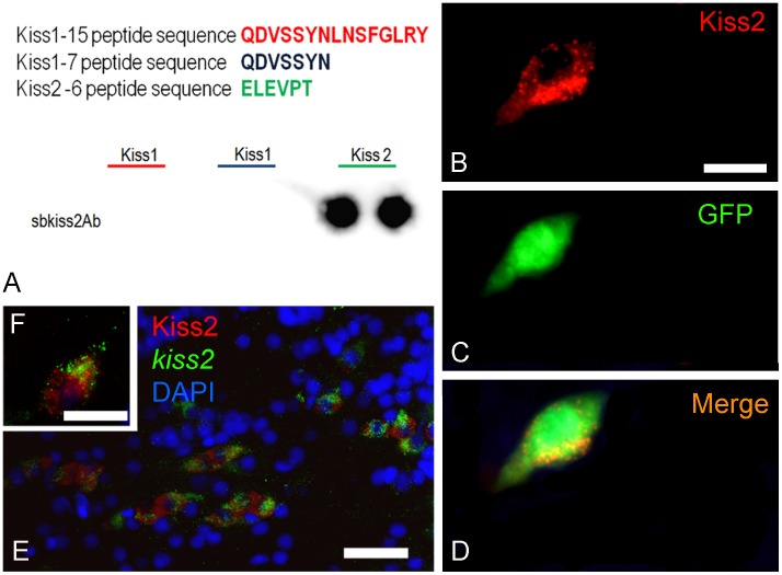
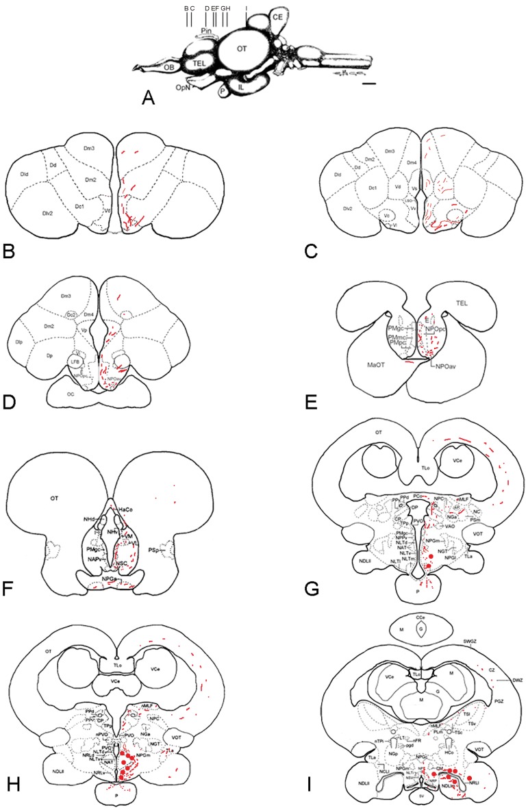
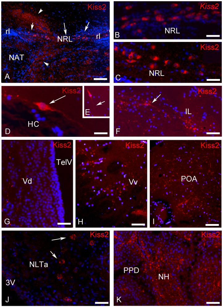
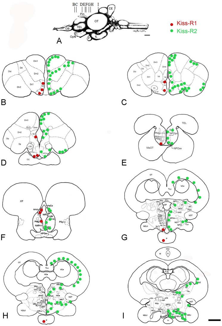
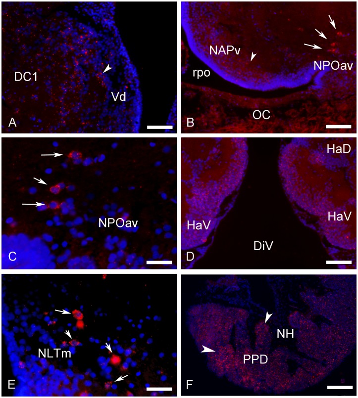
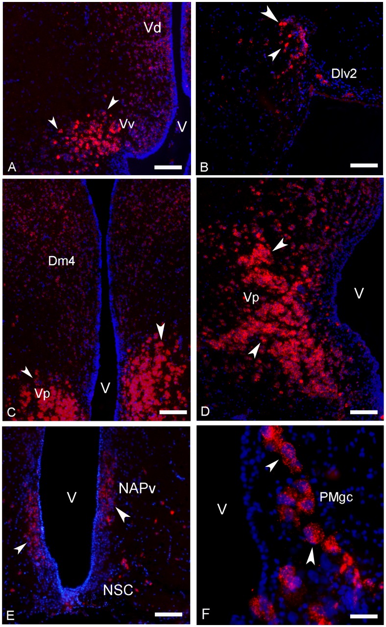
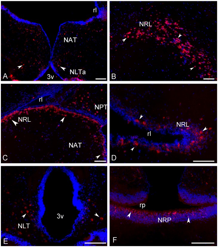
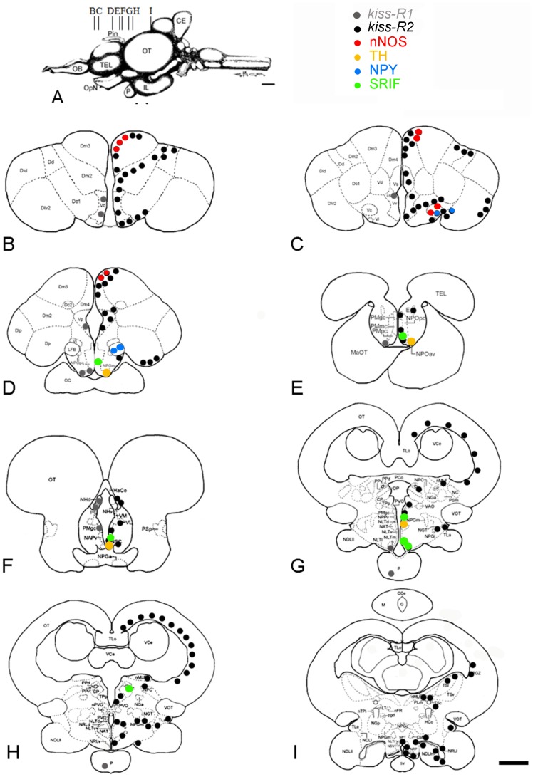
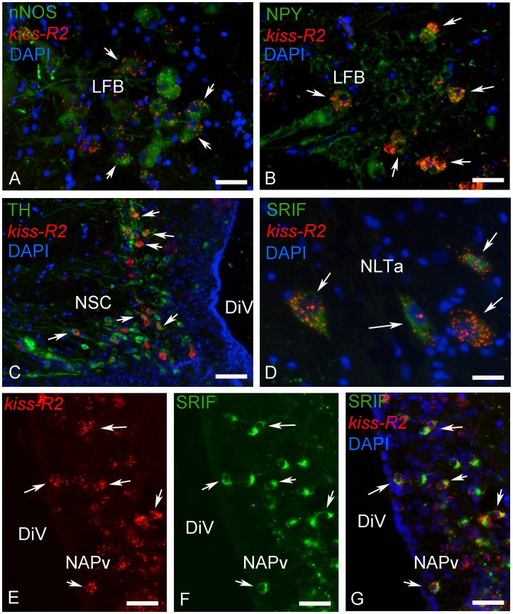
This study, conducted in the brain of a perciform fish, the European sea bass, aimed at raising antibodies against the precursor of the kisspeptins in order to map the kiss systems and to correlate the expression of kisspeptins, kiss1 and kiss2, with that of kisspeptin receptors (kiss-R1 and kiss-R2). Specific antibodies could be raised against the preprokiss2, but not the preoprokiss1. The data indicate that kiss2 neurons are mainly located in the hypothalamus and project widely to the subpallium and pallium, the preoptic region, the thalamus, the pretectal area, the optic tectum, the torus semicircularis, the mediobasal medial and caudal hypothalamus, and the neurohypophysis. These results were compared to the expression of kiss-R1 and kiss-R2 messengers, indicating a very good correlation between the wide distribution of Kiss2-positive fibers and that of kiss-R2 expressing cells. The expression of kiss-R1 messengers was more limited to the habenula, the ventral telencephalon and the proximal pars distalis of the pituitary. Attempts to characterize the phenotype of the numerous cells expressing kiss-R2 showed that neurons expressing tyrosine hydroxylase, neuropeptide Y and neuronal nitric oxide synthase are targets for kisspeptins, while GnRH1 neurons did not appear to express kiss-R1 or kiss-R2 messengers. In addition, a striking result was that all somatostatin-positive neurons expressed-kissR2. These data show that kisspeptins are likely to regulate a wide range of neuronal systems in the brain of teleosts.


Conflict of interest statement

Competing Interests: The authors have declared that no competing interests exist.

Figures

Figure 1
Figure 1. Specificity of the preproKiss2 antibody.
(A) Immunoblotting demonstrating that the antibody directed against the C-terminus of the preproKiss2 recognizes the peptide ELEVPT sequence and does not cross-react with the preproKiss1 derived sequences. (B–D) Cells co-transfected with pCDNA3-Kiss2 (red in B) and pCDNA3-GFP (green) expression vectors exhibit immunoreactivity to the preproKiss2 antibody. Controls transfected with the empty vector are negative. Bar = 15 µm. (E–F) Cells labelled by the preproKiss2 probes (green) using in situ hybridization also exhibit immunoreactivity for preproKiss2 (red). E: Bar = 25 µm; F: Bar = 10 µm.
Figure 2
Figure 2. Distribution of preproKiss2-immunoreactive structures in the brain of the sea bass.
(B,I) Shematic representation of preproKiss2-immunoreactive structures on representative transverse sections taken from the Dicentrarchus labrax brain atlas (Cerda-Reverter et al., 2001a, b; 2008). The level of these sections is shown in panel A taken from the atlas. Red circles represent the cell bodies and red dots the main fiber tracts. CCe corpus of the cerebellum; CE cerebellum; CP central posterior thalamic nucleus; Dlp lateral posterior part of the dorsal telencephalic area; Dm2 subdivision 2 of the medial dorsal telencephalic area; Dm3 subdivision 3 of the medial dorsal telencephalic area; Dp posterior portion of the dorsal telencephalon; E entopeduncular nucleus; HaCo habenular commissure; I intermediate thalamic nucleus; IL inferior lobe of the hypothalamus; MaOT marginal optic tract; NAPv anterior periventricular nucleus; NAT anterior tuberal nucleus; NC cortical nucleus; NDLIl lateral part of the diffuse nucleus; NDLIm medial part of the diffuse nucleus of the inferior lobe; NGT tertiary gustatory nucleus; NHd dorsal habenular nucleus; NHv ventral habenular nucleus; NLTI inferior part of the lateral tuberal nucleus; NLTm medial part of the lateral tuberal nucleus; NLTv ventral part of the lateral tuberal nucleus; nMLF nucleus of the medial longitudinal fasciculus; NPC central pretectal nucleus; NPGa anterior preglomerular nucleus; NPGc commissural preglomerural nucleus; NPGm medial preglomerural nucleus; NPOav anteroventral part of the parvocelullar preoptic nucleus; NPOpc parvocellular part of paraventricular organ; NPPv posterior periventricular nucleus; NPT posterior tuberal nucleus; NRLd dorsal part of the nucleus of the lateral recess; NRLl lateral part of the nucleus of the lateral recces; NRLv ventral part of the nucleus of the lateral recess; NRP nucleus of the posterior recces; NSC suprachiasmatic nucleus; LFB lateral forebrain bundle; OB olfactory bulbs; OC optic chiasm; OpN optic nerve; OT optic tectum; P pituitary; PCo posterior commissure; Pin pineal gland; PMgc gigantocellular part of the magnocellular preoptic nucleus; PMmc magnocellular part of the magnocellular preoptic nuycleus; PMpc parvocellular part of the magnocellular preoptic nucleus; PPd dorsal periventricular pretectal nucleus; PPv ventral periventricular pretectal nucleus; PSp parvocellular superficial pretectal nucleus; PVO paraventricular organ; SV saccus vasculosus; TEG tegmentum; TEL telencephalon; TLa nucleus of the torus lateralis; TLo torus longitudinalis; TPp periventricular nucleus of the posterior tuberculum; VCe valvula of the cerebellum; VL ventrolateral thalamic nucleus; VM ventromedial thalamic nucleus; VOT ventral optic tract; Vp postcommissural part of the ventral telencephalon. Scale bar = 1 mm.
Figure 3
Figure 3. Transverse sections showing examples of preproKiss2-immunoreactive structures in the brain of European sea bass.
(A) Low power view at the level of the hypothalamus showing positive cells in the region of the nucleus of the lateral recess (arrows). Please note the high density of immunoreactive-fibers heading towards the dorsal diencephalon and the ventromedial hypothalamus (arrowheads). Bar = 100 µm; (B–C) High power view at the level of the hypothalamus showing positive cells in the region of the nucleus of the lateral recess showing in (B) many kiss2 mRNAs detected by ISH and in (C), the same area stained with the Kiss2 antibody. D: Bar = 30 µm; D: Bar = 25 µm. (D–F) Examples of isolated bipolar Kiss2-immunoreactive neurons (D) adjacent to the horizontal commissure (HC), (E) above the caudal part of the lateral recess and (F) in the inferior lobe (IL). D: Bar = 10 µm; E: Bar = 40 µm; F: Bar = 40 µm. (G–I) Immunoreactive fibers in the dorsal (G; Vv) and ventral (H; Vv) portions of the subpallium and lateral to the preoptic area (I; POA). G: Bar = 30 µm; H: Bar = 40 µm; D: Bar = 75 µm. (J) Examples of cells receiving immunoreactive innervation in the anterior nucleus lateralis tuberis (aNLT). The cells are completely surrounded by immunoreactive profiles (arrows). (K) Positive fibers in the neurohypophysis (NH) at the level of the proximal pars distalis (PPD). D: Bar = 30 µm.
Figure 4
Figure 4. Distribution of kiss-R1 and kiss-R2 mRNAs in the brain of the sea bass.
Schematic representation of kiss-R1 (red) and kiss-R2 (green) expressing cells on representative transverse sections (B–I) taken from the Dicentrarchus labrax brain atlas (Cerda-Reverter et al., 2001a, b; 2008). The level of these sections is shown in panel A taken from the atlas. See list of abbreviations. Bar = 1 mm.
Figure 5
Figure 5. Transverse sections showing expression of kiss-R1 messengers-expressing cells in the brain and pituitary gland of sea bass.
(A) Small kissr1-positive cells in the ventrodorsal (Vd) part of the telencephalon. Bar = 30 µm. (B–C) kiss-R1 mRNA expression signal in the parvocellular preoptic nucleus (pars anteroventralis, NPOav) at low (B; Bar = 75 µm) and high magnification (C; Bar = 15 µm). (D) In the habenular region numerous kiss-R1 expressing cells are identified especially in the ventral habenula (Hav). Bar = 50 µm. (E) Into the mediobasal hypothalamus, the medial part of the nucleus lateralis tubersi (NLTm) presents small clusters of kiss-R1 mRNA expressing cells, just above the pituitary stalk. Bar = 25 µm. (F) Kiss-R1 mRNA (arrows) are detected in the pituitary gland of sea bass, especially at the level of the proximal pars distalis (PPD). Scale bar = 100 µm.
Figure 6
Figure 6. Transverse sections showing expression of kiss-R2 messengers-expressing cells in the telencephalon, the preoptic area and the pituitary gland of sea bass.
(A–D) A high expression for kiss-R2 mRNAs was detected in the telencephalon, notably in the ventral (Vv in A), post-commissural (Vp in D) parts of the ventral telencephalon and in the dorsal area (Dlv2 in B). However, lower but significant expression of kiss-R2 mRNAs was also observed into the medial dorsal (Dm4 in C) and in the supracommissural parts (Vs). A–E: Bar = 75 µm. (E–F) The preoptic area also contained many cells expressing kiss-R2 messengers notably in the anterior (NAPv in E) and posterior portions of the periventricular preoptic nucleus. Expression was also consistently found in the magnocellular and gigantocellular portions (PMgc in F). In E: Bar = 40 µm.
Figure 7
Figure 7. Transverse sections showing expression of kiss-R2 messengers-expressing cells in the hypothalamus of sea bass.
(A) Kiss-R2 messengers expressing cells (arrowheads) in the nucleus anterioris tuberis (NAT) and the anterior nucleus lateralis tuberis (NLTa). Bar = 100 µm. (B–D) Strong expression of the kiss-R2 mRNAs at different levels of the nucleus of the lateral recess (NRL) surrounding the lateral recess (rl). B: Bar = 75 µm; C: Bar = 75 µm; D Bar = 75 µm. (E–F) Kiss-R2 messengers expressing cells (arrowheads) in the caudal nucleus lateralis tuberis (NLT) and in the nucleus of the posterior recess (NRP) surrounding the posterior recess (rp). E: Bar = 75 µm; F: Bar = 100µm.
Figure 8
Figure 8. Kiss-R2 mRNAs are not expressed in GnRH1 neurons in the brain of sea bass.
Relationships between kiss-R1 (A) or kiss-R2 (B–G) expressing cells (red) and sbGAP (GnRH1)-immunoreactive neurons (green) in the ventral telencephalon (Vv) and the lateral preoptic area (POA). While kiss-R expressing cells (arrowheads) are often found in close vicinity of GnRH1-expressing cells (arrows), no co-expression could be detected. Occasionally, GnRH1 immunoreactive dendrites or varicose fibers were in close apposition to kiss-R2 expressing cells (B,E–G). A: Bar = 50 µm; B: Bar = 30 µm; C: Bar = 30 µm; Bar = 50 µm; D: Bar = 40 µm; E; Bar = 40 µm; F: Bar = 25 µm; G: Bar = 25 µm.
Figure 9
Figure 9. Distribution of kiss-R2 mRNAs in phenotypically-identified neurons in the brain of sea bass.
Shematic representation of phenotypically identified kiss-R2 (black) expressing cells on representative transverse sections (B–I) taken from the Dicentrarchus labrax brain atlas –. The level of these sections is shown in panel A taken from the atlas. For abbreviations, see Figure 2. Bar = 1 mm.
Figure 10
Figure 10. Expression of kiss-R2 mRNAs in chemically identified neurons in the brain of sea bass.
Examples of kiss-R2-expressing cells exhibiting immunoreactivity to nNOS (neuronal nitric oxide synthase in A), NPY (neuropeptide Y in B), TH (tyrosine hydroxylase in C) and SRIF (somatostatin in D–G). A: Bar = 10 µm; B: Bar = 10 µm; C: Bar = 20 µm; D: Bar = 8 µm; D: Bar = 40 µm; E-G: Bar = 15 µm.

References

    1. Roa J, Tena-Sempere M (2007) KiSS-1 system and reproduction: comparative aspects and roles in the control of female gonadotropic axis in mammals. Gen Comp Endocrinol 153: 132–140. - PubMed
    1. Navarro VM, Gottsch ML, Chavkin C, Okamura H, Clifton DK, et al. (2009) Regulation of gonadotropin-releasing hormone secretion by kisspeptin/dynorphin/neurokinin B neurons in the arcuate nucleus of the mouse. J Neurosci 29: 11859–11866. - PMC - PubMed
    1. Tena-Sempere M (2010) Kisspeptins and the metabolic control of reproduction: physiologic roles and physiopathological implications. Ann Endocrinol (Paris) 71: 201–202. - PubMed
    1. Lee YR, Tsunekawa K, Moon MJ, Um HN, Hwang JI, et al. (2009) Molecular evolution of multiple forms of kisspeptins and GPR54 receptors in vertebrates. Endocrinology 150: 2837–2846. - PubMed
    1. Pasquier J, Lafont AG, Jeng SR, Morini M, Dirks R, et al. (2012) Multiple kisspeptin receptors in early osteichthyans provide new insights into the evolution of this receptor family. PLoS One 7: e48931. - PMC - PubMed

Publication types

LinkOut - more resources