. 2013 Jul 23;8(7):e70276.
doi: 10.1371/journal.pone.0070276. Print 2013.

Receptor-associated protein blocks internalization and cytotoxicity of myeloma light chain in cultured human proximal tubular cells

Affiliations

Receptor-associated protein blocks internalization and cytotoxicity of myeloma light chain in cultured human proximal tubular cells

Sule Sengul et al. PLoS One. .

Abstract

Background: Free light chains (LCs) are among the many ligands that bind to cubilin/megalin for endocytosis via the clathrin-dependent endosomal/lysosomal pathway. Receptor associated protein (RAP), is a 39 kDA high-affinity, chaperone-like ligand for megalin that assists in the proper folding and functioning of megalin/cubilin. Although RAP is known to inhibit ligand binding to megalin/cubilin, its effect on LC endocytosis has not been shown directly.

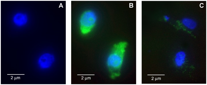
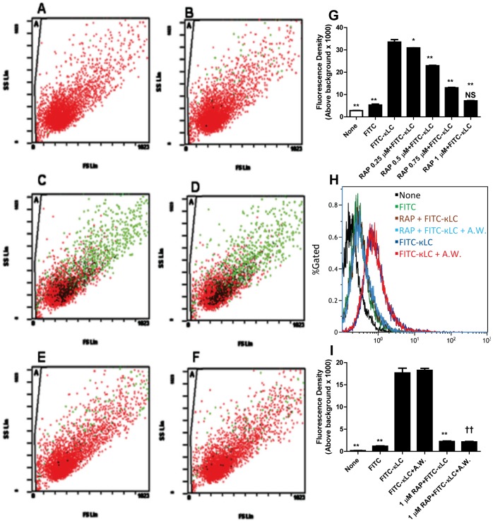
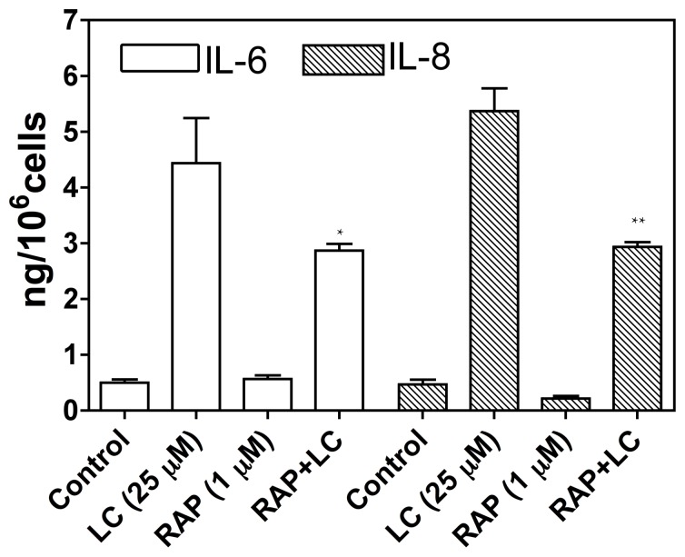
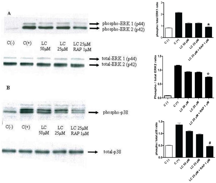
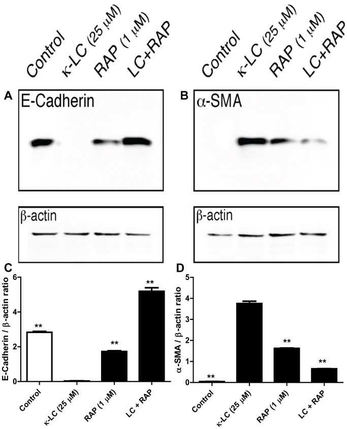
Methods and principal findings: We investigated whether RAP can block the endocytosis of LC in cultured human proximal tubule cells and whether this can prevent LC cytotoxicity. Immunofluorescence microscopy and flow cytometry showed that fluorescently labeled LC endocytosis was markedly inhibited in HK-2 cells pretreated with human RAP. The effect of RAP was dose-dependent, and was predominantly on endocytosis as it had no effect on the small acid-washable fraction of LC bound to cell membrane. RAP significantly inhibited LC induced cytokine production and phosphorylation of ERK1/2 and p38 MAPK. Prolonged exposure to LC for 48 h resulted in epithelial-to-mesenchymal transformation in HK-2 cells as evidenced by marked reduction in the expression of the epithelial cell marker E-cadherin, and increased the expression of the mesenchymal marker α-SMA, which was also prevented by RAP in the endocytosis medium.

Conclusions: RAP inhibited LC endocytosis by ∼88% and ameliorated LC-induced cytokine responses and EMT in human PTCs. The results not only provide additional evidence that LCs endocytosis occurs via the megalin/cubilin endocytic receptor system, but also show that blocking LC endocytosis by RAP can protect proximal tubule cells from LC cytotoxicity.


Conflict of interest statement

Competing Interests: The authors have declared that no competing interests exist.

Figures

Figure 1
Figure 1. Endocytosis of FITC-κLC in cultured human proximal tubule epithelial cells (a representative field with two cells is shown).
HK-2 cells were grown on a glass chamber in 8-well culture slide for 2 days and then observed by fluorescence microscopy. A. FITC without LC in control cells counterstained with DAPI for 30 min. B: after FITC-LC (25 µg/ml) uptake by HK-2 cells grown on slide chamber for 45 min at 37°C after acid wash to strip membrane bound fluorescence and counterstained with DAPI shows distribution of FITC-LC in the cytosol (green fluorescence). C: Pretreatment with 1 uM receptor-associated protein (RAP) markedly inhibits FITC-LC fluorescence in HK-2 cells.
Figure 2
Figure 2. Flow cytometry (A – F) showing the effect of RAP treatment on the association of fluorescently labeled human myeloma light chain (LC) by human renal proximal tubule cells.
Cells with FITC association are represented in green color. (A = cells without LC, B = cells with FITC alone, C = cells with 25 µM FITC-κLC, D = cells with 25 µM FITC-κLC+acid wash, E = cells after 1 µM RAP and with 25 µM FITC-κLC, F = cells after 1 µM RAP +25 µM FITC-κLC+acid wash). Quantitative presentation of flow cytometric events showing the inhibitory effect of RAP on the association of 25 µM FITC-κLC in HK2 cells is dose-dependent G). *p<0.05, **<0.01 compared to 25 µM FITC-κLC, N.S. = Not significant compared to FITC alone. Histograms (H) represent the distributions of events for FITC-LC associated cells. Acid-wash had no discernible effect on the magnitude of FITC-LC association with the cells, suggesting that membrane association is negligible. The percent magnitude of fluorescence above background is shown in the bar graph (I): Control non-FITC conjugated LC = 0.18±0.03%; FITC alone = 1.16±0.10; 25 uM FITC- conjugated κ-LC = 17.59±1.06%; 25 uM κ-LC+A.W = 18.25±0.46%; 1 uM RAP +25 uM FITC-κ-LC = 2.24±0.11%; 1 uM RAP +25 uM FITC- κ-LC+A.W. = 2.14±0.11%. RAP, 1 µM, reduced the association of total FITC-LC with cells by 87.3% compared to cells exposed to FITC-κ-LC (down to 2.24±0.11 from 17.59±1.06%), and reduced acid non-washable fraction (the fraction endocytosed by cells) by 88.3%, (reduced from 18.25±0.46 to 2.14±0.11%; Mean ± SE of three experiments, **p<0.01 compared to 25 µM FITC-κ-LC, †† p<0.01 compared to 25 µM FITC-κ-LC+A.W.; A.W. = Acid Wash).
Figure 3
Figure 3. Effect RAP on myeloma light chain (LC)- stimulated cytokine production.
Cells were pre-incubated for 1-h with or without 1 µM RAP and then exposed to 25 µM myeloma LC for a further 4-h. Interleukins 6 and 8 were then assayed in the cell supernatants. LC-stimulated the production of both IL-6 and IL-8, and both were significantly inhibited by the presence of RAP, 1 µM, in cell culture medium (p<0.05). Bars represent the mean ± SE for each group (n = 3, *p<0.05 vs LC, **p<0.01 vs LC, one-way ANOVA).
Figure 4
Figure 4. Effect of LCs on ERK1/2 and p38 MAPK (immunoblots on the left side of the panel with corresponding bar graphs of densitometric measurements next to each immunoblot panel.
Panel A shows inhibition of light chain (LC)-stimulated phosphorylation of ERK ½, and panel B, inhibition of phosphorylation of p38 MAPK by RAP pretreatment. After 1-h pretreatment with 1 µM RAP human proximal tubule cells were exposed to LC for 4 h (representative blots from 3 different experiments are shown; “C (−)” = negative control; “C (+)” = positive control; bars represent mean ± SE of three densitometric readings). A : Effect of RAP pretreatment on LC-induced activation of ERK1/2. Cells were pretreated with RAP for 1 h and then treated with LC in the continued presence of RAP for 4 h. Top: RAP inhibited LC-induced activation of ERK1/2 (LC 25 µM+RAP 1 µM vs. positive control, C (+), *P<0.001; LC 25 µM+RAP 1 µM vs. LC 25 µM alone, P<0.05; LC 25 µM+RAP 1 µM vs. LC 50 µM alone, P<0.01; LC 25 µM+RAP 1 µM compared to negative control, C (−), P>0.05, Newman-Keuls Multiple Comparison Test). Bottom: Total ERK was not affected. B : Effect of RAP pretreatment on LC-induced activation of p38 MAPK. Top: LC-induced activation of p38 was suppressed by RAP (LC 25 µM+RAP 1 µM vs. C (+), P<0.001; LC 25 µM+RAP 1 µM vs. LC 50 µM alone, # P<0.001. LC 25 µM+RAP 1 µM vs. LC 25 µM alone, P<0.001; LC 25 µM+RAP 1 µM vs. C (−), P>0.05, Newman-Keuls Multiple Comparison Test). Bottom: Total p38 was not affected (representative blots from 3 different experiments are shown).
Figure 5
Figure 5. Effect of receptor-associated protein (RAP) on the expression of E-cadherin and α-SMA in myeloma light chain exposed human renal proximal tubule epithelial cells.
RAP, 1 µM, prevented LC induced decrease in the epithelial cell marker E-cadherin (A), and reduced the increased expression of myofibroblast marker α-SMA (B), demonstrating that RAP protected cells from LC-induced EMT (representative blot from 3 experiments). Quantitative densitometric measurement of the effect of RAP pre-treatment followed by κ-LC in HK2 cells on the production of E-cadherin (C) and α-SMA (D) proteins. **p<0.01 compared to κ-LC alone.

Similar articles

Cited by

References

    1. Klassen RB, Allen PL, Batuman V, Crenshaw K, Hammond TG (2005) Light chains are a ligand for megalin. Journal of applied physiology 98: 257–263. - PubMed
    1. Batuman V, Verroust PJ, Navar GL, Kaysen JH, Goda FO, et al. (1998) Myeloma light chains are ligands for cubilin (gp280). The American journal of physiology 275: F246–254. - PubMed
    1. Batuman V, Guan S (1997) Receptor-mediated endocytosis of immunoglobulin light chains by renal proximal tubule cells. The American journal of physiology 272: F521–530. - PubMed
    1. Batuman V, Dreisbach AW, Cyran J (1990) Light-chain binding sites on renal brush-border membranes. The American journal of physiology 258: F1259–1265. - PubMed
    1. Nakhoul N, Batuman V (2011) Role of proximal tubules in the pathogenesis of kidney disease. Contributions to nephrology 169: 37–50. - PubMed

Publication types

MeSH terms

Substances

LinkOut - more resources