. 2013 Jul 23;8(7):e70278.
doi: 10.1371/journal.pone.0070278. Print 2013.

Two modes of cell death caused by exposure to nanosecond pulsed electric field

Affiliations

Two modes of cell death caused by exposure to nanosecond pulsed electric field

Olga N Pakhomova et al. PLoS One. .

Abstract

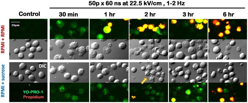
High-amplitude electric pulses of nanosecond duration, also known as nanosecond pulsed electric field (nsPEF), are a novel modality with promising applications for cell stimulation and tissue ablation. However, key mechanisms responsible for the cytotoxicity of nsPEF have not been established. We show that the principal cause of cell death induced by 60- or 300-ns pulses in U937 cells is the loss of the plasma membrane integrity ("nanoelectroporation"), leading to water uptake, cell swelling, and eventual membrane rupture. Most of this early necrotic death occurs within 1-2 hr after nsPEF exposure. The uptake of water is driven by the presence of pore-impermeable solutes inside the cell, and can be counterbalanced by the presence of a pore-impermeable solute such as sucrose in the medium. Sucrose blocks swelling and prevents the early necrotic death; however the long-term cell survival (24 and 48 hr) does not significantly change. Cells protected with sucrose demonstrate higher incidence of the delayed death (6-24 hr post nsPEF). These cells are more often positive for the uptake of an early apoptotic marker dye YO-PRO-1 while remaining impermeable to propidium iodide. Instead of swelling, these cells often develop apoptotic fragmentation of the cytoplasm. Caspase 3/7 activity increases already in 1 hr after nsPEF and poly-ADP ribose polymerase (PARP) cleavage is detected in 2 hr. Staurosporin-treated positive control cells develop these apoptotic signs only in 3 and 4 hr, respectively. We conclude that nsPEF exposure triggers both necrotic and apoptotic pathways. The early necrotic death prevails under standard cell culture conditions, but cells rescued from the necrosis nonetheless die later on by apoptosis. The balance between the two modes of cell death can be controlled by enabling or blocking cell swelling.


Conflict of interest statement

Competing Interests: The authors have declared that no competing interests exist.

Figures

Figure 1
Figure 1. Sucrose inhibits cell swelling and membrane rupture caused by nsPEF.
The bar charts show the frequency distribution of cell diameter values at the indicated time intervals after nsPEF exposure and in sham-exposed controls. Cells were exposed in the RPMI medium and placed immediately afterwards into either RPMI+NaCl or RPMI+sucrose (87 mOsm/kg due to sucrose); see text for more details. 400–600 cells per group were measured at each timepoint. Note fast cell swelling followed by membrane rupture and apparent shrinkage in RPMI+NaCl, but not in the RPMI+sucrose. Representative cell images in the differential interference contrast (DIC) and propidium iodide (PI) fluorescence channels illustrate swelling and eventual membrane rupture in the RPMI+NaCl medium.
Figure 2
Figure 2. Lack of the effect of sucrose on the 24-hr survival of nsPEF-treated cells.
Cells in RPMI were mixed with sucrose (RPMI+sucrose; 60 mOsm/kg due to sucrose) or fresh RPMI (RPMI+RPMI) before nsPEF exposure (600 pulses, 300-ns). At 30 min after the exposure, all samples were diluted tenfold with fresh RPMI and incubated until measuring cell survival by the MTT assay at 24 hr (mean values +/− s.e. for 6 independent experiments).
Figure 3
Figure 3. Inhibition of swelling improves the short-term but not the long-term survival after nsPEF exposure.
Panels A, B, and C represent the data from three independent sets of experiments performed under different exposure conditions and using different protocols. For panel A, cells in RPMI were mixed with sucrose (RPMI+sucrose; 60 mOsm/kg due to sucrose) or fresh RPMI (RPMI+RPMI) before nsPEF exposure (the same protocol as in Fig. 2). For panels B and C, cells were exposed in the RPMI and placed immediately afterwards into either RPMI+NaCl or RPMI+sucrose (87 mOsm/kg due to sucrose), same as in Fig. 2. See graph legends and text for more details. Cell survival was measured by the AO/PI assay and normalized to the pre-exposure value (mean+/− s.e., n = 4–6). Cell survival in sham-exposed controls is shown by dashed lines and open symbols. * p<0.05 for the difference of RPMI+sucrose from RPMI+NaCl (or RPMI+RPMI); # p<0.05 for the difference of RPMI+sucrose from the respective sham-exposed control. Other significant differences are not shown for clarity.
Figure 4
Figure 4. Inhibition of swelling blocks the early cell death after nsPEF.
Dead cells were identified by the AO/PI assay. The total number of cells counted at each timepoint was taken as 100% (mean+/− s.e., n = 4–6). The data in panels A and B are from the same experiments as in Fig. 3, B and C. See text and Fig. 3 for details. PI uptake in sham-exposed controls is shown by dashed lines and open symbols.
Figure 5
Figure 5. Effects of sucrose on cell swelling and membrane permeability.
DIC and fluorescence images of nsPEF-exposed cells incubated in either RPMI+RPMI or RPMI+sucrose. Green: YO-PRO-1; red: PI; yellow: both dyes overlapped. Parameters of exposure and times after it are given in the legend. Cells were handled the same way as in Fig. 2 but without additional dilution at 30 min. The dyes were added 5–10 min prior to taking an image. Note early cell swelling and rupture in the RPMI+RPMI but not in the RPMI+sucrose medium. The survivors show no YO-PRO-1 uptake in the RPMI+RPMI, but remain permeable to the dye in the RPMI+sucrose group. An arrow points to a group of cells that display the apoptotic blebbing and fragmentation.
Figure 6
Figure 6. Inhibition of swelling in nsPEF-exposed cells facilitates caspase 3/7 activation.
The exposure parameters and media are identified in the legend. Growth media were changed the same way as in Fig. 1. Caspase-3/7 was measured by a luminescence assay. For a positive control, apoptosis was induced by 10 µM of staurosporin. Mean values +/− s.e. for n = 3. See text and Fig. 3 for details.
Figure 7
Figure 7. PARP cleavage in nsPEF-exposed cells.
A: The fraction of cleaved PARP is increased when nsPEF-exposed cells are protected with sucrose. Mean values +/− s.e. for n = 4–5. Growth media were changed the same way as in Fig. 1. NsPEF and media conditions are specified in the legend. The numbers in parentheses correspond to the lanes in panel B, which shows representative Western blots for intact and cleaved PARP (116 and 89 kDa, respectively). See text and Fig. 3 for details.
Figure 8
Figure 8. The structure of nsPEF-exposed cell populations with and without blockage of cell swelling with sucrose.
Bars show relative fractions of non-apoptotic, apoptotic, and dead cells at different timepoints after nsPEF (600 pulses, 300 ns, 7 kV/cm). Growth media were changed the same way as in Fig. 1. Dead and live cells were counted by the AO/PI assay. The fraction of apoptotic cells among live cells was considered proportional to the fraction of cleaved PARP. The data were averaged from 4–5 experiments; the error bars are omitted for clarity.
Figure 9
Figure 9. The effect of sucrose on Yo-PRO-1 and PI uptake by nsPEF-exposed cells.
Growth media were changed after nsPEF exposure (600 pulses, 300 ns, 7 kV/cm) the same way as in Fig. 1. The number of cells displaying no dye uptake, YO-PRO-1 uptake, and both YO-PRO-1 and PI uptake were automatically counted in microscope images as described in Methods. The total number of cells counted in each sample was taken as 100%. The PI-positive cells were presumed dead. YO-PRO-1-positive cells could be either apoptotic or just transiently permeabilized to this dye by nsPEF. Cells negative for either dye were regarded as live, non-apoptotic. The data were averaged from 3 experiments; error bars are omitted for clarity.

Similar articles

Cited by

References

    1. Schoenbach KS, Hargrave B, Joshi RP, Kolb J, Osgood C, et al. (2007) Bioelectric Effects of Nanosecond Pulses. IEEE Transactions on Dielectrics and Electrical Insulation 14: 1088–1109.
    1. Ren W, Beebe SJ (2011) An apoptosis targeted stimulus with nanosecond pulsed electric fields (nsPEFs) in E4 squamous cell carcinoma. Apoptosis 16: 382–393. - PMC - PubMed
    1. Ren W, Sain NM, Beebe SJ (2012) Nanosecond pulsed electric fields (nsPEFs) activate intrinsic caspase-dependent and caspase-independent cell death in Jurkat cells. Biochemical and biophysical research communications 421: 808–812. - PubMed
    1. Beebe SJ, Fox PM, Rec LJ, Somers K, Stark RH, et al. (2002) Nanosecond Pulsed Electric Field (nsPEF) Effects on Cells and Tissues: Apoptosis Induction and Tumor Growth Inhibition. IEEE Transactions on Plasma Science 30: 286–292.
    1. Garon EB, Sawcer D, Vernier PT, Tang T, Sun Y, et al. (2007) In vitro and in vivo evaluation and a case report of intense nanosecond pulsed electric field as a local therapy for human malignancies. Int J Cancer 121: 675–682. - PubMed

Publication types