. 2013 Jul 24;8(7):e70417.
doi: 10.1371/journal.pone.0070417. Print 2013.

Combination therapy with local radiofrequency ablation and systemic vaccine enhances antitumor immunity and mediates local and distal tumor regression

Affiliations

Combination therapy with local radiofrequency ablation and systemic vaccine enhances antitumor immunity and mediates local and distal tumor regression

Sofia R Gameiro et al. PLoS One. .

Abstract

Purpose: Radiofrequency ablation (RFA) is a minimally invasive energy delivery technique increasingly used for focal therapy to eradicate localized disease. RFA-induced tumor-cell necrosis generates an immunogenic source of tumor antigens known to induce antitumor immune responses. However, RFA-induced antitumor immunity is insufficient to control metastatic progression. We sought to characterize (a) the role of RFA dose on immunogenic modulation of tumor and generation of immune responses and (b) the potential synergy between vaccine immunotherapy and RFA aimed at local tumor control and decreased systemic progression.

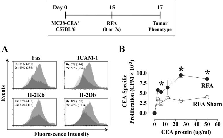
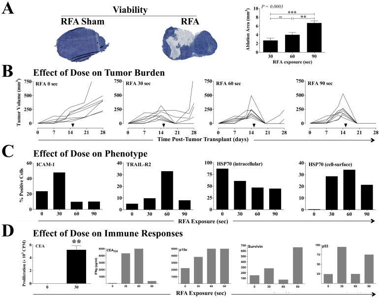
Experimental design: Murine colon carcinoma cells expressing the tumor-associated (TAA) carcinoembryonic antigen (CEA) (MC38-CEA(+)) were studied to examine the effect of sublethal hyperthermia in vitro on the cells' phenotype and sensitivity to CTL-mediated killing. The effect of RFA dose was investigated in vivo impacting (a) the phenotype and growth of MC38-CEA(+) tumors and (b) the induction of tumor-specific immune responses. Finally, the molecular signature was evaluated as well as the potential synergy between RFA and poxviral vaccines expressing CEA and a TRIad of COstimulatory Molecules (CEA/TRICOM).

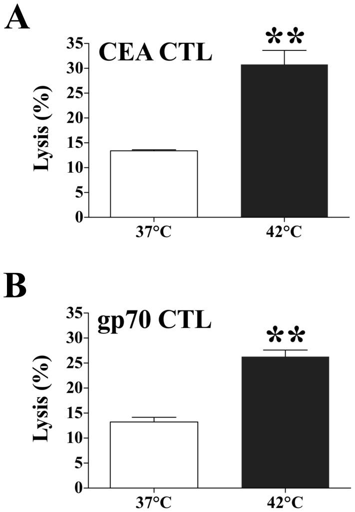
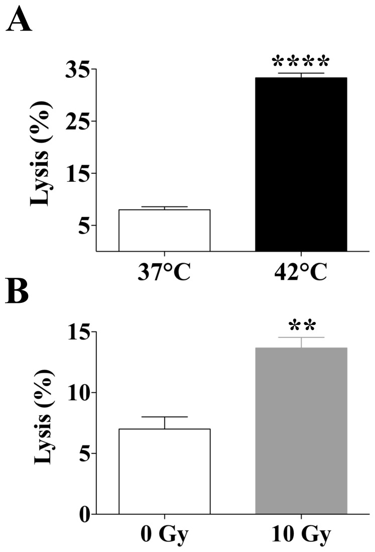
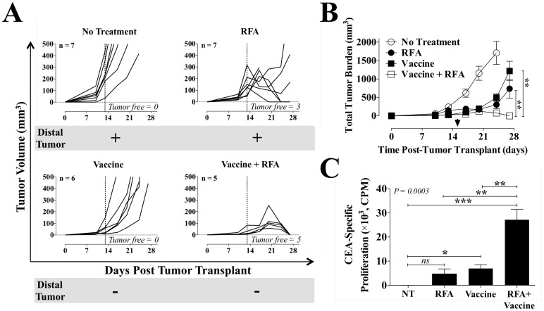
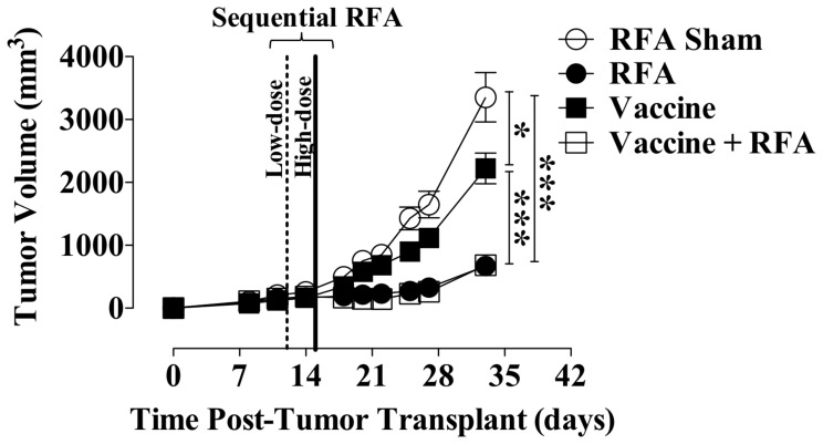
Results: In vitro, sublethal hyperthermia of MC38-CEA(+) cells (a) increased cell-surface expression of CEA, Fas, and MHC class I molecules and (b) rendered tumor cells more susceptible to CTL-mediated lysis. In vivo, RFA induced (a) immunogenic modulation on the surface of tumor cells and (b) increased T-cell responses to CEA and additional TAAs. Combination therapy with RFA and vaccine in CEA-transgenic mice induced a synergistic increase in CD4(+) T-cell immune responses to CEA and eradicated both primary CEA(+) and distal CEA(-) s.c. tumors. Sequential administration of low-dose and high-dose RFA with vaccine decreased tumor recurrence compared to RFA alone. These studies suggest a potential clinical benefit in combining RFA with vaccine in cancer patients, and augment support for this novel translational paradigm.


Conflict of interest statement

Competing Interests: The authors have declared that no competing interests exist.

Figures

Figure 1
Figure 1. Effect of hyperthermia on sensitivity of murine colon carcinoma cells to CTL-mediated lysis.
MC38-CEA+ cells were exposed in vitro to 37°C or 42°C for 1 h. Twenty-four hours after exposure, cells were harvested and labeled with 111In. The sensitivity of MC38-CEA+ target cells to A. CEA– or B. GP70-specific killing was determined after cells were incubated at an effector:target ratio of 80∶1. Results are presented as mean ± S.E.M. from 3 replicate wells. Asterisks denote statistical significance (P<0.004) relative to control cells (2-tailed t test). Data is representative of two independent experiments.
Figure 2
Figure 2. Effect of hyperthermia and gamma radiation on sensitivity of human colon carcinoma cells to CEA-specific CTL-mediated lysis.
SW620 cells were exposed in vitro to A, 37°C or 42°C for 1 h, or to B, 0 or 10 Gy. Forty-eight hours post-exposure, cells were harvested and labeled with 111In. The sensitivity of SW620 target cells to CEA-specific killing was determined after cells were incubated with CEA-specific T cells at an effector:target ratio of 80∶1. Results are presented as mean ± S.E.M. from 3 replicate wells. Asterisks denote statistical significance (**P = 0.008; ****P<0.0001) relative to control cells (2-tailed t test). Radiation exposure data is representative of three independent experiments.
Figure 3
Figure 3. Tumor phenotype and CEA-specific immune responses after ultra low-dose RFA.
Female C57BL/6 mice (n = 7/group) were injected s.c. on day 0 with MC38-CEA+ tumor cells. On day 15, tumors were exposed to RFA sham (0 s) or ultra low-dose RFA (7 s, 60–70°C). A, on day 17, the cell-surface phenotype of pooled excised tumor cells was analyzed by flow cytometry. Histograms depict fluorescence intensity of CEA+ tumor cells expressing Fas, ICAM-1, and MHC class I H-2Kb and H-2Db before (dark histograms) and after (light histograms) RFA. Inset numbers represent % positive cells and MFI (parentheses) for each marker. B, on day 29, purified CD4+ splenic T cells from animals exposed to sham or low-dose RFA were tested by in vitro lymphoproliferation assay for reactivity to CEA protein (0–50 ug/mL). Results are depicted as mean CEA-specific CD4+ proliferation ± S.E.M. after subtraction of background CD4+ reactivity to control beta-galactosidase protein. Asterisks denote statistical significance between treatment groups (P<0.05, 2-tailed t test). Effect of RFA on tumor phenotype data was performed twice yielding similar results.
Figure 4
Figure 4. Effect of RFA dose on tumor phenotype and antigen cascade immune responses.
Female C57BL/6 mice (n = 8–10/group) were injected s.c. on day 0 with MC38-CEA+ tumor cells. On day 15, tumors were exposed to RFA sham or to increasing doses of RFA (30, 60, or 90 s). A, 3 h post-RFA, tumor viability was assessed by tetrazolium staining (n = 4–7) in tumors exposed to sham or 30 s RFA. Histological images are representative of each treatment. Dark blue denotes viable cells. B depicts tumor volumes in individual animals. Arrows denote RFA on day 15. C, on day 17, excised CEA+ tumor cells were evaluated by flow cytometry for percentage of cells with cell-surface expression of ICAM-1, TRAIL-R2, and HSP70, and intracellular expression of HSP70. D, on day 29, purified CD4+ splenic T cells from animals exposed to RFA were tested for specific reactivity to CEA protein (25 ug/mL) in an in vitro lymphoproliferation assay. Results are depicted as mean CD4+ proliferation ± S.E.M. after subtraction of background CD4+ reactivity to control beta-galactosidase protein. Asterisks denote statistical significance (P = 0.001, 2-tailed t test). CD8+ T-cell responses specific for the peptides CEA526, p15e604, p53232, and survivin57 were evaluated in pooled splenocytes through quantification of secreted IFN-g over 24 h of in vitro restimulation. Results are depicted as IFN-g (pg/mL) after subtraction of background IFN-g secretion in response to the control peptide VSV-N52. This experiment was performed twice yielding similar results.
Figure 5
Figure 5. Antitumor efficacy and immune response elicited by combination therapy with RFA and recombinant vaccine.
CEA-Tg mice (n = 5–7/group) received MC38-CEA+ cells on day 0 (right flank s.c.; primary tumor) and CEA MC38 cells on day 5 (left flank s.c.; distal tumor). A, primary tumor growth. MC38-CEA+ tumors received sham or high-dose RFA (30–300 s; 60–70°C) on day 13 (dotted line). Vaccinated animals received rV-CEA/TRICOM+rF-GM-CSF on day 4 and rF-CEA/TRICOM+rF-GM-CSF on days 11 and 18, alone or in combination with RFA. Each graph denotes the number of primary tumor-free mice and the presence (+) or absence (–) of a distal tumor on day 28. B, total tumor burden (primary plus distal). Asterisks denote statistical significance among treatment groups at day 27 (P<0.0001, 1-way ANOVA with Tukey’s multiple comparison test). Arrow indicates RFA treatment on day 15. C, CEA-specific proliferation of CD4+ T cells. CEA-Tg mice were transplanted with MC38-CEA+ tumor cells as before and received sham or 30 s RFA on day 15. Vaccinated mice received rMVA-CEA/TRICOM on day 4 and rF-CEA/TRICOM on days 11 and 18, alone or in combination with 30 s RFA. On day 39, purified CD4+ splenic T cells were tested for reactivity to CEA protein (50 ug/mL) in an in vitro lymphoproliferation assay. Results are depicted as mean CEA-specific CD4+ proliferation ± S.E.M. after subtraction of background CD4+ reactivity to control beta-galactosidase protein. Asterisks denote statistical significance among treatment groups (P = 0.0003, 1-way ANOVA with Tukey’s multiple comparison test). This experiment was performed twice yielding similar results.
Figure 6
Figure 6. Antitumor efficacy of combination therapy with sequential RFA and recombinant vaccine.
MC38-CEA+ cells were transplanted on day 0 on the right flank of CEA-Tg mice (n = 25–29/group). MC38-CEA+ tumors received sham or low-dose RFA (10–30 s at 70°C) on day 12 (dotted line), followed by high-dose ablative RFA (30–300 s at 70°C) on day 15 (solid line). Vaccinated mice received rMVA-CEA/TRICOM s.c. on day 4 and rF-CEA/TRICOM on day 11 and every 7 days thereafter, alone or in combination with sequential RFA. A depicts tumor growth. Data represent tumor volume ± S.E.M. in individual animals. Asterisks denote statistically significant differences among treatment groups (P<0.0001, 1-way ANOVA with Tukey’s multiple comparison test).

References

    1. Pathak S, Jones R, Tang JM, Parmar C, Fenwick S, et al. (2011) Ablative therapies for colorectal liver metastases: a systematic review. Colorectal Dis 13: e252–265. - PubMed
    1. Gervais DA, Goldberg SN, Brown DB, Soulen MC, Millward SF, et al. (2009) Society of Interventional Radiology position statement on percutaneous radiofrequency ablation for the treatment of liver tumors. J Vasc Interv Radiol 20: S342–347. - PubMed
    1. Wood BJ, Ramkaransingh JR, Fojo T, Walther MM, Libutti SK (2002) Percutaneous tumor ablation with radiofrequency. Cancer 94: 443–451. - PMC - PubMed
    1. Haen SP, Pereira PL, Salih HR, Rammensee HG, Gouttefangeas C (2011) More than just tumor destruction: immunomodulation by thermal ablation of cancer. Clin Dev Immunol 2011: 160250. - PMC - PubMed
    1. Zerbini A, Pilli M, Penna A, Pelosi G, Schianchi C, et al. (2006) Radiofrequency thermal ablation of hepatocellular carcinoma liver nodules can activate and enhance tumor-specific T-cell responses. Cancer Res 66: 1139–1146. - PubMed

Publication types