. 2013 Jul 23;8(7):e70518.
doi: 10.1371/journal.pone.0070518. Print 2013.

Mcm10 self-association is mediated by an N-terminal coiled-coil domain

Affiliations

Mcm10 self-association is mediated by an N-terminal coiled-coil domain

Wenyue Du et al. PLoS One. .

Abstract

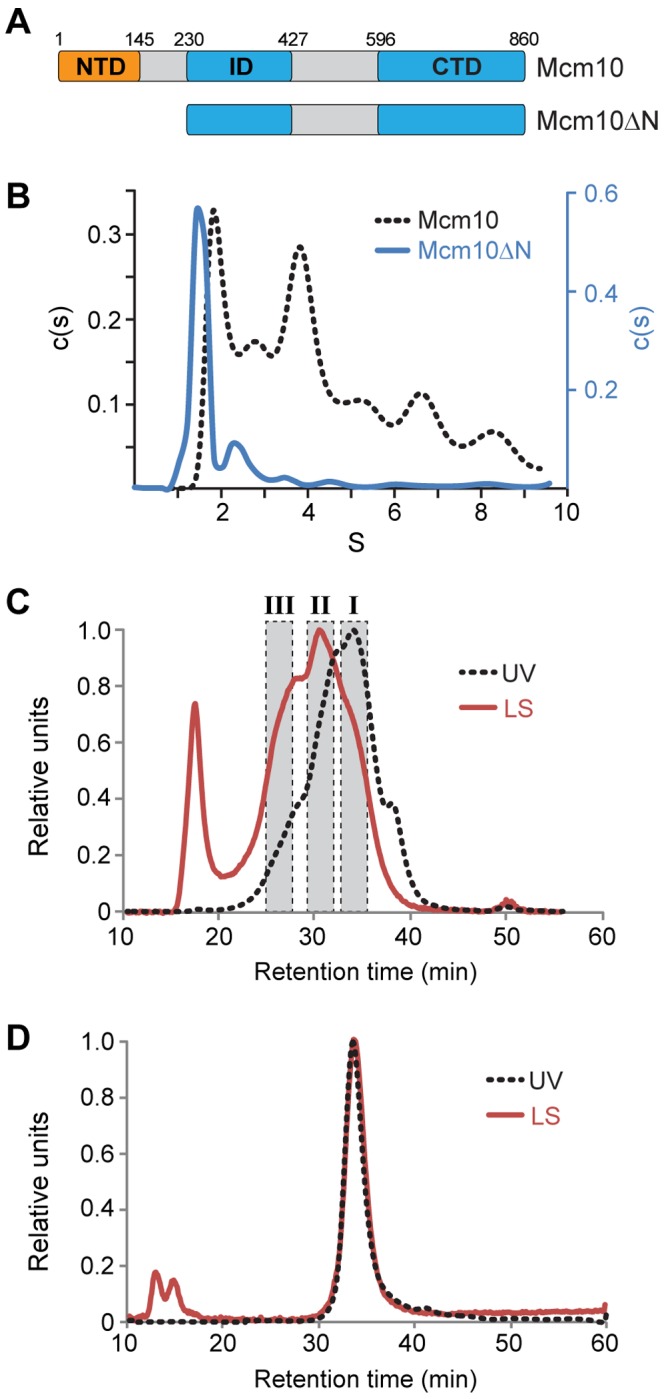
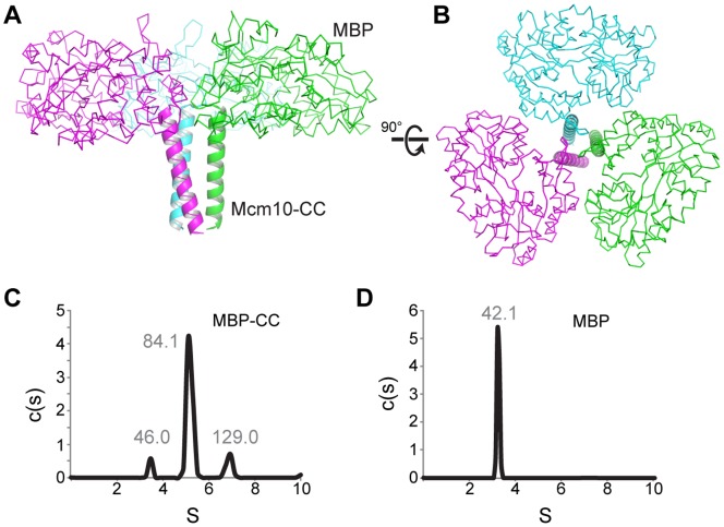
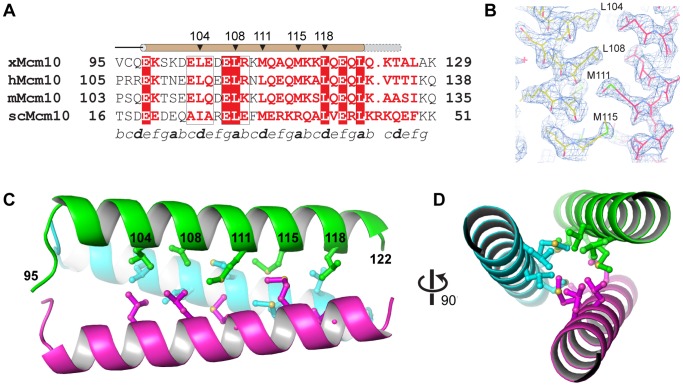
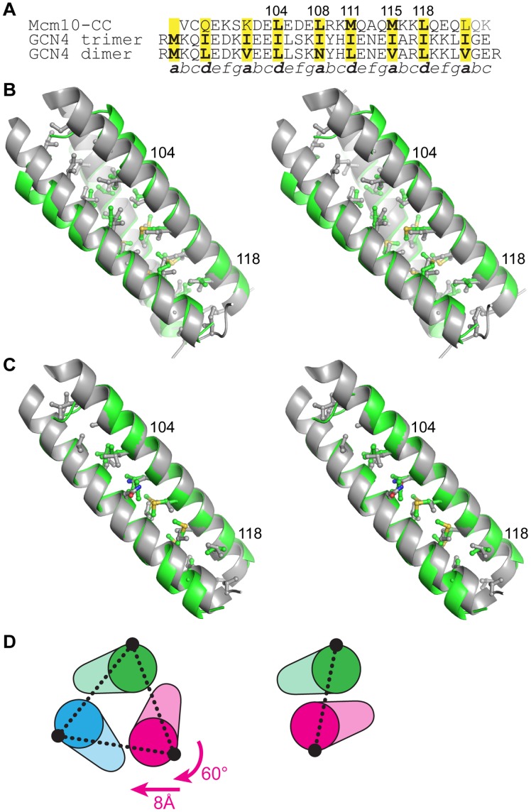
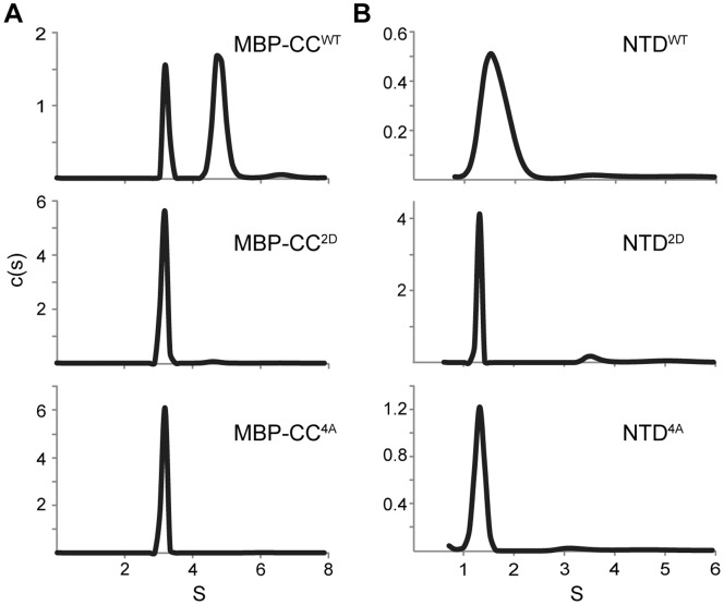
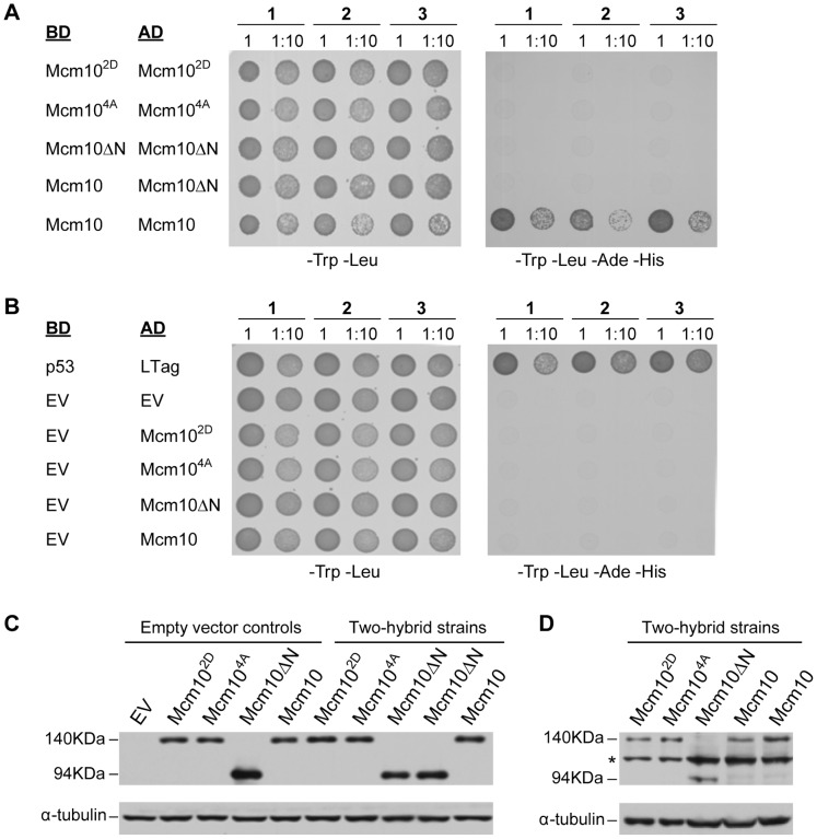
Minichromosome maintenance protein 10 (Mcm10) is an essential eukaryotic DNA-binding replication factor thought to serve as a scaffold to coordinate enzymatic activities within the replisome. Mcm10 appears to function as an oligomer rather than in its monomeric form (or rather than as a monomer). However, various orthologs have been found to contain 1, 2, 3, 4, or 6 subunits and thus, this issue has remained controversial. Here, we show that self-association of Xenopus laevis Mcm10 is mediated by a conserved coiled-coil (CC) motif within the N-terminal domain (NTD). Crystallographic analysis of the CC at 2.4 Å resolution revealed a three-helix bundle, consistent with the formation of both dimeric and trimeric Mcm10 CCs in solution. Mutation of the side chains at the subunit interface disrupted in vitro dimerization of both the CC and the NTD as monitored by analytical ultracentrifugation. In addition, the same mutations also impeded self-interaction of the full-length protein in vivo, as measured by yeast-two hybrid assays. We conclude that Mcm10 likely forms dimers or trimers to promote its diverse functions during DNA replication.


Conflict of interest statement

Competing Interests: The Michigan Economic Development Corporation and the Michigan Technology Tri-Corridor funded the synchrotron beamline that was used to collect X-ray diffraction data for this research. In addition, Anja-Katrin Bielinsky declares that she is a PLOS ONE Editorial Board member. This does not alter the authors’ adherence to all the PLOS ONE policies on sharing data and materials.

Figures

Figure 1
Figure 1. The NTD is necessary for Mcm10 self-association.
(A) Schematic of Xenopus laevis Mcm10 constructs used to study self-association. Theoretical molecular masses are 95.4 kDa (Mcm10) and 70.4 kDa (Mcm10ΔN). (B) Sedimentation velocity data for full-length Mcm10 (black dotted line) and Mcm10ΔN (blue) as described in Materials and Methods. The molecular mass of the Mcm10ΔN major peak was calculated to be 68.8 kDa. Molecular masses could not be accurately determined from the full-length data. (C,D) SEC-MALS analysis of Mcm10 (C) and Mcm10ΔN (D). The UV trace is shown as a black dotted line and the light scattering trace is red. (C) Estimated molecular masses were calculated from the three shaded regions to be 90.4 kDa (I), 189.3 kDa (II), and 322.7 kDa (III). The peak at 18 min corresponds to the void volume. (D) The molecular mass of Mcm10ΔN was calculated to be 75.1±0.8 kDa.
Figure 2
Figure 2. Trimerization of MBP-CC.
(A,B) Crystal structure of the MBP-CC asymmetric unit, with each protomer colored differently. Maltose-binding protein is shown as a Cα-trace, and the xMcm10 coiled-coil is depicted as a cartoon ribbon. (C,D) Sedimentation velocity profiles of MBP-CC95–132 (C) and free MBP (D) at pH 4.7. Molecular masses (kDa) calculated from the sedimentation data are shown above each peak. The molecular mass of a single polypeptide calculated from the amino acid composition are 45.1 kDa (MBP-CC) and 40.4 kDa (MBP).
Figure 3
Figure 3. Crystal structure of the Mcm10 coiled-coil.
(A) Sequence alignment of the coiled-coil region from Xenopus laevis (x), Homo sapiens (h), Mus musculus (m), and Saccharomyces cerevisiae (sc) Mcm10. Red boxes indicate identical amino acids. The predicted heptad repeat pattern is labeled by letters a-g at the bottom. The position of the helix is shown schematically at the top (brown, MBP-CC95–124; grey, MBP-CC95–132). (B) Composite 2Fo−Fc omit electron density map (contoured at 1σ) with carbon atoms colored according to protomer. (C) Crystal structure of xMcm10-CC95–124. Residues at the interface are shown in ball and stick, and the amino acid numbers labeled in black. (D) View down the helical axis, rotated 90° from the view in B.
Figure 4
Figure 4. A model of the dimeric Mcm10 CC.
(A) Structure-based sequence alignment between Mcm10 CC and the trimeric and dimeric forms of GCN4 CC. (B) Stereoview of the Mcm10 CC trimer (green) superimposed onto the isoleucine GCN4 trimer (grey, PDB ID 1 GCM). Side chains at the CC interface are shown, with the exception of the N-terminal methionine in GCN4. Two Mcm10 residues are labeled for orientation. The view is rotated 45° clockwise with respect to Figure 3C. (C) Stereoview of the Mcm10 CC dimer model (green) superimposed on the CGN4 CC (grey, PDB ID 2ZTA). (D) The relationship between trimer (left) and dimer (right) forms of the CC. Schematics are oriented with respect to the green helix. The conformation of the dimer can be constructed from the trimer by a 60° rotation and 8 Å translation of the magenta helix. The interhelical distances (dashed line) are 15.5 Å (trimer) and 10 Å (dimer).
Figure 5
Figure 5. Coiled-coil mutations disrupt CC and NTD dimerization.
Sedimentation velocity data for MBP-CC95–124 (A) and NTD (B) constructs as wild-type (WT) or containing L104D/L108D (2D) or L104A/L108A/M115A/L118A (4A) mutations. Molecular masses corresponding to each peak are reported in Table S1 in the Supporting Information.
Figure 6
Figure 6. Coiled-coil mutations disrupt Mcm10 self-association.
(A, B) Three individual strains (1, 2, 3) harboring yeast two-hybrid plasmids that express the indicated proteins either as a fusion with the Gal4-binding domain (BD) or -activation domain (AD) were spotted onto drop-out plates lacking tryptophan and leucine (-Trp -Leu) or quadruple drop-out plates lacking tryptophan, leucine, adenine and histidine (-Trp -Leu -Ade -His). Cells were spotted at a number of 2×10⁷ (1) or a 10-fold dilution (1∶10) for full-length Xenopus Mcm10, the L104D/L108D mutant (Mcm102D), the L104A/L108A/M115A/L118A mutant (Mcm104A), and the NTD deletion mutant spanning residues 230–860 (Mcm10ΔN). p53 and large T-antigen (LTag) served as a positive control, EV indicates empty vector controls. (C) Western blot showing wild-type and mutant xMcm10 protein expression in representative strains. Gal4-AD fusions were detected by a HA-specific antibody. Strains carrying empty vector controls, or Gal4-AD fusion genes and Gal4-BD empty vectors are shown on the left (Empty vector controls). Strains expressing pair-wise combinations of the Gal4-AD and Gal4-BD fusion genes as indicated in panels A and B are shown on the right (Two-hybrid strains). Full-length xMcm10 and the 2D and 4A mutants ran at an approximate size of 140 kDa, whereas the truncated form of xMcm10 ran at an approximate size of 94 kDa. Tubulin served as a loading control. (D) Gal4-BD fusions were detected by a Myc-specific antibody. Extracts from the identical two-hybrid strains shown in (C) were loaded in the same order. The asterisk denotes a non-specific band.

Similar articles

Cited by

References

    1. Bell SP, Dutta A (2002) DNA replication in eukaryotic cells. Annu Rev Biochem 71: 333–374. - PubMed
    1. MacNeill S (2012) The eukaryotic replisome : a guide to protein structure and function. New York: Springer.
    1. Thu YM, Bielinsky AK (2013) Enigmatic roles of Mcm10 in DNA replication. Trends Biochem Sci 38: 184–194. - PMC - PubMed
    1. Heller RC, Kang S, Lam WM, Chen S, Chan CS, et al. (2011) Eukaryotic origin-dependent DNA replication in vitro reveals sequential action of DDK and S-CDK kinases. Cell 146: 80–91. - PMC - PubMed
    1. Tanaka S, Umemori T, Hirai K, Muramatsu S, Kamimura Y, et al. (2007) CDK-dependent phosphorylation of Sld2 and Sld3 initiates DNA replication in budding yeast. Nature 445: 328–332. - PubMed

Publication types

MeSH terms

Substances

LinkOut - more resources