. 2013 Jun 1;1(6):603-613.
doi: 10.1039/C3BM00201B.

Hydrogels Formed by Oxo-ester Mediated Native Chemical Ligation

Affiliations

Hydrogels Formed by Oxo-ester Mediated Native Chemical Ligation

Iossif Strehin et al. Biomater Sci. .

Abstract

Oxo-ester mediated native chemical ligation (OMNCL) is a variation of the more general native chemical ligation (NCL) reaction that is widely employed for chemoselective ligation of peptide fragments. While OMNCL has been used for a variety of peptide ligations and for biomolecular modification of surfaces, it is typically practiced under harsh conditions that are unsuitable for use in a biological context. In this report we describe the use of OMNCL for polymer hydrogel formation, in-vitro cell encapsulation, and in-vivo implantation. Multivalent polymer precursors containing N-hydroxysuccinimide (NHS) activated oxo-esters and N-cysteine (N-Cys) endgroups were chemically synthesized from branched poly(ethylene glycol) (PEG). Hydrogels formed rapidly at physiologic pH upon mixing of aqueous solutions of NHS and N-Cys functionalized PEGs. Quantitative 1H NMR experiments showed that the reaction proceeds through an OMNCL pathway involving thiol capture to form a thioester intermediate, followed by an S-to-N acyl rearrangement to yield an amide cross-link. pH and temperature were found to influence gelation rate, allowing tailoring of gelation times from a few seconds to a few minutes. OMNCL hydrogels initially swelled before contracting to reach an equilibrium increase in relative wet weight of 0%. This unique behavior impacted the gel stiffness and was attributed to latent formation of disulfide cross-links between network-bound Cys residues. OMNCL hydrogels were adhesive to hydrated tissue, generating a lap shear adhesion strength of 46 kPa. Cells encapsulated in OMNCL hydrogels maintained high viability, and in-situ formation of OMNCL hydrogel by subcutaneous injection in mice generated a minimal acute inflammatory response. OMNCL represents a promising strategy for chemical cross-linking of hydrogels in a biological context and is an attractive candidate for in-vivo applications such as wound healing, tissue repair, drug delivery, and tissue engineering.


Figures

Figure 1
Figure 1
The two polymer precursors P8NHS and P8Cys react in aqueous solution via OMNCL to yield polymer hydrogels with network cross-links as shown at bottom right.
Figure 2
Figure 2
Quantitative 1H NMR analysis of the model reaction between P8NHS and L-Cys in D2O. Chemical structures (A) and relative abundance (B) of polymer species observed during the reaction.
Figure 3
Figure 3
Quantitative 1H NMR analysis of the reaction between P8NHS and L-Cys in buffered D2O. (A,B) Relative abundance of polymer species formed during reaction of P8NHS with L-Cys at (A) pH 6.0 and (B) pH 7.0, indicating that the reaction proceeds more quickly at higher pH. (C) Relative abundance of polymer species formed during the reaction of P8NHS with S-methyl-L-cysteine at pH 7.0, illustrating significantly slower reaction kinetics when the thiol group is protected.
Figure 4
Figure 4
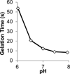
The effect of initial pH on gelation time of 10% w/v hydrogels prepared in 100 mM PBS at room temperature with P8Cys and P8NHS (1:1 w/w).
Figure 5
Figure 5
Physical characterization of OMNCL hydrogels formed by mixing equal volumes of 10% (w/v) P8NHS and 10% (w/v) P8Cys in PBS. (A) Swelling of OMNCL hydrogels in 10mM PBS (closed symbols) or 10 mM PBS substituted with 0.2 M β-ME (open symbols). The two sets of hydrogels (diamonds and circles, n = 5 per set) varied by the sequence in which they were incubated in PBS or β-ME. In one case (circles), the hydrogels were incubated in PBS followed by β-ME and then PBS again. In the second case (diamonds), the hydrogels were incubated in PBS for the first few hours and thereafter in β-ME. (B) Young’s moduli (n=4) at various time points for OMNCL hydrogels incubated in PBS. * p < 0.05, *** p < 0.001.
Figure 6
Figure 6
In-vitro cytocompatibility of OMNCL hydrogels. (A) Quantitative analysis of 3T3 fibroblast viability after 24 hours in conditioned medium, conducted in accordance with ISO standards 10993-05 and 10993-12. Cell culture medium included either extract from P8NHS/P8Cys hydrogel or 5% w/v P8NHS. (B) 3T3 fibroblasts encapsulated in OMNCL hydrogels and stained with calcein AM (green, live cells) and ethidium homodimer-1 (red, dead cells). Image analysis indicated 87 ± 7% of cells remained viable after 24 hours of encapsulation.
Figure 7
Figure 7
In-vivo subcutaneous characterization of OMNCL hydrogel. A. H&E stained tissue section at 20x magnification with gel associated with the outer skin. The gel is stained blue and surrounding tissue stained blue and red (an overview of the skin-gel injection area is shown on the right at 4× magnification). B. H&E stained tissue section at 40× magnification from sequentially obtained tissue sections, showing the gel (blue body at the bottom), subtle fibrous capsule (marked with black arrows) and supra-capsular muscle layer (purple-red, marked with green arrows). C. Picro-Sirius Red stained section (40× magnification) obtained from the same area as B. Hydrogel is at the bottom; the capsule surrounding the hydrogel is a bright-red fibrous structure (marked with white arrows) and muscle mass shown in brown-red (marked with green arrows). The scale bars indicate length in micrometers (mc).
Scheme 1
Scheme 1
Generalized reaction schemes for native chemical ligation (NCL) and oxo-ester mediated native chemical ligation (OMNCL).
Scheme 2
Scheme 2
OMNCL crosslinking of P8Cys and P8NHS. Fast reaction pathways are indicated by solid arrows, slow pathways by dashed arrows. Thiol capture followed by S-to-N acyl rearrangement results in polymer cross-linking. Secondary cross-links arise through the formation of disulfide bonds among network-bound Cys residues. P1 = P8NHS; P2 = P8Cys.

References

    1. Schnolzer M, Kent SB. Science. 1992;256:221. - PubMed
    1. Kent SB. Chemical Society reviews. 2009;38:338. - PubMed
    1. Bang D, Kent SB. Angew Chem Int Ed Engl. 2004;43:2534. - PubMed
    1. Komarov AG, Linn KM, Devereaux JJ, Valiyaveetil FI. ACS chemical biology. 2009;4:1029. - PMC - PubMed
    1. Kajihara Y, Yamamoto N, Okamoto R, Hirano K, Murase T. Chem Rec. 2010;10:80. - PubMed