. 2013 Jun 1;2(6):e24962.
doi: 10.4161/onci.24962. Epub 2013 May 15.

HLA Class II tetramers reveal tissue-specific regulatory T cells that suppress T-cell responses in breast carcinoma patients

Affiliations

HLA Class II tetramers reveal tissue-specific regulatory T cells that suppress T-cell responses in breast carcinoma patients

Hans-Henning Schmidt et al. Oncoimmunology. .

Abstract

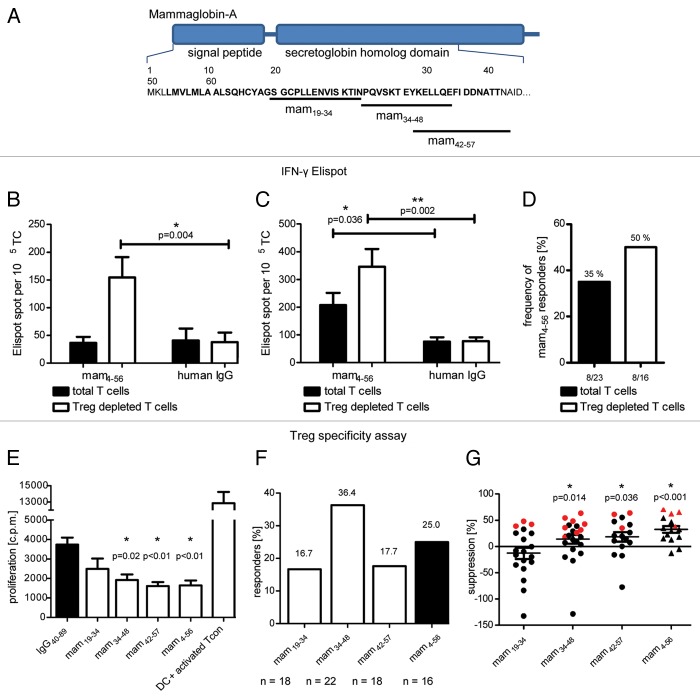
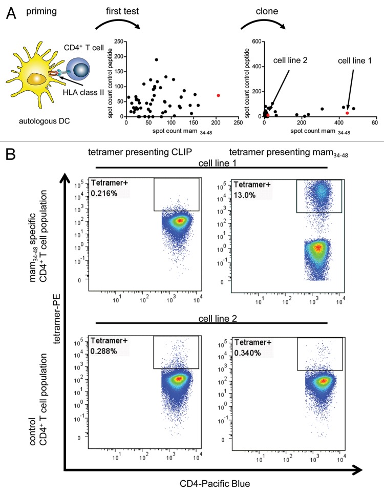
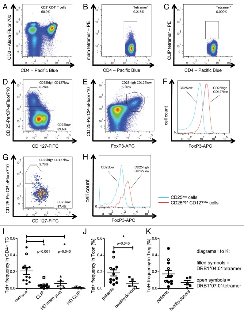
Regulatory T cells (Tregs) play an important role in controlling antitumor T-cell responses and hence represent a considerable obstacle for cancer immunotherapy. The abundance of specific Treg populations in cancer patients has been poorly analyzed so far. Here, we demonstrate that in breast cancer patients, Tregs often control spontaneous effector memory T-cell responses against mammaglobin, a common breast tissue-associated antigen that is overexpressed by breast carcinoma. Using functional assays, we identified a HLA-DRB1*04:01- and HLA-DRB1*07:01-restricted epitope of mammaglobin (mam34-48) that was frequently recognized by Tregs isolated from breast cancer patients. Using mam34-48-labeled HLA Class II tetramers, we quantified mammaglobin-specific Tregs and CD4+ conventional T (Tcon) cells in breast carcinoma patients as well as in healthy individuals. Both mammaglobin-specific Tregs and Tcon cells were expanded in breast cancer patients, each constituting approximately 0.2% of their respective cell subpopulations. Conversely, mammaglobin-specific Tregs and CD4+ Tcon cells were rare in healthy individuals (0.07%). Thus, we provide here for the first time evidence supporting the expansion of breast tissue-specific Tregs and CD4+ Tcon cells in breast cancer patients. In addition, we substantiate the potential implications of breast tissue-specific Tregs in the suppression of antitumor immune responses in breast cancer patients. The HLA Class II tetramers used in this study may constitute a valuable tool to elucidate the role of antigen-specific Tregs in breast cancer immunity and to monitor breast cancer-specific CD4+ T cells.

Keywords: HLA Class II; Tregs; breast cancer; mammaglobin; multimer; regulatory T cells; suppression; tetramer; tumor-specific T cells.


Figures

None
Figure 1. Functional assays of mammaglobin-specific conventional and regulatory T cells. (A) Position of tested peptides within the mammaglobin sequence. (B andC) Total T cells or T cells depleted of regualtory T cells (Tregs) were analyzed in an interferon γ (IFNγ) ELISPOT assay upon stimulation with the mammaglobin-derived peptide mam4–56 or human IgG (negative control). The assay was conducted in triplicate instances. Representative results from two patients are shown (p values as per two-tailed Student's t-tests are indicated). (D) Frequency of patients demonstrating a significant mammaglobin-specific activation of T cells in ELISPOT assays. Response rate of total T cells (23 patients tested) and Treg-depleted T cells (16 patients tested) are shown. (E and F) Analysis of mammaglobin-specific circulating Tregs from breast carcinoma patients. Treg cells were stimulated with dendritic cells pulsed with the peptides of interest (or with IgG40–89, as a negeative control) and polyclonally activated conventional T (Tcon) cells were added after 18 h. The proliferation of Tcon cells was measured by thymidine uptake assays. As a control, the proliferation of Tcon cells in the absence of Tregs is shown. Assays were conducted in triplicate instances. Results are reported as means of triplicate assays ± SEM (p values as per two-tailed Student's t-tests are indicated). In (E), representative results from one patient exhibiting Tregs specific for mam34–48, mam42–57 and mam4–56 are shown. (F) illustrates the percentage of patients exhibiting a significant activation of Tregs upon stimulation with the indicated peptide (n = number of patients tested). (G) Peptide-specific Treg-mediated suppression of Tcon proliferation, calculated as a percent reduction in the proliferation of polyclonally activated Tcon cells incubated with Tregs stimulated with mammaglobin-derived vs. control peptides. Red symbols represent a significant activation in Treg specificity assays (p values as per Wilcoxon signed-rank tests comparing the median to zero are indicated).
None
Figure 2. Tetramer staining of tumor-specific T cells. (A) T cells specific for the mammaglobin-derived peptide mam34–48 were induced from the CD4+ T cells of a HLA-DRB1*04:01-positive subjected by means of peptide-pulsed, autologous dendritic cells. In a first test, a cell population showing strong mam34–48-specific responses in interferon γ (IFNγ) ELISPOT assays upon mam34–48-specific activation was selected. This population was subcloned and further expanded. In a second test, a cell line showing strong mam34–48-specific IFNγ ELISPOT responses (Cell line 1) and one showing no response (Cell line 2) were selected for tetramer staining (red dots indicate selected T-cell populations). (B) Cell lines 1 (upper panels) and 2 (lower panels) were stained with a control tetramer presenting the CLIP peptide (left panels) or a tetramer presenting mam34–48 (right panels). The percentage of tetramer-positive cells is reported in each dot plot.
None
Figure 3. Tetramer staining of conventional and regulatory T cells from breast cancer patients. (A–G) Peripheral blood mononuclear cells (PBMCs) of a breast cancer patient were analyzed, upon gating on living CD3+CD4+ T cells. The patient sample was stained with tetramers presenting either mam34–48 (B) or the CLIP peptide (C). Numbers indicate the percentage of cells in the respective gate, referring to lymphocytes (A) or CD3+CD4+ T cells (B) and (C). Within CD3+CD4+ T cells, regulatory T cells (Tregs) were identified as CD25highCD127low (D) and (G) and CD25highCD127lowFOXP3+ (E), (F) and (H). (G) reports the percentage of Tregs within tetramer-positive cells in a representative patient. (I) Frequency of mam34–48- and CLIP-presenting tetramer-positive cells among CD3+CD4+ T cells of breast cancer patients and healthy donors (HD) (p values as per Mann–Whitney U tests are indicated). (J and K) Frequency of mam34–48-specific Tcon (J) cells and Tregs (K) of breast cancer patients and HDs.

References

    1. Domschke C, Schuetz F, Ge Y, Seibel T, Falk C, Brors B, et al. Intratumoral cytokines and tumor cell biology determine spontaneous breast cancer-specific immune responses and their correlation to prognosis. Cancer Res. 2009;69:8420–8. doi: 10.1158/0008-5472.CAN-09-1627. - DOI - PubMed
    1. Domschke C, Schuetz F, Sommerfeldt N, Rom J, Scharf A, Sohn C, et al. Effects of distant metastasis and peripheral CA 15-3 on the induction of spontaneous T cell responses in breast cancer patients. Cancer Immunol Immunother. 2010;59:479–86. doi: 10.1007/s00262-009-0801-9. - DOI - PMC - PubMed
    1. Sommerfeldt N, Schütz F, Sohn C, Förster J, Schirrmacher V, Beckhove P. The shaping of a polyvalent and highly individual T-cell repertoire in the bone marrow of breast cancer patients. Cancer Res. 2006;66:8258–65. doi: 10.1158/0008-5472.CAN-05-4201. - DOI - PubMed
    1. Feuerer M, Beckhove P, Bai L, Solomayer EF, Bastert G, Diel IJ, et al. Therapy of human tumors in NOD/SCID mice with patient-derived reactivated memory T cells from bone marrow. Nat Med. 2001;7:452–8. doi: 10.1038/86523. - DOI - PubMed
    1. Schuetz F, Ehlert K, Ge Y, Schneeweiss A, Rom J, Inzkirweli N, et al. Treatment of advanced metastasized breast cancer with bone marrow-derived tumour-reactive memory T cells: a pilot clinical study. Cancer Immunol Immunother. 2009;58:887–900. doi: 10.1007/s00262-008-0605-3. - DOI - PMC - PubMed

Publication types