. 2013 Dec;18(6):433-43.
doi: 10.1111/hel.12084. Epub 2013 Jul 29.

Contribution of Helicobacter hepaticus cytolethal distending toxin subunits to human epithelial cell cycle arrest and apoptotic death in vitro

Affiliations

Contribution of Helicobacter hepaticus cytolethal distending toxin subunits to human epithelial cell cycle arrest and apoptotic death in vitro

Namal P M Liyanage et al. Helicobacter. 2013 Dec.

Abstract

Background: Cytolethal distending toxin (CDT) is the only known virulence factor found in H. hepaticus, the cause of chronic typhlocolitis and hepatitis leading to colonic and hepatocellular carcinomas in mice. Interaction of the tripartite polypeptide CdtA, CdtB, and CdtC subunits produced by H. hepaticus CDT (HhepCDT) causes cell cycle arrest and apoptotic death of cultured cells; however, the contribution of individual subunit to these processes has not been investigated.

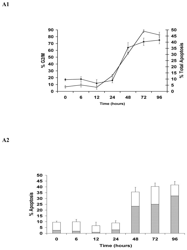
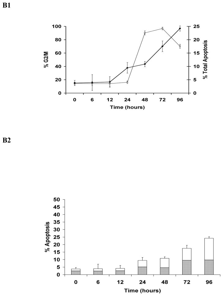
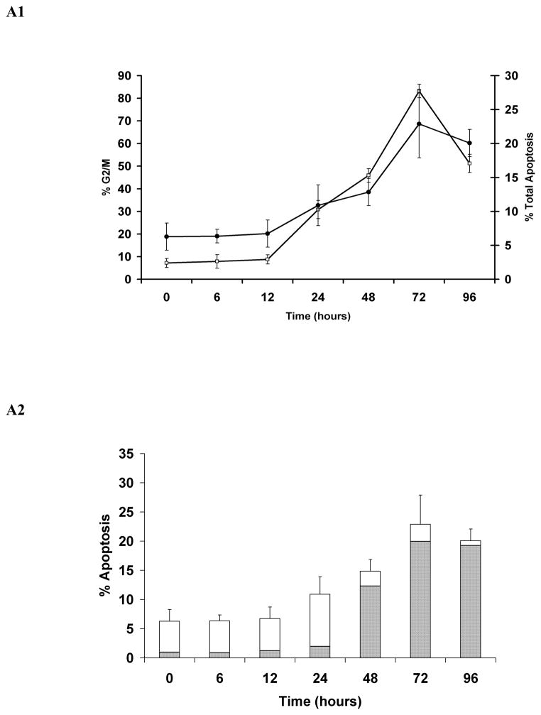
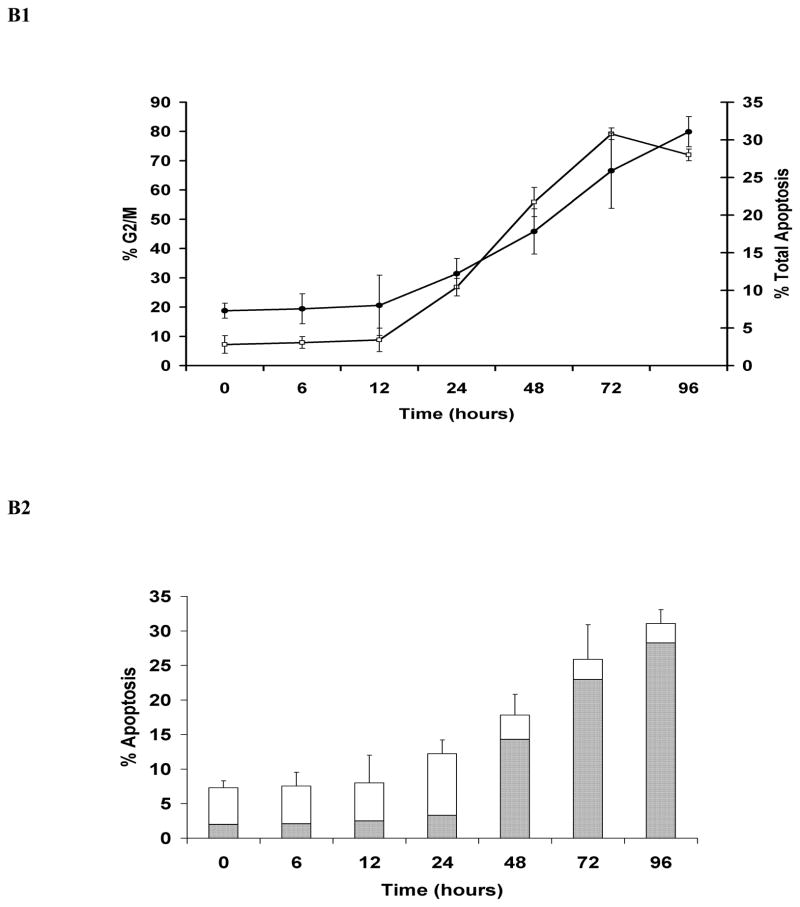
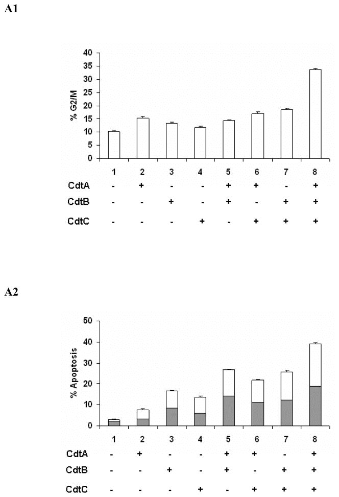
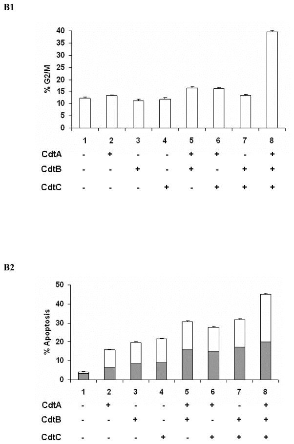
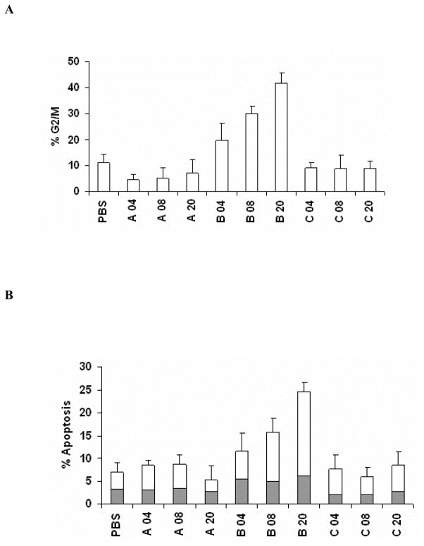
Materials and methods: The temporal relationship between cell cycle and apoptotic death of human epithelial HeLa and INT407 cells intoxicated with HhepCDT holotoxin or reconstituted recombinant HhepCDT was compared by flow cytometry. The genotoxic activity of individual and combinations of recombinant HhepCDT protein subunits or increasing concentrations of individual recombinant HhepCDT protein subunits transfected into HeLa cells was assessed at 72 hours post-treatment by flow cytometry.

Results: Similar time course of HhepCDT-induced G2 /M cell cycle arrest and apoptotic death was found with both cell lines which reached a maximum at 72 hours. The presence of all three HhepCDT subunits was required for maximum cell cycle arrest and apoptosis of both cell lines. Transfection of HeLa cells with HhepCdtB, but not with HhepCdtA or HhepCdtC, resulted in a dose-dependent G2 /M arrest and apoptotic death.

Conclusion: All three subunits of HhepCDT are required for maximum epithelial cell cycle arrest and progression to apoptotic death, and HhepCdtB subunit alone is necessary and sufficient for epithelial cell genotoxicity.

Keywords: CDT; Helicobacter hepaticus; apoptosis; cell cycle arrest.


Figures

Fig. 1
Fig. 1. Kinetics of HeLa and INT407 cell cycle arrest and apoptosis induced by Helicobacter hepaticus CDT holotoxin
Simultaneous quantitative flow cytometry analysis of HeLa (A) and INT407 (B) G2/M cell cycle arrest (open squares) and apoptosis (solid circles in A1 and B1; solid bars in A2 and white bars in B2 represent early and late apoptosis, respectively) over 96 hours post-treatment with HhepCDT holotoxin (50 μg/mL final total protein concentration). Each point and bar represents mean percent and standard deviation of 1.0 × 10⁴ cells in three independent experiments.
Fig. 2
Fig. 2. Kinetics of HeLa and INT407 cell cycle arrest and apoptosis induced by reconstituted recombinant Helicobacter hepaticus CDT
Simultaneous quantitative flow cytometry analysis of HeLa (A) and INT407 (B) G2/M cell cycle arrest (open squares) and apoptosis (solid circles in A1 and B1; solid bars in A2 and white bars in B2 represent early and late apoptosis, respectively) over 96 hours post-treatment with reconstituted recombinant HhepCDT (12 μg/mL final total protein concentraton). Each point and bar represents mean percent and standard deviation of 1.0 × 10⁴ cells in three independent experiments.
Fig. 3
Fig. 3. All three Helicobacter hepaticus CDT subunits are required for maximum HeLa and INT407 cell cycle arrest and apoptotic cell death
Quantitative flow cytometry analysis of HeLa (A) and INT407 (B) G2/M cell cycle arrest (A1 and B1 open bars) and apoptosis (solid bars and white bars in A2 and B2 represent early and late apoptosis, respectively) 72 hours post-treatment with individual and combinations of recombinant HhepCDT fusion protein subunits (12 μg/mL final total protein concentration). Each bar represents mean percent and standard deviation of 1.0 × 10⁴ cells in three independent experiments.
Fig. 4
Fig. 4. Helicobacter hepaticus CdtB alone is necessary and sufficient for HeLa cell cycle arrest and apoptotic cell death
Quantitative flow cytometry analysis of HeLa G2/M cell cycle arrest (A open bars) and apoptosis (solid bars and white bars in B represent early and late apoptosis, respectively) 72 h post-transfection with increasing concentrations from 4, 8 and 20 μg/mL of recombinant HhepCDT fusion protein subunits. Each bar represents mean percent and standard deviation of 1.0 × 10⁴ cells in three independent experiments.

References

    1. Jinadasa RN, Bloom SE, Weiss RS, Duhamel GE. Cytolethal distending toxin: a conserved bacterial genotoxin that blocks cell cycle progression, leading to apoptosis of a broad range of mammalian cell lineages. Microbiology. Jul;157(Pt 7):1851–75. - PMC - PubMed
    1. Guerra L, Cortes-Bratti X, Guidi R, Frisan T. The biology of the cytolethal distending toxins. Toxins (Basel) Mar;3(3):172–90. - PMC - PubMed
    1. Smith JL, Bayles DO. The contribution of cytolethal distending toxin to bacterial pathogenesis. Critical Reviews in Microbiology. 2006 Oct-Dec;32(4):227–48. - PubMed
    1. den Bakker HC, Moreno Switt AI, Govoni G, Cummings CA, Ranieri ML, Degoricija L, et al. Genome sequencing reveals diversification of virulence factor content and possible host adaptation in distinct subpopulations of Salmonella enterica. BMC Genomics. 12:425. - PMC - PubMed
    1. Haghjoo E, Galan JE. Salmonella typhi encodes a functional cytolethal distending toxin that is delivered into host cells by a bacterial-internalization pathway. Proceedings of the National Academy of Sciences of the United States of America. 2004 Mar 30;101(13):4614–9. - PMC - PubMed

Publication types

MeSH terms

Substances

Associated data

LinkOut - more resources