

Progesterone and vitamin D: Improvement after traumatic brain injury in middle-aged rats
- PMID: 23896206
- PMCID: PMC3833454
- DOI: 10.1016/j.yhbeh.2013.06.009
Progesterone and vitamin D: Improvement after traumatic brain injury in middle-aged rats
Abstract
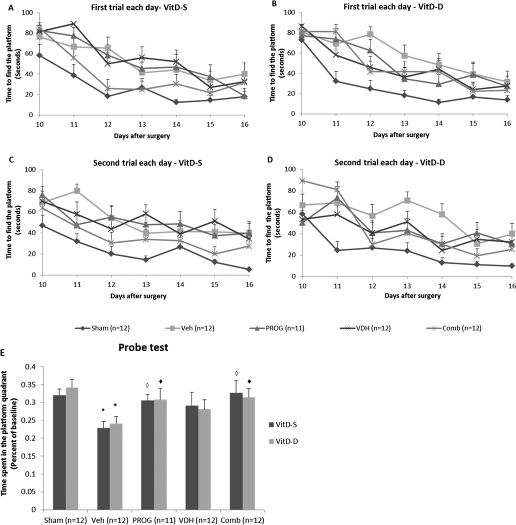
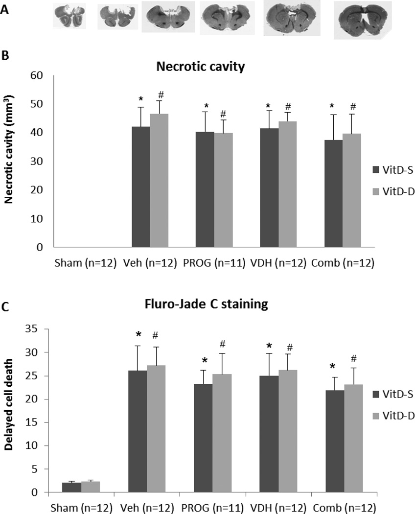
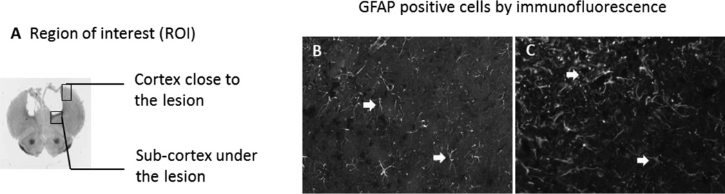
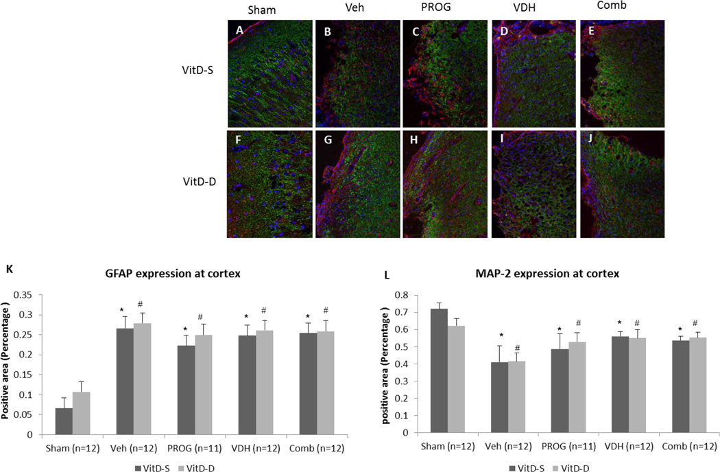
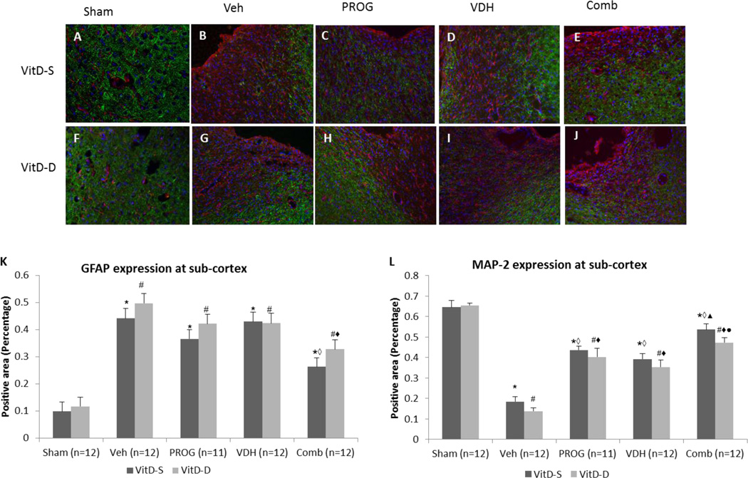
Progesterone (PROG) and vitamin D hormone (VDH) have both shown promise in treating traumatic brain injury (TBI). Both modulate apoptosis, inflammation, oxidative stress, and excitotoxicity. We investigated whether 21 days of VDH deficiency would alter cognitive behavior after TBI and whether combined PROG and VDH would improve behavioral and morphological outcomes more than either hormone alone in VDH-deficient middle-aged rats given bilateral contusions of the medial frontal cortex. PROG (16 mg/kg) and VDH (5 μg/kg) were injected intraperitoneally 1 h post-injury. Eight additional doses of PROG were injected subcutaneously over 7 days post-injury. VDH deficiency itself did not significantly reduce baseline behavioral functions or aggravate impaired cognitive outcomes. Combination therapy showed moderate improvement in preserving spatial and reference memory but was not significantly better than PROG monotherapy. However, combination therapy significantly reduced neuronal loss and the proliferation of reactive astrocytes, and showed better efficacy compared to VDH or PROG alone in preventing MAP-2 degradation. VDH+PROG combination therapy may attenuate some of the potential long-term, subtle, pathophysiological consequences of brain injury in older subjects.
Keywords: Aging; Combination treatments; Functional repair; Progesterone; Traumatic brain injury; Vitamin D deficiency; Vitamin D3 hormone.
© 2013 Elsevier Inc. All rights reserved.
Figures
Similar articles
-
Progesterone and low-dose vitamin D hormone treatment enhances sparing of memory following traumatic brain injury.Horm Behav. 2012 Apr;61(4):642-51. doi: 10.1016/j.yhbeh.2012.02.017. Horm Behav. 2012. PMID: 22570859 Free PMC article.
-
Progesterone with vitamin D affords better neuroprotection against excitotoxicity in cultured cortical neurons than progesterone alone.Mol Med. 2009 Sep-Oct;15(9-10):328-36. doi: 10.2119/molmed.2009.00016. Epub 2009 Jun 26. Mol Med. 2009. PMID: 19603099 Free PMC article.
-
Vitamin D deficiency reduces the benefits of progesterone treatment after brain injury in aged rats.Neurobiol Aging. 2011 May;32(5):864-74. doi: 10.1016/j.neurobiolaging.2009.04.017. Epub 2009 May 30. Neurobiol Aging. 2011. PMID: 19482377 Free PMC article.
-
A review of the neuroprotective role of vitamin D in traumatic brain injury with implications for supplementation post-concussion.Brain Inj. 2016;30(8):960-8. doi: 10.3109/02699052.2016.1147081. Epub 2016 May 16. Brain Inj. 2016. PMID: 27185224 Review.
-
Progesterone and vitamin d hormone as a biologic treatment of traumatic brain injury in the aged.PM R. 2011 Jun;3(6 Suppl 1):S100-10. doi: 10.1016/j.pmrj.2011.03.010. PM R. 2011. PMID: 21703565 Free PMC article. Review.
Cited by
-
Combination Therapies for Traumatic Brain Injury: Retrospective Considerations.J Neurotrauma. 2016 Jan 1;33(1):101-12. doi: 10.1089/neu.2014.3855. Epub 2015 Aug 6. J Neurotrauma. 2016. PMID: 25970337 Free PMC article.
-
Neuroprotective effect of paricalcitol in a rat model of transient global cerebral ischemia.Int J Emerg Med. 2020 Jun 10;13(1):30. doi: 10.1186/s12245-020-00289-7. Int J Emerg Med. 2020. PMID: 32522270 Free PMC article.
-
Sex-related responses after traumatic brain injury: Considerations for preclinical modeling.Front Neuroendocrinol. 2018 Jul;50:52-66. doi: 10.1016/j.yfrne.2018.03.006. Epub 2018 May 18. Front Neuroendocrinol. 2018. PMID: 29753798 Free PMC article. Review.
-
Neuro-Inflammation Modulation and Post-Traumatic Brain Injury Lesions: From Bench to Bed-Side.Int J Mol Sci. 2022 Sep 23;23(19):11193. doi: 10.3390/ijms231911193. Int J Mol Sci. 2022. PMID: 36232495 Free PMC article. Review.
-
Multi-Mechanistic Approaches to the Treatment of Traumatic Brain Injury: A Review.J Clin Med. 2023 Mar 11;12(6):2179. doi: 10.3390/jcm12062179. J Clin Med. 2023. PMID: 36983181 Free PMC article. Review.
References
-
- Annweiler C, Schott AM, Allali G, Bridenbaugh SA, Kressig RW, Allain P, Herrmann FR, Beauchet O. Association of vitamin D deficiency with cognitive impairment in older women: cross-sectional study. Neurology. 2010;74:27–32. - PubMed
-
- Atalay B, Caner H, Can A, Cekinmez M. Attenuation of microtubule associated protein-2 degradation after mild head injury by mexiletine and calpain-2 inhibitor. Br. J. Neurosurg. 2007;21(3):281–287. - PubMed
-
- Atif F, Yousuf S, Sayeed I, Ishrat T, Hua F, Stein DG. Combination treatment with progesterone and vitamin D hormone is more effective than monotherapy in ischemic stroke: The role of BDNF/TrkB/Erk1/2 signaling in neuroprotection. Neuropharmacology. 2013;67:78–87. [Epub ahead of print]. - PMC - PubMed
Publication types
MeSH terms
Substances
Grants and funding
LinkOut - more resources
Full Text Sources
Other Literature Sources
Medical
