Review
. 2013 Dec;23(6):1073-9.
doi: 10.1016/j.conb.2013.07.002. Epub 2013 Jul 26.

Probing the enigma: unraveling glial cell biology in invertebrates

Affiliations
Review

Probing the enigma: unraveling glial cell biology in invertebrates

Jaeda Coutinho-Budd et al. Curr Opin Neurobiol. 2013 Dec.

Abstract

Despite their predominance in the nervous system, the precise ways in which glial cells develop and contribute to overall neural function remain poorly defined in any organism. Investigations in simple model organisms have identified remarkable morphological, molecular, and functional similarities between invertebrate and vertebrate glial subtypes. Invertebrates like Drosophila and Caenorhabditis elegans offer an abundance of tools for in vivo genetic manipulation of single cells or whole populations of glia, ease of access to neural tissues throughout development, and the opportunity for forward genetic analysis of fundamental aspects of glial cell biology. These features suggest that invertebrate model systems have high potential for vastly improving the understanding of glial biology. This review highlights recent work in Drosophila and other invertebrates that reveal new insights into basic mechanisms involved in glial development.


Figures

Figure 1
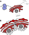
Figure 1. Variation in complexity of Drosophila glia
A) The Drosophila wing blade provides a simple environment where approximately 130 glia migrate along three sensory nerves (costa, L1, and L3), then mature to wrap and insulate the nerves. B) Three segments of the Drosophila embryonic ventral nerve cord, depicting the ladder-like array of axons that are separated and insulated by midline glia, and peripheral glia that insulate the peripheral nerves. C-D) The Drosophila larval CNS provides even more complexity and glial diversity. (C) depicts the structure of the larval CNS and proximal portions of the nerves that stretch from the VNC to the larval body wall. (D) shows a close-up of the brain lobe outlined in (C), illustrating the diverse morphology of glial cells found within this sophisticated invertebrate model organism.
Figure 2
Figure 2. Glial morphogenesis, proliferation, and regulation of neuroblast reactivation through insulin and FGF signaling
A) Regulation of neuroblast proliferation and animal growth. The fat body is activated by amino acid nutrient signaling, which then releases a fat body derived mitogen (FBDM) to glial cells, which in turn release dILP6, causing neuroblast reactivation and proliferation into neurons and glia. Surface glia also release a secreted decoy of insulin receptor (SDR) to block dILP activity in the brain where it can regulate neuroblast proliferation, or to the hemolymph to regulate animal body growth. FMRP signals non-autonomously in glia to inhibit neuroblast proliferation. B) Glia respond to drosophila Insulin-Like Peptides (dILPs) through the insulin receptor (InR, purple) to regulate proliferation through PI3K/tor signaling in PGs, and Ras/MAPK signaling in CGs. Likewise, glia respond to pyramus (pyr) through the FGF receptor, Heartless (green) from nearby self-type specific glial cells to regulate morphology and proliferation. CGs, but not PGs, can respond to neuronally-derived pyr as well.

Similar articles

Cited by

References

    1. Buchanan RL, Benzer S. Defective glia in the Drosophila brain degeneration mutant drop-dead. Neuron. 1993;10:839–850. - PubMed
    1. Hidalgo A, Booth GE. Glia dictate pioneer axon trajectories in the Drosophila embryonic CNS. Development. 2000;127:393–402. - PubMed
    1. Spindler SR, Ortiz I, Fung S, Takashima S, Hartenstein V. Drosophila cortex and neuropile glia influence secondary axon tract growth, pathfinding, and fasciculation in the developing larval brain. Dev Biol. 2009;334:355–368. - PMC - PubMed
    1. Stork T, Thomas S, Rodrigues F, Silies M, Naffin E, Wenderdel S, Klambt C. Drosophila Neurexin IV stabilizes neuron-glia interactions at the CNS midline by binding to Wrapper. Development. 2009;136:1251–1261. - PubMed
    1. Doherty J, Logan MA, Tasdemir OE, Freeman MR. Ensheathing glia function as phagocytes in the adult Drosophila brain. J Neurosci. 2009;29:4768–4781. - PMC - PubMed

Publication types

LinkOut - more resources