

Hyperexcitability of bladder afferent neurons associated with reduction of Kv1.4 α-subunit in rats with spinal cord injury
- PMID: 23896350
- PMCID: PMC3920734
- DOI: 10.1016/j.juro.2013.07.058
Hyperexcitability of bladder afferent neurons associated with reduction of Kv1.4 α-subunit in rats with spinal cord injury
Abstract
Purpose: To clarify the functional and molecular mechanisms inducing hyperexcitability of C-fiber bladder afferent pathways after spinal cord injury we examined changes in the electrophysiological properties of bladder afferent neurons, focusing especially on voltage-gated K channels.
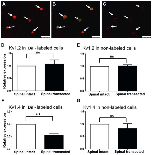
Materials and methods: Freshly dissociated L6-S1 dorsal root ganglion neurons were prepared from female spinal intact and spinal transected (T9-T10 transection) Sprague Dawley® rats. Whole cell patch clamp recordings were performed on individual bladder afferent neurons. Kv1.2 and Kv1.4 α-subunit expression levels were also evaluated by immunohistochemical and real-time polymerase chain reaction methods.
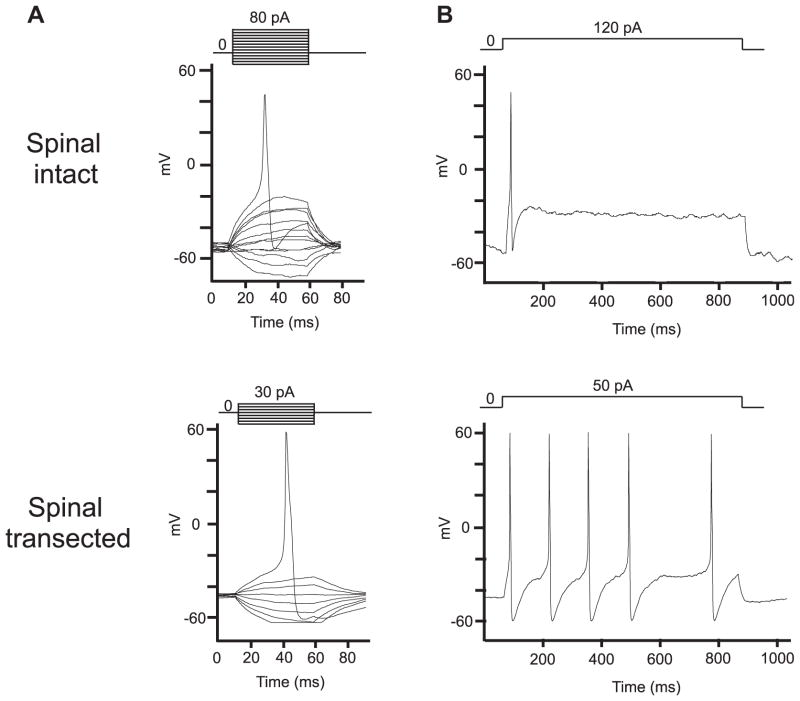
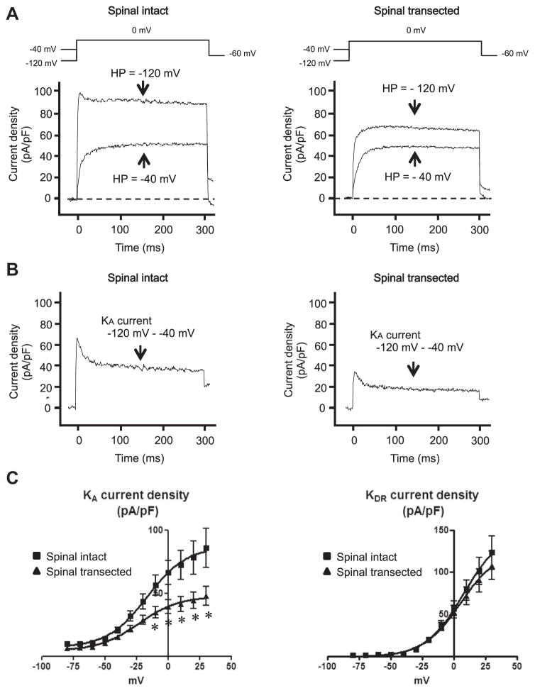
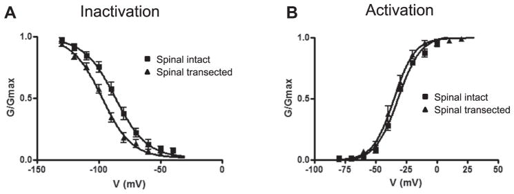
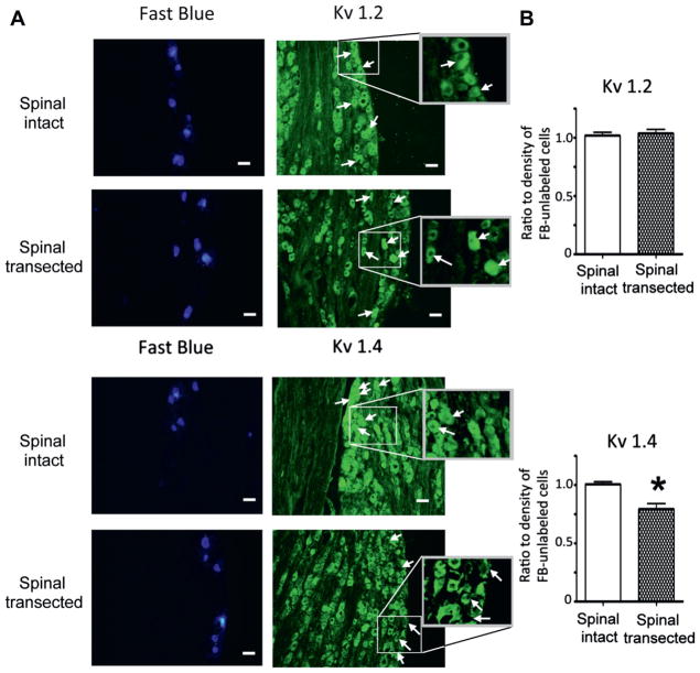
Results: Capsaicin sensitive bladder afferent neurons from spinal transected rats showed increased cell excitability, as evidenced by lower spike activation thresholds and a tonic firing pattern. The peak density of transient A-type K+ currents in capsaicin sensitive bladder afferent neurons from spinal transected rats was significantly less than that from spinal intact rats. Also, the KA current inactivation curve was displaced to more hyperpolarized levels after spinal transection. The protein and mRNA expression of Kv1.4 α-subunits, which can form transient A-type K+ channels, was decreased in bladder afferent neurons after spinal transection.
Conclusions: Results indicate that the excitability of capsaicin sensitive C-fiber bladder afferent neurons is increased in association with reductions in transient A-type K+ current density and Kv1.4 α-subunit expression in injured rats. Thus, the Kv1.4 α-subunit could be a molecular target for treating overactive bladder due to neurogenic detrusor overactivity.
Keywords: 1,1′-dioctadecyl-3,3,3′,3′-tetramethylindocarbocyanine perchlorate; A-type K(+); DRG; DTX; DiI; FITC; GAPDH; K(A); K(DR); Kv; NDO; PBS; PCR; SCI; TTX; V(h); afferent pathways; delayed rectifier-type K(+); dorsal root ganglion; fluorescein isothiocyanate; glyceraldehyde-3-phosphate dehydrogenase; half-maximal conductance; nerve fibers; neurogenic detrusor overactivity; overactive; phosphate buffered saline; polymerase chain reaction; potassium channels; spinal cord injuries; spinal cord injury; tetrodotoxin; unmyelinated; urinary bladder; voltage-gated; voltage-gated K(+); α-dendrotoxin.
Copyright © 2013 American Urological Association Education and Research, Inc. Published by Elsevier Inc. All rights reserved.
Figures
References
-
- de Groat WC, Yoshimura N. Mechanisms underlying the recovery of lower urinary tract function following spinal cord injury. Prog Brain Res. 2006;152:59. - PubMed
-
- Gold MS, Shuster MJ, Levine JD. Characterization of six voltage-gated K+ currents in adult rat sensory neurons. J Neurophysiol. 1996;75:2629. - PubMed
-
- Everill B, Kocsis JD. Reduction in potassium currents in identified cutaneous afferent dorsal root ganglion neurons after axotomy. J Neurophysiol. 1999;82:700. - PubMed
Publication types
MeSH terms
Substances
Grants and funding
LinkOut - more resources
Full Text Sources
Other Literature Sources
Medical
Research Materials
