Review
. 2013 Oct;750(1-2):15-22.
doi: 10.1016/j.mrfmmm.2013.07.004. Epub 2013 Jul 27.

Structural mechanisms underlying signaling in the cellular response to DNA double strand breaks

Affiliations
Review

Structural mechanisms underlying signaling in the cellular response to DNA double strand breaks

Inbal Mermershtain et al. Mutat Res. 2013 Oct.

Abstract

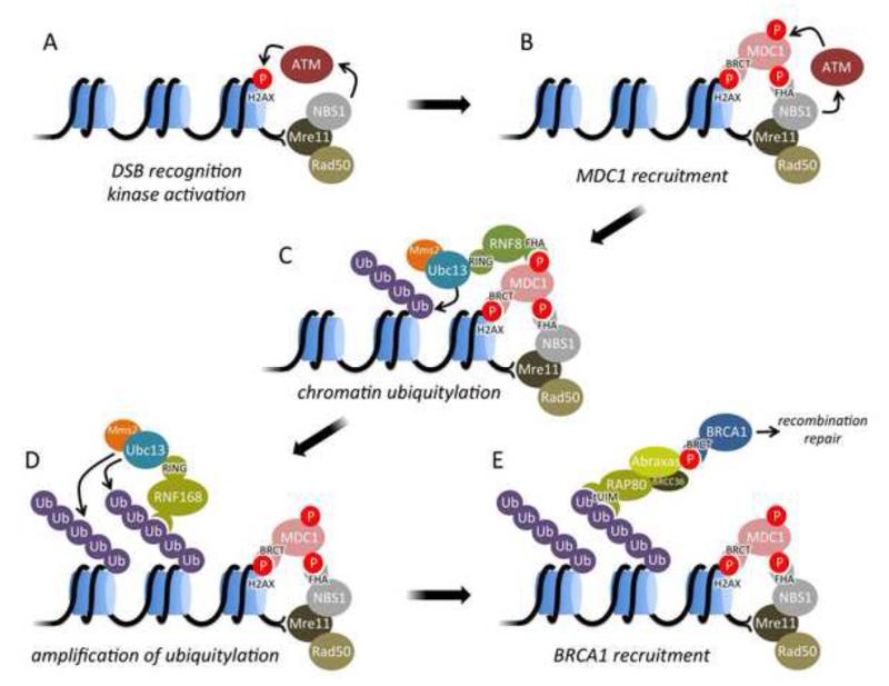
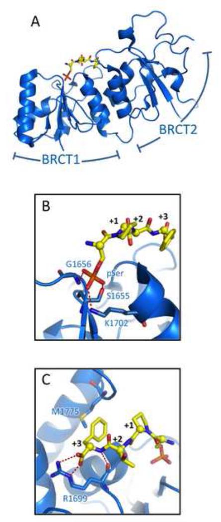
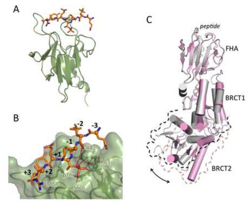
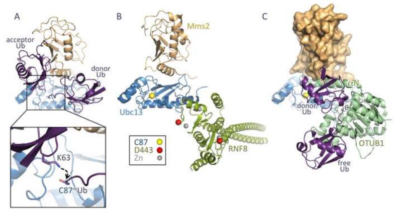
DNA double strand breaks (DSBs) constitute one of the most dangerous forms of DNA damage. In actively replicating cells, these breaks are first recognized by specialized proteins that initiate a signal transduction cascade that modulates the cell cycle and results in the repair of the breaks by homologous recombination (HR). Protein signaling in response to double strand breaks involves phosphorylation and ubiquitination of chromatin and a variety of associated proteins. Here we review the emerging structural principles that underlie how post-translational protein modifications control protein signaling that emanates from these DNA lesions.

Keywords: BRCA1 C-terminal domain; BRCT; BRCT domains; DSB; Double-strand break signaling; FHA domains; FHAf; HR; MRN; Mre11–Rad50–NBS1; OTUB; Phosphorylation signaling; RIDDLE syndrome; UIM; Ubc13; Ubiquitin; double strand break; homologous recombination; orkhead-associated domain.; otubain or ovarian tumor domain protein; radiosensitivity, immunodeficiency, dysmorphic features and learning difficulties; ubiquitin interaction motif.


Figures

Fig. 1
Fig. 1
Overview of signaling from DNA double strand breaks (DSBs) to initiate repair by homologous recombination (HR). Phosphorylation events are indicated by red circles, ubiquitylation by purple circles and the domains mediating protein-protein interactions (BRCT, FHA, tUIM, RING) are highlighted.
Fig. 2
Fig. 2
Phospho-peptide recognition by BRCT domains. (A) Structure of the BRCA1 BRCT domain (blue) bound to a pSer-x-x-Phe target peptide (yellow) with the two tandem BRCT domains indicated. (B) Details of pSer recognition by the BRCA1 BRCT. (C) Details of recognition of the Phe +3 residue and C-terminal carboxylate. Note that the MDC1 BRCT uses a similar mechanism to recognize the C-terminus of γH2AX.
Fig. 3
Fig. 3
Phospho-peptide recognition by FHA domains. (A) Structure of the FHA domain of RNF8 (green) bound to its pThr-containing target peptide (orange). (B) Detailed view of the recognition of the pThr and neighboring residues by the RNF8 FHA. (C) Phospho-peptide binding by the NBS1 FHA domain drives a conformational change that leads to a rearrangement of BRCT-BRCT packing. Structure of the NBS1 FHA-BRCT-BRCT domain bound to a CtIP peptide is in pink, while the apo structure is in grey. The structures were aligned on their FHA domains to reveal the resulting rotation of the C-terminal BRCT with respect to the rest of the structure.
Fig. 4
Fig. 4
Structure and regulation of Mms2-Ubc13. (A) Mms2 (orange) binds an acceptor ubiquitin (purple) such that its Lys63 is positioned to attack the thioester of the donor ubiquitin bound to the active site Cys87 of Ubc13 (blue). (B) RNF8 RING domain exists as a coiled-coil – stabilized dimer (green) and binds to Ubc13. Highlighted in red is Asp443, which regulates the ability of the RING domain to target nucleosomes for ubiquitination. (C) OTUB1 (green) binds Ubc13~Ub, blocking RNF8 access and interfering with Mms2 binding to Ubc13. In this figure, Mms2 (orange surface) is docked onto Ubc13 to illustrate the predicted clash with the N-terminal OTUB1 helix (αN). OTUB1 binding is allosterically regulated through the binding of free ubiquitin.
Fig. 5
Fig. 5
Recognition of Lys63-linked di-ubiquitin by the RAP80 tandem UIM (tUIM). The tUIM is shown in green and grey and the sequence of the human RAP80 tUIM is displayed below. Conserved acidic residues in the tUIM and the positively charged residues in ubiquitin with which they interact are shown as sticks. The conserved Ala-Ser residues in the tUIM and Ile44 in ubiquitin that make hydrophobic contact are displayed as spheres.

References

    1. Kasparek TR, Humphrey TC. DNA double-strand break repair pathways, chromosomal rearrangements and cancer. Semin Cell Dev Biol. 2011;22:886–897. - PubMed
    1. Chapman JR, Taylor MR, Boulton SJ. Playing the end game: DNA double-strand break repair pathway choice. Mol Cell. 2012;47:497–510. - PubMed
    1. Caestecker KW, Van de Walle GR. The role of BRCA1 in DNA double-strand repair: past and present. Exp Cell Res. 2013;319:575–587. - PubMed
    1. Dever SM, White ER, Hartman MC, Valerie K. BRCA1-directed, enhanced and aberrant homologous recombination: mechanism and potential treatment strategies. Cell Cycle. 2012;11:687–694. - PMC - PubMed
    1. Zhang J. The role of BRCA1 in homologous recombination repair in response to replication stress: significance in tumorigenesis and cancer therapy. Cell Biosci. 2013;3:11. - PMC - PubMed

Publication types

MeSH terms