. 2013 Sep 5;122(10):1822-32.
doi: 10.1182/blood-2013-01-482315. Epub 2013 Jul 29.

Thrombospondin-1 modulates VEGF signaling via CD36 by recruiting SHP-1 to VEGFR2 complex in microvascular endothelial cells

Affiliations

Thrombospondin-1 modulates VEGF signaling via CD36 by recruiting SHP-1 to VEGFR2 complex in microvascular endothelial cells

Ling-Yun Chu et al. Blood. .

Abstract

Thrombospondin-1 (TSP-1) inhibits growth factor signaling at the receptor level in microvascular endothelial cells (MVEC), and CD36 has been suggested to be involved in this inhibition, but the mechanisms are not known. We hypothesized that CD36-TSP-1 interaction recruits Src homology 2 domain-containing protein tyrosine phosphatase (SHP)-1 to the vascular endothelial growth factor receptor 2 (VEGFR2) signaling complex and attenuates vascular endothelial growth factor (VEGF) signaling. Western blots of anti-CD36 and anti-VEGFR2 immunoprecipitates from VEGF-treated MVEC showed that exposure of the cells to a recombinant protein containing the CD36 binding domain of thrombospondin-1 (known as the TSR domain) induced association of SHP-1 with the VEGFR2/CD36 signaling complex and thereby suppressed VEGFR2 phosphorylation. SHP-1 phosphatase activity was increased in immunoprecipitated VEGFR2 complexes from TSR-treated cells. Silencing CD36 expression in MVEC by small interfering RNA (siRNA) or genetic deletion of cd36 in mice showed that TSR-induced SHP-1/VEGFR2 complex formation required CD36 in vitro and in vivo. Silencing SHP-1 expression in MVEC by siRNA abrogated TSR-mediated inhibition of VEGFR2 phosphorylation as well as TSR-mediated inhibition of VEGF-induced endothelial cell migration and tube formation. These studies reveal a SHP-1-mediated antiangiogenic pathway induced by CD36-TSP-1 interaction that inhibits VEGFR2 signaling and they provide a novel target to modulate angiogenesis therapeutically.


Figures
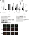
Figure 1
Figure 1
TSR induces SHP-1 association with CD36 and VEGFR2 in MVEC. (A) MVEC were cultured to confluence and then treated with TSP-1, recombinant TSR, or thioredoxin (Thio) control for 4 hours before exposure to 50 ng/mL VEGF for 5 minutes. VEGFR2 phosphorylation was analyzed by western blot with an antibody to VEGFR2 pTyr1175. Blots were reprobed with anti-VEGFR2 as a loading control. VEGFR2 phosphorylation levels are expressed as the ratio of phosphorylated VEGFR2 to total VEGFR2. (B-C) MVEC were treated with TSR (10 nmol/L) in low-serum medium for 4 hours before exposure to VEGF (50 ng/mL) for 15 minutes. Cell lysates were then immunoprecipitated with anti-VEGFR2 (B) or anti-CD36 (C), and coprecipitated proteins were detected by western blot analysis with antibodies to SHP-1, SHP-2, phospho-SHP-2, phospho-VEGFR2, VEGFR2, or CD36. Blots are representative of 3 experiments. (D) Immunofluorescence images of MVEC treated with 10 nmol/L TSR and/or 50 ng/mL VEGF and then probed with mouse monoclonal anti–SHP-1 and rabbit polyclonal anti-CD36, followed by Alexa Fluor 488–conjugated anti-mouse IgG and Alexa Fluor 594–conjugated anti-rabbit IgG. The top row shows Alexa Fluor 488 images, the middle row shows Alexa Fluor 594 images, and the bottom row shows the merged images.
Figure 2
Figure 2
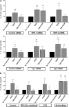
Inhibition of VEGF-induced VEGFR2 phosphorylation by TSR is mediated through SHP-1. MVEC transfected with control siRNA or siRNA directed against SHP-1 or SHP-2 (A) or against Fyn or Syk (B) were exposed to 10 nmol/L TSR in low-serum medium for 4 hours and then to 50 ng/mL VEGF for 5 minutes. Phosphorylated VEGFR2 in these samples were analyzed by western blot with pTyr1175 antibody. Blots were reprobed for VEGFR2 and phosphorylation levels expressed as a ratio of phosphorylated to total VEGFR2 (n = 3). The images above the bar graphs are representative western blots showing the efficiency of protein knock-down 48 hours after transfection. Anti-VEGFR2 or anti-IgG control immunoprecipitates of MVEC (C) or MVEC silenced with respective siRNA (D) were incubated with the phosphatase substrate pNPP for 1 hour at room temperature, and phosphatase activity was detected by absorbance at 405 nm.
Figure 3
Figure 3
CD36 is required for TSR-induced SHP-1 association with VEGFR2 and VEGFR2 dephosphorylation in MVEC. (A) MVEC were transfected with CD36 or control siRNA for 6 hours and then cultured for 48 hours before analysis of CD36 expression by immunoprecipitation and western blot. (B) MVEC transfected as in (A) were treated with 10 nmol/L TSR in low-serum medium for 4 hours and then exposed to 50 ng/mL VEGF for 5 minutes. VEGFR2 phosphorylation was analyzed by western blot with an antibody to pTyr1175. Blots were reprobed with an antibody to VEGFR2. The level of phosphorylation was shown as the ratio of phosphorylated to total VEGFR2 (B). (C) MVEC transfected as in (B) were treated with 10 nmol/L TSR in low-serum medium for 4 hours and then either exposed (left) or not exposed (right) to 50 ng/mL VEGF for 15 minutes. VEGFR2 was then immunoprecipitated and the precipitates were analyzed by western blot with anti–SHP-1 antibody. Blots were reprobed with an antibody to VEGFR2. Blots are representative of 3 experiments.
Figure 4
Figure 4
SHP-1 is required for TSR-mediated inhibition of VEGF-induced MVEC migration. (A) MVEC transfected with specific siRNA as in Figure 2 were allowed to migrate toward VEGF for 15 hours through an 8.0-μm membrane in a Transwell insert in the presence or absence of 10 nmol/L TSR. Within each group, siRNA transfected MVEC not exposed to VEGF or TSR served as controls. The number of migrated cells was counted and the result expressed as percent of cells migrated compared with control (n = 8). (B) Migration assays as in panel A were performed with MVEC treated with Src inhibitor (PP2) or Syk inhibitor (Piceatannol) (n = 6). PP3 served as a negative control for PP2.
Figure 5
Figure 5
CD36 and SHP-1 are required for TSR inhibition of MVEC tubelike structure formation. (A) MVEC were transfected with CD36 or control siRNA as in Figure 3 and then treated with 10 nmol/L TSR in low-serum medium for 4 hours before being transferred onto Matrigel-coated tissue culture wells. Cells were then exposed to 50 ng/mL VEGF for 6 hours, fixed with 4% paraformaldehyde, and then stained with 30 nmol/L fluorescence-labeled Phalloidin for 1 hour. Images from 4 randomly chosen areas were obtained with a fluorescence microscope, and the number of branches and average branch length of tubelike structures were quantified using NIH ImageJ software. The scale bar represents 200 μm. (B) MVEC were transfected with control siRNA and treated with TSR as in panel A. Cells were then exposed to 50 ng/mL VEGF for 6 hours in the presence of 100 μmol/L SHP inhibitor NSC-87877 or vehicle control. The number of branches and the average branch length of tubelike structures were analyzed as in panel A.
Figure 6
Figure 6
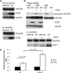
Genetic deletion of cd36 increases VEGFR2 phosphorylation and decreases SHP-1 association with VEGFR2 after in vivo infusion of VEGF. (A) Lysates of lung tissue removed from cd36 null or wild-type mice were analyzed by western blot with anti-VEGFR2, anti-CD36, or antitubulin (top) or by immunoprecipitation with anti-VEGFR2 followed by western blot (bottom). (B) Lung tissue from wild-type or cd36 null mice was harvested 5 minutes after injection of 1 μg VEGF via the jugular vein. The top panel shows VEGFR2 Tyr1175 phosphorylation detected by western blot analysis of tissue lysates. Blots were reprobed with antibodies to total VEGFR2, CD36, and tubulin. The bottom panel shows western blot analysis of anti-VEGFR2 immunoprecipitates of the tissue lysates probed with antibodies to SHP-1 and VEGFR2. Blots are representative of 3 experiments. (C) VEGFR2 phosphorylation levels in panel B were expressed as the ratio of phosphorylated VEGFR2 to total VEGFR2.
Figure 7
Figure 7
Model depicting CD36-dependent antiangiogenic pathways. VEGFR2 forms a complex with CD36. When CD36 is not engaged by its ligand, TSP-1, VEGF binding to VEGFR2 induces VEGFR2 phosphorylation at Tyr1175 and promotes MVEC migration and tube formation. Engagement of CD36 by TSP-1 leads to inhibition of migration and tube formation by 2 independent processes. Recruitment of SHP-1 to the VEGFR2-CD36 complex leads to dephosphorylation of VEGFR2 (Tyr1175), thus dampening VEGF signaling, and activation of Fyn leads to apoptosis via activation of MAP kinases and caspases. Syk may participate in these pathways, although the mechanisms remain undefined.

Similar articles

Cited by

References

    1. Silverstein RL, Febbraio M. CD36, a scavenger receptor involved in immunity, metabolism, angiogenesis, and behavior. Sci Signal. 2009;2(72):re3. - PMC - PubMed
    1. Clemetson KJ, Pfueller SL, Luscher EF, Jenkins CS. Isolation of the membrane glycoproteins of human blood platelets by lectin affinity chromatography. Biochim Biophys Acta. 1977;464(3):493–508. - PubMed
    1. Silverstein RL, Baird M, Lo SK, Yesner LM. Sense and antisense cDNA transfection of CD36 (glycoprotein IV) in melanoma cells. Role of CD36 as a thrombospondin receptor. J Biol Chem. 1992;267(23):16607–16612. - PubMed
    1. Dawson DW, Pearce SF, Zhong R, Silverstein RL, Frazier WA, Bouck NP. CD36 mediates the In vitro inhibitory effects of thrombospondin-1 on endothelial cells. J Cell Biol. 1997;138(3):707–717. - PMC - PubMed
    1. Asch AS, Barnwell J, Silverstein RL, Nachman RL. Isolation of the thrombospondin membrane receptor. J Clin Invest. 1987;79(4):1054–1061. - PMC - PubMed

Publication types

MeSH terms

Substances