. 2013 Aug;4(8):1253-65.
doi: 10.18632/oncotarget.1141.

Combination therapy with anti-ErbB3 monoclonal antibodies and EGFR TKIs potently inhibits non-small cell lung cancer

Affiliations

Combination therapy with anti-ErbB3 monoclonal antibodies and EGFR TKIs potently inhibits non-small cell lung cancer

Alessia Noto et al. Oncotarget. 2013 Aug.

Abstract

Personalized therapy of advanced non-small cell lung cancer (NSCLC) has been improved by the introduction of EGFR tyrosine kinase inhibitors (TKIs), gefitinib and erlotinib. EGFR TKIs induce dramatic objective responses and increase survival in patients bearing sensitizing mutations in the EGFR intracytoplasmic tyrosine kinase domain. However, virtually all patients develop resistance, and this is responsible for disease relapse. Hence several efforts are being undertaken to understand the mechanisms of resistance in order to develop combination treatments capable to sensitize resistant cells to EGFR TKIs. Recent studies have suggested that upregulation of another member of the EGFR receptor family, namely ErbB3 is involved in drug resistance, through increased phosphorylation of its intracytoplasmic domain and activation of PI3K/AKT signaling. In this paper we first show, by using a set of malignant pleural effusion derived cell cultures (MPEDCC) from patients with lung adenocarcinoma, that surface ErbB3 expression correlates with increased AKT phosphorylation. Antibodies against ErbB3, namely A3, which we previously demonstrated to induce receptor internalization and degradation, inhibit growth and induce apoptosis only in cells overexpressing surface ErbB3. Furthermore, combination of anti-ErbB3 antibodies with EGFR TKIs synergistically affect cell proliferation in vitro, cause cell cycle arrest, up-regulate p21 expression and inhibit tumor growth in mouse xenografts. Importantly, potentiation of gefitinib by anti-ErbB3 antibodies occurs both in de novo and in ab initio resistant cells. Anti-ErbB3 mAbs strongly synergize also with the dual EGFR and HER2 inhibitor lapatinib. Our results suggest that combination treatment with EGFR TKI and antibodies against ErbB3 should be a promising approach to pursue in the clinic.


Figures

Figure 1
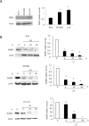
Figure 1. ErbB3 expression correlates with enhanced AKT signaling in primary and stable lung cancer cells
(a) Percentage of positive cells expressing surface ErbB3 was determined by FACS analysis in the indicated cell lines. Data represent the mean ± SD of three independent experiments. (b) Western Blot analysis of basal level of ErbB3 and pErbB3 and its downstream signalling in 7 representative primary MEPDCCs and stable cell lines PC9 and PC9ZD. At the right panel relative densitometry was evaluated. Data represent the mean ± SD of three independent experiments. (c) Graphic correlation between pAKT and surface ErbB3. Spearman's correlation index=0.88, p=0.003. pAKT was also strongly correlated with pErbB3 (Spearman's correlation index=0.82, p=0.011).
Figure 2
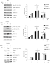
Figure 2. A3 mAb induces a time dependent down-modulation of ErbB3
(a) Representative Western blot of Heregulin expression in PC9, PC9ZD and Pe e/10 cell lines and relative densitometry were illustrated. (b) Cell cultures were treated at the indicated times with A3 at 50 μg/ml. Total ErbB3 was evaluated by western blot. Data are the mean ± SD of three independent experiments. *p<0.01, **p<0.05 versus untreated cells.
Figure 3
Figure 3. A3 inhibits HRG-induced signaling
Phosphorylation of AKT, ERK1/2 and ErbB3 was determined in PC9, PC9ZD and Pe e/10 treated with 50 μg/ml of A3 for 6 hrs and stimulated with 50 ng/ml of HRG for 10 min. At the right panel densitometries were calculated for phosphorylated proteins with respect to each total proteins. Data shown represent the mean ± SD of three independent experiments. *p<0.01 versus untreated cells.
Figure 4
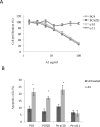
Figure 4. A3 affects proliferation and induces apoptosis in high surface expressing ErbB3 cells
(a) Clonogenic assay was performed in different cell cultures with different doses of A3, as described in Materials and Methods. (b) Apoptosis induction was evaluated by FACS analysis using Annexin V staining after 72 hr of treatment with 50 μg/ml of A3. Results are the mean ± SD of three independent experiments.* p<0.01 versus untreated cells.
Figure 5
Figure 5. A3 potentiates the effect of TK inhibitors in gefitinib resistant lung cancer cell cultures both in a clonogenic assay and in apoptosis induction
(a) PC9ZD and Pe e/10 cell cultures were incubated with various concentration of gefitinib and/or A3 at 25 μg/ml for 10 days in a clonogenic assay and analyzed as described in Material and Methods. Results are the mean ±SD of three independent experiments. (b) Clonogenic assay was performed on PC9ZD incubated with lapatinib and or/A3 at 25 μg/ml for 10 days as described above. Results are the mean ±SD of three independent experiments. (c) Apoptosis induction was evaluated in PC9ZD and Pe/e10 cell cultures after 72 hrs of treatment with A3 at 50 μg/ml and/or 1 μM gefitinib. Apoptosis was analyzed using Annexin V as described in Material and Methods. Results are the mean of three independent experiments ±SD. *p<0.01 in respect of untreated cells.
Figure 6
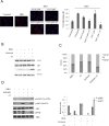
Figure 6. The combination A3 plus gefitinib reduces the proliferation, impairs cell cycle and reduces pAKT and pERK signaling
(a) PC9ZD cells were pre-treated with 50 μg/ml A3, then incubated with HRG for 24 hrs and stained with anit-Ki67 antibodies to identify cycling cells. Quantitative analysis of the percentage of cells presenting Ki67-positive nuclei was performed as reported in Material and Methods. Values are the mean ±SD. *p<0.05 versus HRG treated cells. (b) PC9ZD cells were treated for 24 hrs with indicated compounds and p53, p27 and p21 protein level were evaluated by western blotting. (c) Cell cycle analysis was evaluated by FACS analysis on PC9ZD treated with A3, gefitinib or the combination. (d) Phosphorylation of AKT and ERK1/2 was evaluated by Western blotting in PC9ZD cell line treated as above. For densitometric analysis results are expressed as mean values ± SD from three independent experiments.
Figure 7
Figure 7. A3 increases the efficacy of gefitinib in vivo
NOD/SCID mice xenografted with Pe e/10 primary cultures were treated with either gefitinib (100 mg/Kg) or A3 (20 mg/Kg) alone or with the combination of both. After 4 weeks mice were sacrificed and tumors weight were determined. *p<0.05 versus vehicle.

Similar articles

Cited by

References

    1. Jemal A, Bray F, Center MM, Ferlay J, Ward E, Forman D. Global cancer statistics. CA Cancer J Clin. 2011;61(2):69–90. - PubMed
    1. Heffernan KS, Wanke CA, Dong K, Warner PJ, Anskat PE, Karas RH, Kuvin JT. Lung cancer stem cells: tumor biology and clinical implications. Asia Pac J Clin Oncol. 2012;8:217–22. - PubMed
    1. Saintigny P, Burger JA. Recent advances in non-small cell lung cancer biology and clinical management. Discov Med. 2012;13:287–97. - PubMed
    1. Chunhacha P, Chanvorachote P. Roles of caveolin-1 on anoikis resistance in non small cell lung cancer. Int J Physiol Pathophysiol Pharmacol. 2012;4:149–55. - PMC - PubMed
    1. Dienstmann R, Martinez P, Felip E. Personalizing therapy with targeted agents in non-small cell lung cancer. Oncotarget. 2011;2(3):165–77. - PMC - PubMed

Publication types

MeSH terms