. 2013 Sep 15;537(2):217-24.
doi: 10.1016/j.abb.2013.07.015. Epub 2013 Jul 26.

A solution NMR investigation into the impaired self-assembly properties of two murine amelogenins containing the point mutations T21→I or P41→T

Affiliations

A solution NMR investigation into the impaired self-assembly properties of two murine amelogenins containing the point mutations T21→I or P41→T

Garry W Buchko et al. Arch Biochem Biophys. .

Abstract

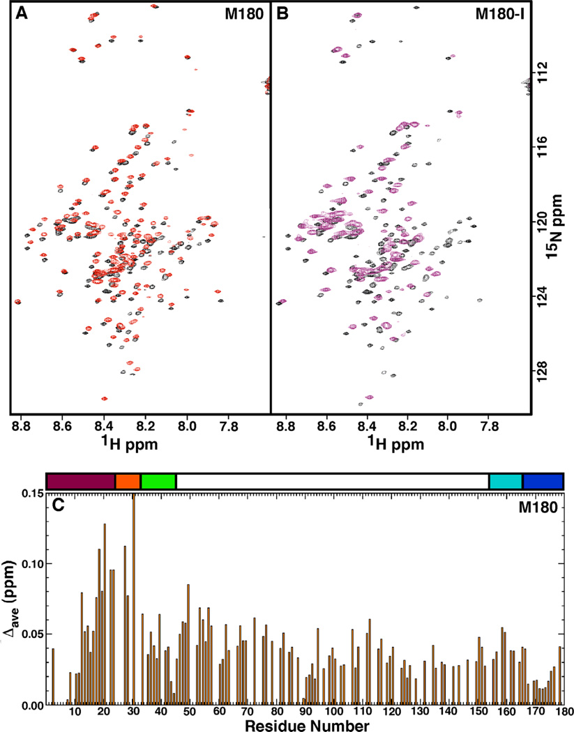
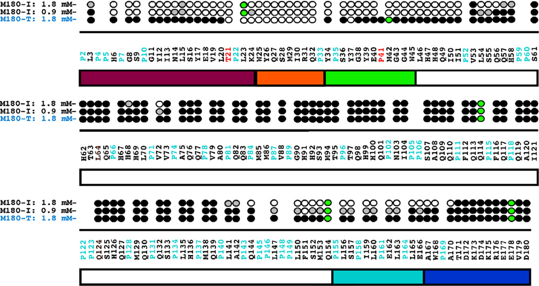
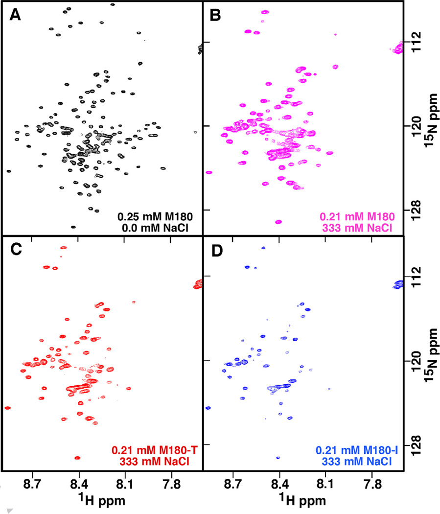
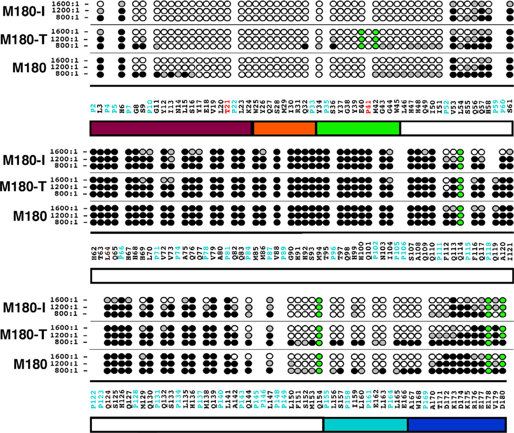
Amelogenesis imperfecta describes a group of inherited disorders that results in defective tooth enamel. Two disorders associated with human amelogenesis imperfecta are the point mutations T21→I or P40→T in amelogenin, the dominant protein present during the early stages of enamel biomineralization. The biophysical properties of wildtype murine amelogenin (M180) and two proteins containing the equivalent mutations in murine amelogenin, T21→I (M180-I) and P41→T (M180-T), were probed by NMR spectroscopy. At low protein concentration (0.1mM), M180, M180-I, and M180-T are predominately monomeric at pH 3.0 in 2% acetic acid and neither mutation produces a major structural change. Chemical shift perturbation studies as a function of protein (0.1-1.8mM) or NaCl (0-400mM) concentrations show that the mutations affect the self-association properties by causing self-assembly at lower protein or salt concentrations, relative to wildtype amelogenin, with the largest effect observed for M180-I. Under both conditions, the premature self-assembly is initiated near the N-terminus, providing further evidence for the importance of this region in the self-assembly process. The self-association of M180-I and M180-T at lower protein concentrations and lower ionic strengths than wildtype M180 may account for the clinical phenotypes of these mutations, defective enamel formation.

Keywords: Amelogenesis imperfecta; Amelogenin; Biomineralization; Enamel; Intrinsic disorder; Nanospheres.


Figures

Fig. 1
Fig. 1
(A) Overlay of the 1H-15N HSQC spectrum of M180 (black) and M180-T (red). Data collected under similar experimental conditions (0.1 mM protein concentration, 2% acetic acid, pH 3.0, 20 °C) at a 1H resonance frequency of 750 MHz. Residues with significantly perturbed chemical shifts are labeled. (B) Plot of the average combined chemical shift perturbation in the 1H and 15N resonances of M180- (blue) and M180-T (yellow) relative to M180 at a protein concentration of 0.1 mM. Significant chemical shift perturbations were only observed in the N-terminal region. The positions of the point mutations are shown with a solid black arrow. Amide resonances that could not be unambiguously tracked in the mutant proteins are identified with a red circle. The average chemical shift change = Δave = [((Δ1HN)2 + (Δ15N/5)2)/2)1/2 Above the plot is a schematic illustration of identified regions in the protein [16] (see Fig. 2 caption for description). Part of the murine amelogenin sequence is shown with the proline residues highlighted in cyan and the residues numbered sequentially starting at P2 (P2 -D180) after the 12-residue, N-terminal poly-histidine tag.
Fig. 2
Fig. 2
(A) Overlay of the 1H-15N HSQC spectrum of M180 at a protein concentration of 0.1 mM (black) and 1.8 mM (red). (B) Overlay of the 1H-15N HSQC spectrum of M180-I at a protein concentration of 0.1 mM (black) and 1.8 mM (purple). Data collected under similar experimental conditions (0.1 mM protein concentration, 2% acetic acid, pH 3.0, 20 °C) at a 1H resonance frequency of 750 MHz. (C) Plot of the average combined chemical shift perturbation in the 1H and 15N resonances of M180 between a protein concentration of 0.1 mM and 1.8 mM. The average chemical shift change = Δave = [((Δ1HN)2 + (Δ15N/5)2)/2)]1/2. Above the plot is a schematic illustration of the various regions of the protein [16]. The majority of the protein, colored white, is composed of the HQP-rich region due to the many histidine, glutamine, and proline residues. The first N-terminal 45-residues consists of the tyrosine-rich amelogenin polypeptide (TRAP), a proteolytic fragment of the hydrolysis of amelogenin by metalloproteinase 20 (enamelysin). The TRAP region has been further sub-divided into the protein-protein interaction region (magenta), linker region (orange), and lectin-like binding tri-tyrosine region (green). The C-terminal contains a hydrophobic section that is also cleaved by enamelysin (cyan) and a hydrophilic mineral-binding region (blue).
Fig. 3
Fig. 3
Summary of the amide resonances that partially disappear (grey-filled circles) or completely disappear (open circles) in the 1H-15N HSQC spectra of 15N-labelled M180-I and M180-T as a function of increasing protein concentration. Amide cross peaks whose intensity change little over the concentration range are indicated by solid circles and cross peaks that could not be tracked unambiguously are indicated by green-filled circles. The full murine amelogenin sequence is shown with the proline residues highlighted in cyan and the site of the point mutations highlighted in red.
Fig. 4
Fig. 4
The 1H-15N HSQC spectra of M180, M180-I, and M180-T at various NaCl concentrations illustrating the different effects NaCl has on the self-association properties of each protein. (A) Spectrum of M180 (0.25 mM) with no NaCl. (B – D) Spectra of 0.21 mM M180 (B), M180-T (C), and M180-I (D) collected at a salt:protein molar ratio of ~1600:1. All the data were collected under identical conditions (1H resonance frequency of 750 MHz, 20 °C, 2% acetic acid (pH 3.0)) and similarly processed. The spectra in B, C, and D are shown at identical contour levels.
Fig. 5
Fig. 5
Summary of the amide resonances that partially disappear (grey-filled circles) or completely disappear (open circles) in the 1H-15N HSQC spectrum of 15N-labelled M180, M180-I, and M180-T as a function of increasing NaCl concentration. Amide cross peaks whose intensity change little over the concentration range are indicated by solid circles and cross peaks that could not be tracked unambiguously are indicated by green-filled circles. The full murine amelogenin sequence is shown with the proline residues highlighted in cyan and the site of the point mutations highlighted in red. Underneath the primary sequence is a schematic illustration of the various regions of the protein (see Fig. 2 caption for description).

Similar articles

Cited by

References

    1. Ten Cate AR. Oral histology: developement, structure, and function. Mosby: St. Louis; 1994.
    1. Shore RC, Robinson J, Kirkham J, Brookes SJ. In: Dental Enamel from Formation to Destruction. Robinson C, Kirkham J, Shore RC, editors. Boca Raton: CRC Press; 1995. p. 151.
    1. White SN, Luo W, Paine ML, Fong H, Sarikaya M, Snead ML. J. Dent. Res. 2001;80:321–326. - PubMed
    1. Hunter G. Curr. Opin. Mat. Sci. 1996;1:430–435.
    1. Margolis HC, Beniash E, Fowler CE. Crit. Rev. Oral Biol. Med. 2006;85:775–793. - PubMed

Publication types

LinkOut - more resources