. 2013 Oct;61(10):706-18.
doi: 10.1369/0022155413501677. Epub 2013 Jul 29.

Laminin and type IV collagen isoform substitutions occur in temporally and spatially distinct patterns in developing kidney glomerular basement membranes

Affiliations

Laminin and type IV collagen isoform substitutions occur in temporally and spatially distinct patterns in developing kidney glomerular basement membranes

Dale R Abrahamson et al. J Histochem Cytochem. 2013 Oct.

Abstract

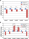
Kidney glomerular basement membranes (GBMs) undergo laminin and type IV collagen isoform substitutions during glomerular development, which are believed to be required for maturation of the filtration barrier. Specifically, GBMs of earliest glomeruli contain laminin α1β1γ1 and collagen α1α2α1(IV), whereas mature glomeruli contain laminin α5β2γ1 and collagen α3α4α5(IV). Here, we used confocal microscopy to simultaneously evaluate expression of different laminin and collagen IV isoforms in newborn mouse GBMs. Our results show loss of laminin α1 from GBMs in early capillary loop stages and continuous linear deposition of laminin bearing the α5 chain thereafter. In contrast, collagen α1α2α1(IV) persisted in linear patterns into late capillary loop stages, when collagen α3α4α5(IV) first appeared in discontinuous, non-linear patterns. This patchy pattern for collagen α3α4α5(IV) continued into maturing glomeruli where there were lengths of linear, laminin α5-positive GBM entirely lacking either isoform of collagen IV. Relative abundance of laminin and collagen IV mRNAs in newborn and 5-week-old mouse kidneys also differed, with those encoding laminin α1, α5, β1, β2, and γ1, and collagen α1(IV) and α2(IV) chains all significantly declining at 5 weeks, but α3(IV) and α4(IV) were significantly upregulated. We conclude that different biosynthetic mechanisms control laminin and type IV collagen expression in developing glomeruli.

Keywords: glomerular endothelial cells; glomerular filtration barrier; podocytes.


Conflict of interest statement

Declaration of Conflicting Interests: The author(s) declared no potential conflicts of interest with respect to the research, authorship, and/or publication of this article.

Figures

Figure 1.
Figure 1.
Dual label scanning confocal immunofluorescence microscopy of newborn mouse kidney sections showing the distribution of laminin α1 (Lamα1, red), laminin α5 (Lamα5, green), and both proteins (Merge), in developing glomerular basement membranes (GBMs). (A) Laminin α1 is present throughout the vascular cleft basement membrane (arrows) of comma- and S-shaped glomeruli. In contrast, only short stretches of these same basement membranes also contain laminin α5 (double arrows in B and C). GBMs of early capillary loop stage glomeruli contain both laminin α chains simultaneously (arrowheads in A–C), but the signal for laminin α1 appears to fade as glomerular development progresses (compare arrowed sites in D). (G–I) As additional capillary loops form, GBMs contain abundant laminin α5 (arrows) but laminin α1 is absent. Bars = 20 µm.
Figure 2.
Figure 2.
Maturing stage glomerulus from a newborn mouse (A–C) and fully mature glomerulus from an 8-week-old mouse (D–F), dually immunolabeled for laminin α1 and laminin α5, as indicated. In both cases, peripheral loop glomerular basement membranes contain only laminin α5 (arrows), whereas mesangial matrices contain laminin α1 and α5 (yellow in merged images C and F). Bars = 20 µm.
Figure 3.
Figure 3.
Newborn mouse kidney sections dually immunolabeled with anti-collagen α1α2α1(IV) [Colα1α2α1(IV), red] and anti-laminin α5 (green). (A–C) The earliest glomerular basement membranes (GBMs) of vascular clefts contain collagen α1α2α1(IV) (arrows) but no laminin α5 (compare with Figs. 1A–1C). (D–F) At the early capillary loop stage, there is overlapping distribution of collagen α1α2α1(IV) with laminin α5 (arrows). (G–I) As capillary loop stage glomeruli continue to progress, collagen α1α2α1(IV) codistributes with laminin α5 in GBMs (arrows). Collagen α1α2α1(IV) can also be seen in developing mesangial matrices (*). Bars = 20 µm.
Figure 4.
Figure 4.
Laminin α5 is distributed in linear, continuous patterns in early and later stage capillary loop glomerular basement membranes (GBMs) (A–G), as well as throughout GBMs and mesangial matrices of maturing stage glomeruli (H–J). Collagen α3α4α5(IV) is first observed in early capillary loop GBMs in focal, discontinuous patterns (B–C, arrows) that are interrupted by GBM segments that are positive for laminin α5 (B–C, arrowheads). As more capillary loops are added to developing glomeruli, more collagen α3α4α5(IV) is deposited, but lengths of laminin α5 positive GBM lack collagen α3α4α5(IV) (G, arrowheads). GBMs of maturing stage glomeruli of newborn kidneys are still not fully labeled with anti-collagen α3α4α5(IV) (H–J). Bars = 20 µm.
Figure 5.
Figure 5.
Localization of collagen α1α2α1(IV) and α3α4α5(IV) in a capillary loop stage glomerulus. Dually immunolabeled section shows collagen α1α2α1(IV) in loop glomerular basement membranes (A, arrows) and developing mesangial matrix (*); collagen α3α4α5(IV) is seen only in fragmentary, discontinuous patterns in a few loops (arrows, B, C). Bars = 20 µm.
Figure 6.
Figure 6.
Localization of collagen α1α2α1(IV) and α3α4α5(IV) in maturing stage glomeruli of newborn (A–H) and 8-week-old mice (I–K). In maturing stage glomeruli of newborns, there is very little signal overlap in glomerular basement membranes (GBMs) or mesangial matrices for these two type IV collagen isoforms (C and G). Boxed areas in C and G are shown in D and H, respectively, where there are overt discontinuities or gaps in labeling for either isoform of type IV collagen in peripheral loop GBMs (arrows). In fully mature glomeruli of 8-week-old kidneys, anti-collagen α1α2α1(IV) immunolabels only mesangial matrices, whereas anti-collagen α3α4α5(IV) labels GBMs exclusively in a continuous, linear pattern. Unlike the case for laminin α1 and laminin α5, there is essentially no signal overlap for these different collagen IV networks in mesangial areas (compare with Figs. 2C and 2F). Bars = 20 µm.
Figure 7.
Figure 7.
Immunoelectron microscopic localization of collagen α3α4α5(IV) in newborn mouse kidney. (A) Glomerular basement membranes (GBMs) of early capillary loop stage glomeruli are unlabeled (arrows). (B) In later stage capillary loop GBMs, discontinuous, uneven deposits of peroxidase reaction product (arrowheads) can be seen in GBMs that also contain unlabeled segments (arrows). (C) Maturing stage glomeruli contain GBMs that appear more uniformly labeled (arrowheads), although there are areas that remain spotty (double arrow). Note that reaction product is absent from mesangial matrices (arrows). En, endothelium; Po, podocyte; Me, mesangium. Bars A and B = 10 µm; bar C = 3 µm.
Figure 8.
Figure 8.
Histograms showing relative expression levels for mRNAs encoding laminin (A) and type IV collagen (B) chains in kidneys from 3-day-old (blue) and 5-week-old (red) mice. As seen in A, compared with newborns, all of the laminin chain mRNAs are significantly downregulated in mature mice. In contrast, mRNAs encoding the α3 and α4 chains of type IV collagen are significantly more abundant in mature kidneys, whereas those encoding collagen α1(IV), α2(IV), and α6(IV) chains are downregulated (B). *p<0.05, **p<0.005, and ***p<0.0005. Light red bars = alternate (alt) primers for COL4A3 and COL4A4 (Table 2), confirming increase for these mRNAs at 5 weeks of age.

References

    1. Abrahamson DR. 2012. Role of the podocyte (and glomerular endothelium) in building the GBM. Semin Nephrol. 32:342–349 - PMC - PubMed
    1. Abrahamson DR, Hudson BG, Stroganova L, Borza DB, St.John PL. 2009. Cellular origins of type IV collagen networks in developing glomeruli. J Am Soc Nephrol. 20:1471–1479 - PMC - PubMed
    1. Abrahamson DR, Irwin MH, St.John PL, Perry EW, Accavitti MA, Heck LW, Couchman JR. 1989. Selective immunoreactivities of kidney basement membranes to monoclonal antibodies against laminin: localization of the end of the long arm and the short arms to discrete microdomains. J Cell Biol. 109:3477–3491 - PMC - PubMed
    1. Abrahamson DR, St.John PL. 1992. Loss of laminin epitopes during glomerular basement membrane assembly in developing mouse kidneys. J Histochem Cytochem. 40:1943–1953 - PubMed
    1. Abrahamson DR, St.John PL, Isom K, Robert B, Miner JH. 2007. Partial rescue of glomerular laminin α5 mutations by wild-type endothelia produce hybrid glomeruli. J Am Soc Nephrol. 18:2285–2293 - PubMed

Publication types