. 2013 Sep;23(9):1119-32.
doi: 10.1038/cr.2013.102. Epub 2013 Jul 30.

Highly efficient induction and long-term maintenance of multipotent cardiovascular progenitors from human pluripotent stem cells under defined conditions

Affiliations

Highly efficient induction and long-term maintenance of multipotent cardiovascular progenitors from human pluripotent stem cells under defined conditions

Nan Cao et al. Cell Res. 2013 Sep.

Abstract

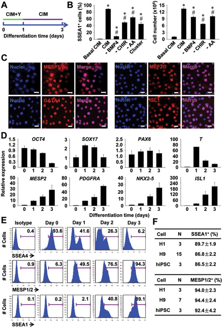
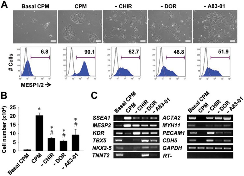
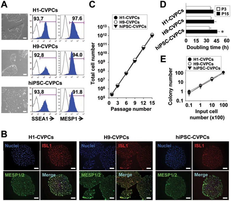
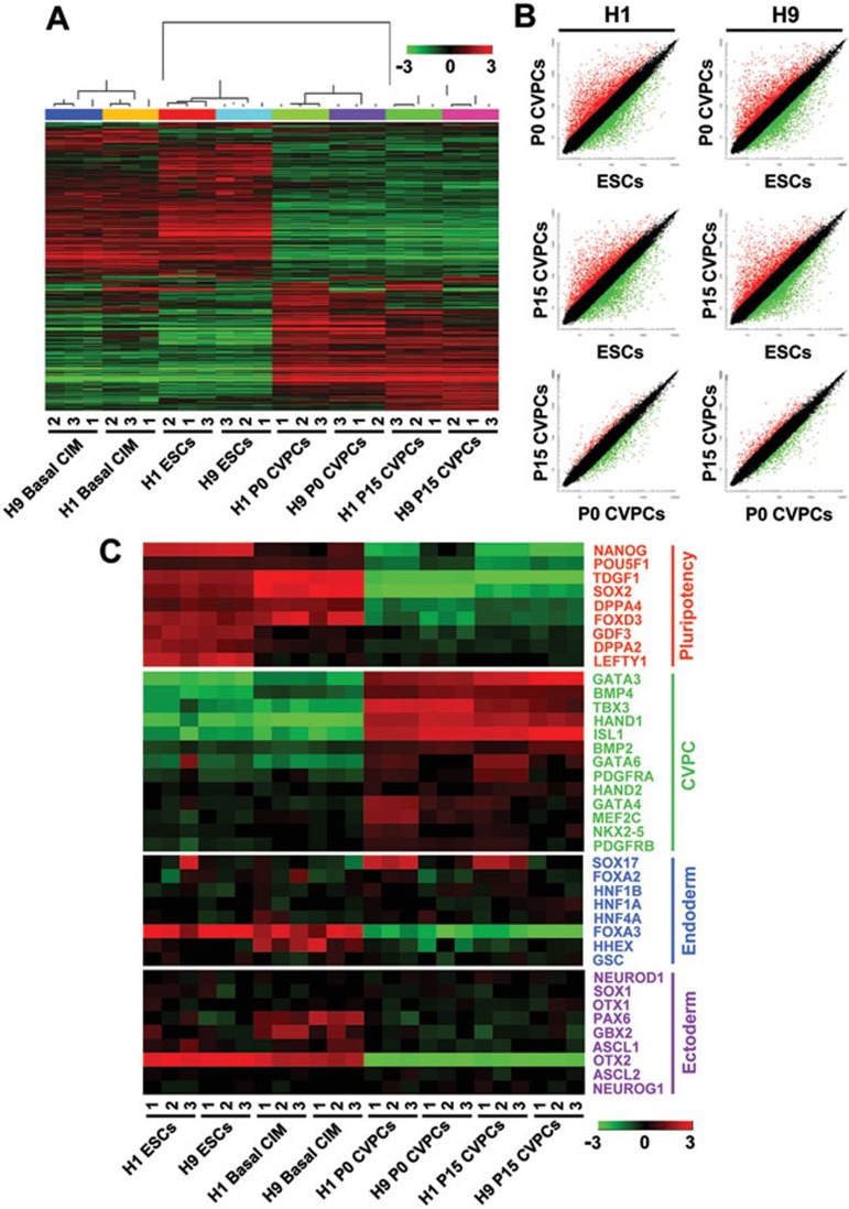
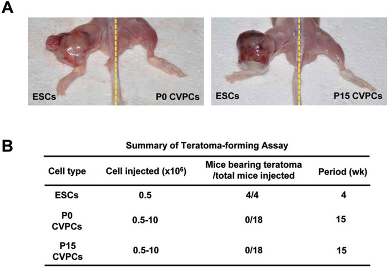
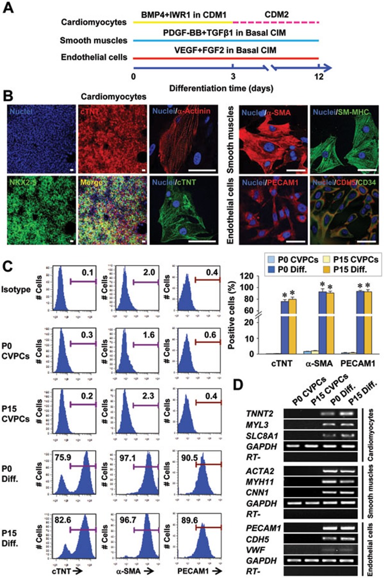
Cardiovascular progenitor cells (CVPCs) derived from human pluripotent stem cells (hPSCs), including human embryonic stem cells (hESCs) and human induced pluripotent stem cells (hiPSCs), hold great promise for the study of cardiovascular development and cell-based therapy of heart diseases, but their applications are challenged by the difficulties in their efficient generation and stable maintenance. This study aims to develop chemically defined systems for robust generation and stable propagation of hPSC-derived CVPCs by modulating the key early developmental pathways involved in human cardiovascular specification and CVPC self-renewal. Herein we report that a combination of bone morphogenetic protein 4 (BMP4), glycogen synthase kinase 3 (GSK3) inhibitor CHIR99021 and ascorbic acid is sufficient to rapidly convert monolayer-cultured hPSCs, including hESCs and hiPSCs, into homogeneous CVPCs in a chemically defined medium under feeder- and serum-free culture conditions. These CVPCs stably self-renewed under feeder- and serum-free conditions and expanded over 10(7)-fold when the differentiation-inducing signals from BMP, GSK3 and Activin/Nodal pathways were simultaneously eliminated. Furthermore, these CVPCs exhibited expected genome-wide molecular features of CVPCs, retained potentials to generate major cardiovascular lineages including cardiomyocytes, smooth muscle cells and endothelial cells in vitro, and were non-tumorigenic in vivo. Altogether, the established systems reported here permit efficient generation and stable maintenance of hPSC-derived CVPCs, which represent a powerful tool to study early embryonic cardiovascular development and provide a potentially safe source of cells for myocardial regenerative medicine.


Figures

Figure 1
Figure 1
Induction of CVPCs from hPSCs. (A) An outline of the differentiation protocol. CIM, CVPC induction medium; Y, Y27632. (B) Percentage of SSEA1+ cells and total cell number at differentiation day 3 under various conditions (n = 3). CHIR, CHIR99021; AA, ascorbic acid. Basal CIM, CIM without CHIR, BMP4 and AA; –, indicates withdrawal. *P < 0.01 vs Basal CIM; #P < 0.01 vs CIM. (C) Immunofluorescence analysis showing the expression of CVPC markers in cells at differentiation day 3 following CIM treatment. Scale bars = 25 μm. (D, E) qRT-PCR (D) and FACS (E) analysis showing the downregulation of pluripotency markers and upregulation of CVPC markers during CVPC induction (n = 3). (F) Induction efficiency of CVPCs from various hPSC lines.
Figure 2
Figure 2
Conditions for CVPC maintenance. (A) Phase contrast images (upper panels); percentages of MESP1/2+ cells (lower panels), (B) total cell numbers (n = 3), and (C) the expression of CVPC, CM, SM and EC markers of H9 hESC-derived CVPCs at differentiation day 3 cultured in various conditions as indicated for another 5 days. CPM, CVPC propagation medium; DOR, dorsomorphin; Basal CPM, CPM without CHIR, DOR and A83-01. *P < 0.01 vs Basal CPM; #P < 0.01 vs CPM.
Figure 3
Figure 3
Long-term maintenance of hPSC-derived CVPCs. (A) Representative images showing the typical morphology (left panels), the SSEA1 and MESP1 expression analyzed by flow cytometry (middle and right panels). The control sample used in this assay was a 1:1:1 mixture from all three types of parental cells examined (H1-, H9-hESCs and hiPSCs). (B) Representative images of the ISL1 and MESP1 expression analyzed by immunostaining of CVPC colonies at passage 15. Scale bar = 100 μm. (C, D) Growth curves (C) and mean doubling time (D, n = 3) of CVPCs. *P < 0.05 vs P3. (E) Cell dose response experiments showing the lineage relationship between the number of cells plated and the number of colonies developed (n = 3). Scale bars = 100 μm.
Figure 4
Figure 4
Genome-wide transcriptional profiling of CVPCs. (A, B) Hierarchical clustering (A) and scatter plot (B) analysis of global gene expression patterns in hESCs cultured in the basal CIM, ESCs, and CVPCs at passage 0 (P0) or 15 (P15), derived from either H1 or H9 hESCs. The expression values in log2 scale in A were calculated and presented on the heat map with red representing upregulated genes and green representing downregulated genes. Black dots in B indicate the population with less than two-fold differences in gene expression levels between both samples. (C) Heat map showing expression levels of key marker genes related to pluripotency, CVPC, definitive endoderm, and neuronal ectoderm.
Figure 5
Figure 5
In vivo tumorigenicity analysis of CVPCs. (A) Representative images showing teratoma formation in left leg of the mouse injected with H9 hESCs (left panels) and lack of tumor formation in right leg of the mouse injected with P0 or P15 H9 hESC-derived CVPCs (right panels). (B) A summary of teratoma-forming ability of H9 hESCs and P0 or P15 CVPCs.
Figure 6
Figure 6
In vitro differentiation potential of CVPCs. (A) An outline of the conditions for inducing CVPC differentiation. (B) Differentiation potential of P15 CVPCs into cardiomyocytes, smooth muscle cells and endothelial cells determined by immunostaining analyses for NKX2-5 (green), cTNT (red or green), α-Actinin (red) and merged NKX2-5 with cTnT in cardiomyocytes, α-SMA (red) and SM-MHC (green) in smooth muscle cells, and PECAM1 (red), CDH5 (red), and CD34 (green) in endothelial cells after treatment in respective patterning conditions for 12 days. Scale bars = 50 μm. (C) Representative (left panels) and averaged FACS population analysis of cTNT+ (CM marker), α-SMA+ (SMC cell marker) and PECAM1+ (EC marker) cells from P0 and P15 CVPCs before or after treatment in respective patterning conditions for 12 days. n = 3. *P < 0.01 vs CVPC. (D) RT-PCR analysis showing the expression of cardiovascular derivate markers in various groups.

Similar articles

Cited by

References

    1. Burridge PW, Keller G, Gold JD, Wu JC. Production of de novo cardiomyocytes: human pluripotent stem cell differentiation and direct reprogramming. Cell Stem Cell. 2012;10:16–28. - PMC - PubMed
    1. Mummery CL, Zhang J, Ng ES, et al. Differentiation of human embryonic stem cells and induced pluripotent stem cells to cardiomyocytes: a methods overview. Circ Res. 2012;111:344–358. - PMC - PubMed
    1. Blin G, Neri T, Stefanovic S, Puceat M. Human embryonic and induced pluripotent stem cells in basic and clinical research in cardiology. Curr Stem Cell Res Ther. 2010;5:215–226. - PubMed
    1. Murry CE, Keller G. Differentiation of embryonic stem cells to clinically relevant populations: lessons from embryonic development. Cell. 2008;132:661–680. - PubMed
    1. Kattman SJ, Witty AD, Gagliardi M, et al. Stage-specific optimization of activin/nodal and BMP signaling promotes cardiac differentiation of mouse and human pluripotent stem cell lines. Cell Stem Cell. 2011;8:228–240. - PubMed

Publication types

MeSH terms