. 2013 Sep;23(9):1133-46.
doi: 10.1038/cr.2013.101. Epub 2013 Jul 30.

Drosophila Myc integrates multiple signaling pathways to regulate intestinal stem cell proliferation during midgut regeneration

Affiliations

Drosophila Myc integrates multiple signaling pathways to regulate intestinal stem cell proliferation during midgut regeneration

Fangfang Ren et al. Cell Res. 2013 Sep.

Abstract

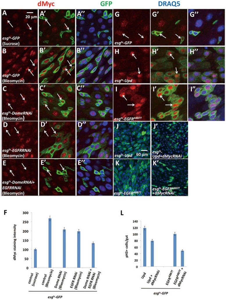
Intestinal stem cells (ISCs) in the Drosophila adult midgut are essential for maintaining tissue homeostasis, and their proliferation and differentiation speed up in order to meet the demand for replenishing the lost cells in response to injury. Several signaling pathways including JAK-STAT, EGFR and Hippo (Hpo) pathways have been implicated in damage-induced ISC proliferation, but the mechanisms that integrate these pathways have remained elusive. Here, we demonstrate that the Drosophila homolog of the oncoprotein Myc (dMyc) functions downstream of these signaling pathways to mediate their effects on ISC proliferation. dMyc expression in precursor cells is stimulated in response to tissue damage, and dMyc is essential for accelerated ISC proliferation and midgut regeneration. We show that tissue damage caused by dextran sulfate sodium feeding stimulates dMyc expression via the Hpo pathway, whereas bleomycin feeding activates dMyc through the JAK-STAT and EGFR pathways. We provide evidence that dMyc expression is transcriptionally upregulated by multiple signaling pathways, which is required for optimal ISC proliferation in response to tissue damage. We have also obtained evidence that tissue damage can upregulate dMyc expression post-transcriptionally. Finally, we show that a basal level of dMyc expression is required for ISC maintenance, proliferation and lineage differentiation during normal tissue homeostasis.


Figures

Figure 1
Figure 1
dMyc is required for ISC proliferation and midgut regeneration in response to tissue damage. (A-C') Adult midguts expressing esgtsF/O without (A-C) or with UAS-dMyc-RNAi (A'-C') were treated with sucrose (A-A'), DSS (B-B'), or bleomycin (C-C'), and immunostained with a GFP (green) antibody and a nuclear dye DRAQ5 (blue). (D-F') Adult midguts expressing UAS-GFP without (D-F) or with UAS-dMyc-RNAi (D'-F') using the esgts system were treated with sucrose (D-D'), DSS (E-E'), or bleomycin (F-F'), and immunostained with GFP (green), PH3 (red) antibodies, and a nuclear dye DRAQ5 (blue). Adult flies were shifted to non-permissive temperature (29 °C) for 3-5 days and then fed with different reagents for another 2 days before dissection. The scale bar shown in A is also applied to A-F'. (G) Quantification of PH3+ cells in midguts of the indicated genotypes (n = 15 for each genotype). (H-M”) Adult midguts expressing esgts-GFP (H-J”) or esgts-GFP + dMycRNAi (K-M”) were treated with sucrose (H-H”, K-K”), DSS (I-I”, L-L”), or bleomycin (J-J”, M-M”), and immunostained with GFP (green) and dMyc (red) antibodies, and a nuclear dye DRAQ5 (blue). Arrows indicate precursor cells whereas asterisks indicate ECs. The scale bar shown in H is also applied to H-M”.
Figure 2
Figure 2
dMyc functions downstream of Hpo signaling pathway. (A-E”) Adult midguts expressing UAS-GFP without (A-A”, B-B”, D-D”) or with UAS-Yki-RNAi (C-C”, E-E”) using the esg-Gal4ts system were treated with sucrose (A-A”), DSS (B-B”, C-C”), or bleomycin (D-D”, E-E”), and immunostained with GFP (green) and dMyc (red) antibodies, and a nuclear dye DRAQ5 (blue). Arrows indicate precursor cells. The scale bar shown in A is also applied to A-E”. (F-H”) Adult midguts expressing UAS-GFP without (F-F”), or with UAS-Yki (G-G”) or UAS-Wts-RNAi (H-H”) using esgts were immunostained with GFP (green) and dMyc (red) antibodies, and a nuclear dye DRAQ5 (blue). Arrows indicate precursor cells. The scale bar shown in F is also applied to F-H”. (I-J') Adult midguts expressing UAS-GFP with UAS-Yki (I), UAS-Yki + dMyc-RNAi (I'), UAS-Wts-RNAi (J), or UAS-Wts-RNAi + dMyc-RNAi (J') using esgts were immunostained with a GFP (green) antibody and a nuclear dye DRAQ5 (blue). The scale bar shown in I is also applied to I-J'. (K) Quantification of PH3+ cells in midguts of the indicated genotypes (n = 15 for each genotype).
Figure 3
Figure 3
dMyc functions downstream of both JAK-STAT and EGFR pathways. (A-E”) Adult midguts expressing UAS-GFP without (A-B”), or with UAS-Dome-RNAi (C-C”), UAS-EGFR-RNAi (D-D”), or UAS-Dome-RNAi plus UAS-EGFR-RNAi (E-E”) using esgts were treated with sucrose (A-A”) or bleomycin (B-E”) and immunostained with GFP (green) and dMyc (red) antibodies, and a nuclear dye DRAQ5 (blue). Arrows indicate precursor cells. (F) Quantification of dMyc staining in precursor cells shown in A-E (n > 40 for each genotype). (G-I”) Adult midguts expressing UAS-GFP without (G-G”), or with UAS-Upd (H-H”) or UAS- EGFRA887T (I-I”) using esgts were immunostained with GFP (green) and dMyc (red) antibodies, and a nuclear dye DRAQ5 (blue). Arrows indicate precursor cells. (J-K') Adult midguts expressing UAS-GFP together with UAS-Upd (J), UAS-Upd + dMyc-RNAi (J'), UAS-EGFRA887T (K), or UAS-EGFRA887T+ dMyc-RNAi (K') using the esgts system were immunostained with GFP (green) antibody and a nuclear dye DRAQ5 (blue). 3- to 5-day-old females were shifted to 29 °C for 2 days before dissection and immunostaining. (L) Quantification of PH3+ cells in midguts of the indicated genotypes (n = 15 for each genotype).
Figure 4
Figure 4
dMyc is regulated at transcriptional level by multiple signaling pathways. (A-G”) Adult midguts expressing dMyc-lacZ and UAS-GFP without (A-A”), or with UAS-Yki (B-B”), UAS-Wts-RNAi (C-C”), UAS-Upd (D-D”), UAS- EGFRA887T (E-E”), UAS-StatΔNΔC (F-F”), or UAS-Pnt2-Vp16 (G-G”) using the esgts system were immunostained with GFP (green) and lacZ (red) antibodies and a nuclear dye DRAQ5 (blue). Arrows indicate precursor cells. (H) Quantification of dMyc-LacZ expression in precursor cells shown in A-G (n > 50 for each genotype).
Figure 5
Figure 5
dMyc promoter/enhancer region contains consensus Sd-, Stat- and Pnt-binding sites. (A) Schematic representation of the dm locus showing the first three exons. Black rectangles represent coding regions, white rectangles represent noncoding regions; lines denote introns. The blowup indicates the 1.0 kb enhancer region with black bars indicating consensus Stat-binding sites, blue bars indicating consensus Sd-binding sites and red bars indicating consensus Pnt-binding sites, respectively. W: A or T; N: any nucleotide; D: G, A or T; H: A, C or T; S: C or G; V: A, C or G. The DNA sequences for individual putative binding sites are listed. (B) (Top) Diagram of the dMyc1.0-luc reporter gene. The dMyc1.0 enhancer region was placed upstream of the heat shock basal promoter followed by the luciferase-coding sequence. (Bottom) S2 cells were transfected by the indicated expression constructs plus the luciferase reporter gene, and the cell lysates were subject to the dual-luciferase reporter assay. dMyc1.0m-luc has all the Stat-binding sites mutated. (C) ChIP experiment to detect the direct binding of exogenously expressed HA-Sd or HA-Stat to the dMyc1.0 enhancer region. Transgenic flies expressing UAS-N-RNAi alone or together with UAS-HA-Sd plus UAS-Myc-Yki or UAS-HA-StatΔNΔC with esgts were subject to ChIP experiment using an anti-HA antibody. The enhancer regions encompassed by different primers were indicated in A. (D) dMyc integrates multiple signaling pathways to drive ISC proliferation in response to tissue damage.
Figure 6
Figure 6
Transcriptional upregulation of dMyc is required for optimal ISC proliferation in response to injury. (A) Quantification of PH3+ cells in midguts of dm4/+; tub-dMyc or dm4/dm4; tub-dMyc adult flies fed with sucrose, DSS or bleomycin (n = 20 for each genotype). Statistical significance was determined by Student's t test (*P < 0.05, ***P < 0.001). (B) Quantification of dMyc staining in precursor cells shown in C-K (n > 50 for each genotype). (C-K') Adult midguts with indicated genotype were treated with sucrose (C-C', F-F', I-I'), DSS (D-D', G-G', J-J') or bleomycin (E-E', H-H', K-K') and immunostained with a rabbit anti-dMyc (red) antibody and a nuclear dye DRAQ5 (blue). Arrows indicate precursor cells marked by small nuclear size.
Figure 7
Figure 7
dMyc is essential for midgut homeostasis. (A-B”) Adult midguts expressing esgtsF/O without (A-A”) or with UAS-dMyc-RNAi (B-B”) were cultured at non-permissive temperature (29 °C) for 15 days, followed by immunostaining with a GFP (green) antibody and a nuclear dye DRAQ5 (blue). (C-H”) Adult midguts containing GFP-positive WT (wt) clones (C-C”, E-E”, G-G”), or dm4 clones (D-D”, F-F”, H-H”) were immunostained to show the expression of GFP (green), Dl (red in C-D”), Pdm1 (red in E-F”), Pros (red in G-H”), and DRAQ5 (blue). dm4 mutant clones grew slower and failed to differentiate into ECs (Pdm1+) or EEs (Pros+). The control and mutant clones were generated using the MARCM system. Guts were dissected out from adult flies grown at 18 °C for 20 days after clone induction. (I) Quantification of ISC lineage clone size for the control (wt) and dm4 clones (n > 50 for each genotype). ****P < 0.0001. (J) 2- to 5-day-old adult females expressing UAS-GFP without (blue curve) or with UAS-dMyc-RNAi (red curve) or with UAS-dMyc-RNAi + UAS-dMyc (green curve) using the esgts system were cultured at non-permissive temperature (29 °C) for various periods of time. Their midguts were dissected at the indicated time points after temperature shift and immunostained with Dl and GFP antibodies. The number of ISCs in the posterior midguts was quantified for different time points.

Similar articles

Cited by

References

    1. Biteau B, Hochmuth CE, Jasper H. Maintaining tissue homeostasis: dynamic control of somatic stem cell activity. Cell Stem Cell. 2011;9:402–411. - PMC - PubMed
    1. Jiang H, Edgar BA. Intestinal stem cell function in Drosophila and mice. Curr Opin Genet Dev. 2012;22:354–60. - PMC - PubMed
    1. Micchelli CA, Perrimon N. Evidence that stem cells reside in the adult Drosophila midgut epithelium. Nature. 2006;439:475–479. - PubMed
    1. Ohlstein B, Spradling A. The adult Drosophila posterior midgut is maintained by pluripotent stem cells. Nature. 2006;439:470–474. - PubMed
    1. Jiang H, Patel PH, Kohlmaier A, Grenley MO, McEwen DG, Edgar BA. Cytokine/Jak/Stat signaling mediates regeneration and homeostasis in the Drosophila midgut. Cell. 2009;137:1343–1355. - PMC - PubMed

Publication types

MeSH terms

Substances