. 2013 Nov;65(11):2876-86.
doi: 10.1002/art.38099.

Transplantation of Achilles tendon treated with bone morphogenetic protein 7 promotes meniscus regeneration in a rat model of massive meniscal defect

Affiliations
Free PMC article

Transplantation of Achilles tendon treated with bone morphogenetic protein 7 promotes meniscus regeneration in a rat model of massive meniscal defect

Nobutake Ozeki et al. Arthritis Rheum. 2013 Nov.
Free PMC article

Abstract

Objective: This study was undertaken to examine whether bone morphogenetic protein 7 (BMP-7) induces ectopic cartilage formation in the rat tendon, and whether transplantation of tendon treated with BMP-7 promotes meniscal regeneration. Additionally, we analyzed the relative contributions of host and donor cells on the healing process after tendon transplantation in a rat model.

Methods: BMP-7 was injected in situ into the Achilles tendon of rats, and the histologic findings and gene profile were evaluated. Achilles tendon injected with 1 μg of BMP-7 was transplanted into a meniscal defect in rats. The regenerated meniscus and articular cartilage were evaluated at 4, 8, and 12 weeks. Achilles tendon from LacZ-transgenic rats was transplanted into the meniscal defect in wild-type rats, and vice versa.

Results: Injection of BMP-7 into the rat Achilles tendon induced the fibrochondrocyte differentiation of tendon cells and changed the collagen gene profile of tendon tissue to more closely approximate meniscal tissue. Transplantation of the rat Achilles tendon into a meniscal defect increased meniscal size. The rats that received the tendon treated with BMP-7 had a meniscus matrix that exhibited increased Safranin O and type II collagen staining, and showed a delay in articular cartilage degradation. Using LacZ-transgenic rats, we determined that the regeneration of the meniscus resulted from contribution from both donor and host cells.

Conclusion: Our findings indicate that BMP-7 induces ectopic cartilage formation in rat tendons. Transplantation of Achilles tendon treated with BMP-7 promotes meniscus regeneration and prevents cartilage degeneration in a rat model of massive meniscal defect. Native cells in the rat Achilles tendon contribute to meniscal regeneration.


Figures

Figure 1
Figure 1
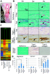
Effect of in situ bone morphogenetic protein 7 (BMP-7) injection into the rat Achilles tendon. A, Procedure used to inject BMP-7 into the rat Achilles tendon. B, Histologic examination of rat Achilles tendon stained with Safranin O and immunostained with type I and type II collagen. Arrows indicate ectopic chondrocyte formation. C, Dendrogram generated from microarray analysis of native rat tendon, rat tendon treated with 1 μg of BMP-7 4 weeks before analysis, native rat meniscus, and native rat articular cartilage (AC) with subchondral bone. D, Results of polymerase chain reaction analysis of aggrecan, Col1a1, and Col2a1. Bars show the mean ± SD expression level relative to the expression level in native tendon (n = 4 rats per group). ∗ = P < 0.05 by Kruskal-Wallis test.
Figure 2
Figure 2
Macroscopic analyses of transplantation of Achilles tendon treated with bone morphogenetic protein 7 (BMP-7) into meniscal defects in rats. A, Study scheme. B, Procedure used for the surgery. The anterior half of the medial meniscus was resected (untreated group), then transplantation of Achilles tendon was performed (tendon group). In the third group, Achilles tendon was transplanted just after injection of BMP-7 (tendon + BMP-7 group). BMP-7 solution with dying agent is indicated in blue. C, Macroscopic features. Arrows indicate the resected site; arrowheads indicate integration sites of regenerated tissue and native meniscus. D, Method of determining the “meniscus covering ratio,” defined as the ratio of medial meniscus area to medial plateau area. E, Meniscus covering ratio. Bars show the mean ± SD (n = 6 rats per group). ∗ = P < 0.05 by non–repeated-measures analysis of variance. NS = not significant. Color figure can be viewed in the online issue, which is available at http://onlinelibrary.wiley.com/doi/10.1002/acr.38099/abstract.
Figure 3
Figure 3
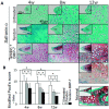
Histologic analysis of regenerated rat meniscus. A, Sections from untreated rats, rats that received transplantation of untreated Achilles tendon (tendon), and rats that received transplantation of bone morphogenetic protein 7 (BMP-7)–treated Achilles tendon (tendon + BMP-7), stained with Safranin O 4 weeks, 8 weeks, and 12 weeks after surgery. The boxed areas in the insets are shown at a higher-magnification view in the larger panels. Broken lines indicate the outer zone of the regenerated meniscus in a rat that received BMP-7–treated tendon. B, Modified Pauli scores for histology. Bars show the mean ± SD (n = 6 rats per group). ∗ = P < 0.05 by Kruskal-Wallis test. Color figure can be viewed in the online issue, which is available at http://onlinelibrary.wiley.com/doi/10.1002/acr.38099/abstract.
Figure 4
Figure 4
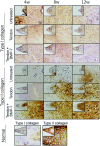
Immunohistochemical analysis for type I collagen and type II collagen in the regenerated meniscus of normal rats, rats that received transplantation of bone morphogenetic protein 7 (BMP-7)–treated tendon (tendon + BMP-7), rats that received transplantation of untreated tendon (tendon), and untreated rats, 4 weeks, 8 weeks, and 12 weeks after surgery. The boxed areas in the insets are shown at a higher-magnification view in the larger panels. Color figure can be viewed in the online issue, which is available at http://onlinelibrary.wiley.com/doi/10.1002/acr.38099/abstract.
Figure 5
Figure 5
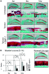
Histologic analysis of rat articular cartilage at the medial tibial plateau. A, Sagittal sections from untreated rats, rats that received transplantation of untreated Achilles tendon (tendon), and rats that received transplantation of bone morphogenetic protein 7 (BMP-7)–treated Achilles tendon (tendon + BMP-7), stained with Safranin O 4 weeks, 8 weeks, and 12 weeks after surgery. A section from a 22-week-old normal rat was used as a control. Boxed areas in the top panels are shown at a higher-magnification view in the bottom panels. B, Mankin scores. Bars show the mean ± SD (n = 6 rats per group). ∗ = P < 0.05 by Kruskal-Wallis test. NS = not significant. Color figure can be viewed in the online issue, which is available at http://onlinelibrary.wiley.com/doi/10.1002/acr.38099/abstract.
Figure 6
Figure 6
A, Illustration of transplantation of Achilles tendon from a LacZ-transgenic rat into the meniscus of a wild-type rat. B, Macroscopic (left) and histologic (right) analysis of sections from a wild-type rat that received transplantation of Achilles tendon from a LacZ-transgenic rat. Macroscopic features of the tibial plateau with medial and lateral menisci 4 weeks after transplantation are shown. Whole tissue was stained with X-Gal. Arrow indicates the LacZ-positive area. Histologic analysis showed LacZ-positive cells (blue) in transplanted tendon at 4 weeks. The boxed area in the inset is shown at a higher-magnification view in the larger panel. C, Illustration of transplantation of Achilles tendon from a wild-type rat into the meniscus of a LacZ-transgenic rat. D, Macroscopic (left and middle) and histologic (right) analysis of sections from a LacZ-transgenic rat that received transplantation of Achilles tendon from a wild-type rat. Macroscopic features of the medial tibial plateau left unstained (left) or stained with X-Gal (middle) 4 weeks after transplantation are shown. Arrows indicate the transplantation site. Histologic analysis of transplanted tendon stained with X-Gal was performed at 4 weeks. The boxed area in the inset is shown at a higher-magnification view in the larger panel. E, Possible mechanism of meniscus regeneration by transplantation of Achilles tendon treated with bone morphogenetic protein 7 (BMP-7) in a rat model of massive meniscal defect. In the untreated group, only a small amount of synovium filled the space of the meniscal defect, and it did not mature into meniscus. In the rats that received transplantation of untreated tendon, the transplanted tendon played the role of scaffold, synovium covered the tendon, and both promoted meniscus regeneration. In the rats that received transplantation of tendon treated with BMP-7, in addition to the effect of tendon transplantation, BMP-7 induced chondrocyte differentiation of tendon cells, and consequently menicsus regeneration was enhanced. Color figure can be viewed in the online issue, which is available at http://onlinelibrary.wiley.com/doi/10.1002/acr.38099/abstract.

Similar articles

Cited by

References

    1. Walker PS, Erkman MJ. The role of the menisci in force transmission across the knee. Clin Orthop Relat Res. 1975:184–92. - PubMed
    1. Walsh CJ, Goodman D, Caplan AI, Goldberg VM. Meniscus regeneration in a rabbit partial meniscectomy model. Tissue Eng. 1999;5:327–37. - PubMed
    1. Burks RT, Metcalf MH, Metcalf RW. Fifteen-year follow-up of arthroscopic partial meniscectomy. Arthroscopy. 1997;13:673–9. - PubMed
    1. Englund M, Roos EM, Roos HP, Lohmander LS. Patient-relevant outcomes fourteen years after meniscectomy: influence of type of meniscal tear and size of resection. Rheumatology (Oxford) 2001;40:631–9. - PubMed
    1. Kraus T, Heidari N, Svehlík M, Schneider F, Sperl M, Linhart W. Outcome of repaired unstable meniscal tears in children and adolescents. Acta Orthop. 2012;83:261–6. - PMC - PubMed

Publication types

MeSH terms