

Skin gene expression correlates of severity of interstitial lung disease in systemic sclerosis
- PMID: 23897225
- PMCID: PMC3898704
- DOI: 10.1002/art.38101
Skin gene expression correlates of severity of interstitial lung disease in systemic sclerosis
Abstract
Objective: We undertook this hypothesis-generating study to identify skin transcripts correlating with severity of interstitial lung disease (ILD) in systemic sclerosis (SSc).
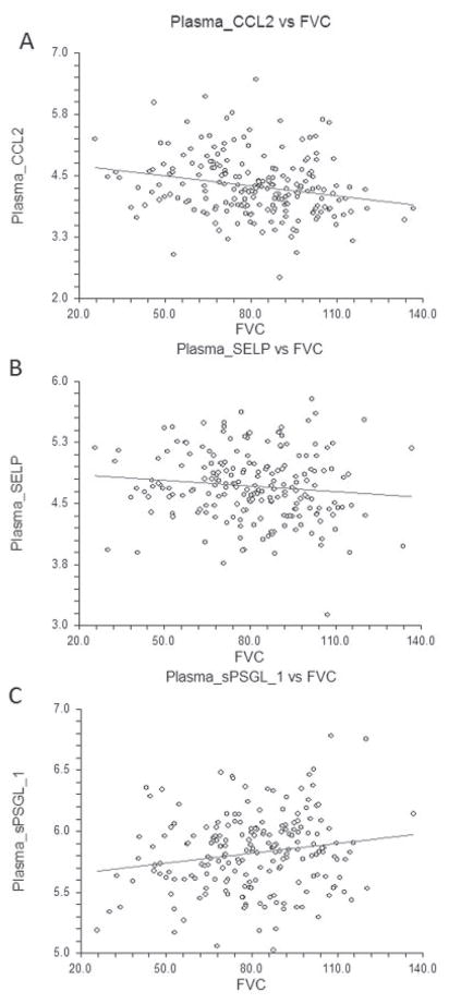
Methods: Skin biopsy samples from 59 patients enrolled in the Genetics versus Environment in Scleroderma Outcome Study (GENISOS) cohort or an open-label imatinib study (baseline visit) were examined by global gene expression analysis using Illumina HT-12 arrays. Skin transcripts correlating with concomitantly obtained forced vital capacity (FVC) values and the modified Rodnan skin thickness score (MRSS) were identified by quantitative trait analysis. Also, immunofluorescence staining for selected transcripts was performed in affected skin and lung tissue. Plasma levels of CCL2, soluble SELP, and soluble P-selectin glycoprotein ligand 1 (sPSGL-1) were examined in all patients enrolled in the GENISOS cohort (n = 266).
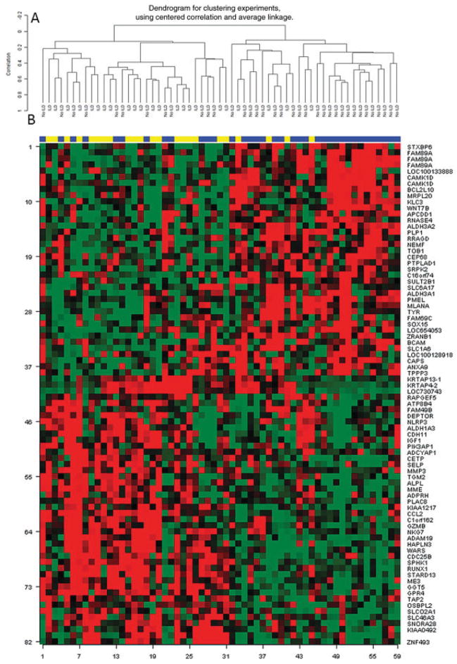
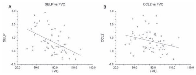
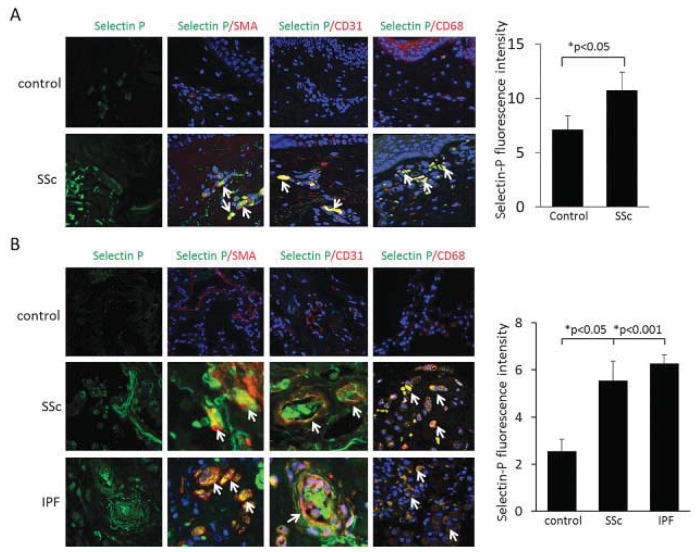
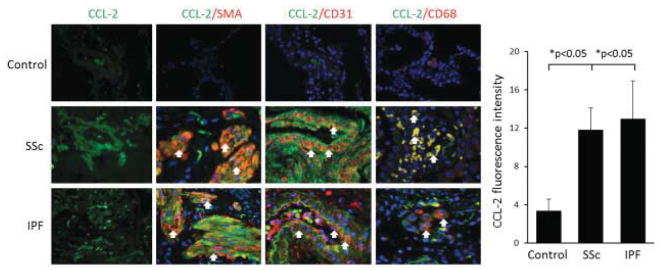
Results: Eighty-two skin transcripts correlated significantly with FVC. This gene list distinguished patients with more severe ILD (FVC <70% predicted) in unsupervised hierarchical clustering analysis (P < 0.001). These genes included SELP, CCL2, and matrix metalloproteinase 3, which are involved in extravasation and adhesion of inflammatory cells. Among the FVC correlates, 8 genes (CCL2, HAPLN3, GPR4, ADCYAP1, WARS, CDC25B, PLP1, and STXBP6) also correlated with the MRSS. Immunofluorescence staining revealed that SELP and CCL2 were also overexpressed in affected skin and lung tissue from SSc patients compared to those from controls. Plasma levels of CCL2 and sPSGL-1 correlated with concomitantly obtained FVC values (r = -0.22, P = 0.001 and r = 0.17, P = 0.015, respectively). This relationship was independent of potential confounders (age, sex, ethnicity, smoking status, anti-topoisomerase I positivity, treatment with immunosuppressive agents, MRSS, disease type, and disease duration).
Conclusion: A limited number of skin transcripts including genes involved in extravasation and adhesion of inflammatory cells correlate with severity of ILD.
Copyright © 2013 by the American College of Rheumatology.
Figures
References
-
- Tyndall AJ, Bannert B, Vonk M, Airo P, Cozzi F, Carreira PE, et al. Causes and risk factors for death in systemic sclerosis: a study from the EULAR Scleroderma Trials and Research (EUSTAR) database. Ann Rheum Dis. 2010;69:1809–15. - PubMed
-
- Tashkin DP, Elashoff R, Clements PJ, Goldin J, Roth MD, Furst DE, et al. Cyclophosphamide versus placebo in scleroderma lung disease. N Engl J Med. 2006;354:2655–66. - PubMed
Publication types
MeSH terms
Substances
Grants and funding
LinkOut - more resources
Full Text Sources
Other Literature Sources
Medical
Molecular Biology Databases
Research Materials
Miscellaneous
