. 2013 Oct 15;591(20):5071-81.
doi: 10.1113/jphysiol.2013.260042. Epub 2013 Jul 29.

Permeability and contractile responses of collecting lymphatic vessels elicited by atrial and brain natriuretic peptides

Affiliations

Permeability and contractile responses of collecting lymphatic vessels elicited by atrial and brain natriuretic peptides

Joshua P Scallan et al. J Physiol. .

Abstract

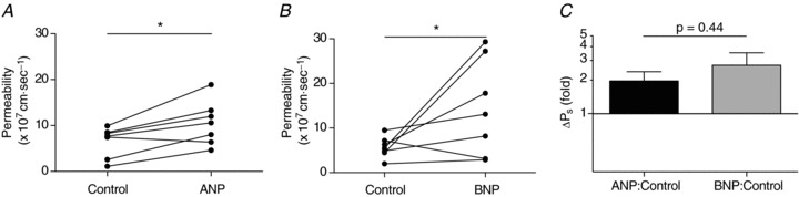
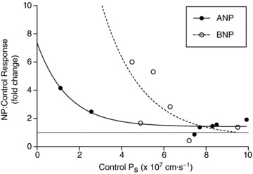
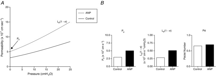
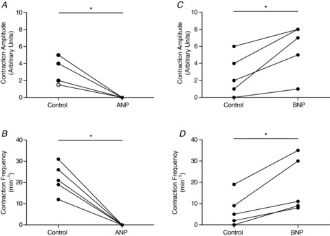
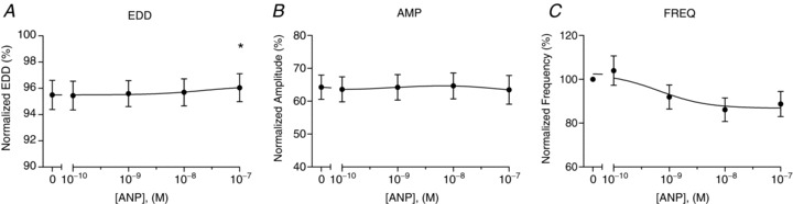
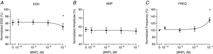
Atrial and brain natriuretic peptides (ANP and BNP, respectively) are cardiac hormones released into the bloodstream in response to hypervolaemia or fluid shifts to the central circulation. The actions of both peptides include natriuresis and diuresis, a decrease in systemic blood pressure, and inhibition of the renin-angiotensin-aldosterone system. Further, ANP and BNP elicit increases in blood microvessel permeability sufficient to cause protein and fluid extravasation into the interstitium to reduce the vascular volume. Given the importance of the lymphatic vasculature in maintaining fluid balance, we tested the hypothesis that ANP or BNP (100 nM) would likewise elevate lymphatic permeability (Ps) to serum albumin. Using a microfluorometric technique adapted to in vivo lymphatic vessels, we determined that rat mesenteric collecting lymphatic Ps to rat serum albumin increased by 2.0 ± 0.4-fold (P = 0.01, n = 7) and 2.7 ± 0.8-fold (P = 0.07, n = 7) with ANP and BNP, respectively. In addition to measuring Ps responses, we observed changes in spontaneous contraction amplitude and frequency from the albumin flux tracings in vivo. Notably, ANP abolished spontaneous contraction amplitude (P = 0.005) and frequency (P = 0.006), while BNP augmented both parameters by ∼2-fold (P < 0.01 each). These effects of ANP and BNP on contractile function were examined further by using an in vitro assay. In aggregate, these data support the theory that an increase in collecting lymphatic permeability opposes the absorptive function of the lymphatic capillaries, and aids in the retention of protein and fluid in the interstitial space to counteract volume expansion.


Figures

Figure 1
Figure 1. Collecting lymphatic vessel permeability in vivo increases upon exposure to either atrial or brain natriuretic peptide
A and B, collecting lymphatic vessels underwent a significant increase in Ps to rat serum albumin versus control when perfused with either 100 nm ANP (P= 0.01) or BNP (P= 0.07). For each peptide there are n= 7 paired measures. One vessel from each data set did not respond to the natriuretic peptide. C, Ps responses are graphed as the ratio of Ps during natriuretic peptide infusion to that measured during control conditions. The mean ± SEM fold changes for ANP and BNP are 2.0 ± 0.4 and 2.7 ± 0.8, respectively. Note the logarithmic y-scale. At y= 1 there is no change from control, and lies where the x-axis is drawn. *P < 0.10.
Figure 2
Figure 2. Sensitivity of the in vivo Ps response to natriuretic peptide infusion as a function of the control Ps
The fold increase in Ps is plotted on the y-axis for vessels exposed to either 100 nm ANP (filled circles, n= 7) or BNP (open circles, n= 6). The general trend is that vessels with a low basal Ps are more responsive to perfusion with natriuretic peptides. The gray line drawn at y= 1 marks where there is no change in Ps during perfusion with natriuretic peptides relative to control. The two curves are significantly different (P < 0.05).
Figure 3
Figure 3. ANP increases the diffusion-mediated solute flux and convective (water-driven) coupling of solute flux to water flux
A, the continuous curve represents individual in vivo measures of control Ps, while the dashed curve represents the same vessels perfused with 100 nm ANP (n= 6 measures per group). Net pressure, on the x-axis, is defined as the difference between the hydrostatic and effective oncotic pressures. The y-intercept is equal to the diffusional permeability (Pd), and the limiting slope of each fitted line is equal to Lp(1 −σ). The fitted curves were not statistically different, but were used to obtain the information in panel B. B, values derived from the graph in A are shown and include estimates of the diffusive permeability to albumin (Pd), hydraulic conductivity (Lp), and the Péclet number (Pé) at the average in vivo collecting lymphatic hydrostatic pressure (7 cmH2O; Scallan & Huxley, 2010).
Figure 4
Figure 4. Perfusion of 100 nm ANP and BNP appear to differentially regulate lymphatic spontaneous contractions in vivo
Luminal exposure to ANP (n= 6) apparently inhibits collecting lymphatic vessel contraction amplitude (A) and frequency (B). Exposure to BNP, however, enhances both contraction amplitude (C) and frequency (D) by approximately 2-fold each (n= 5). Amplitude and frequency were measured from the fluorescence intensity data tracing obtained prior to or during solute flux measurements made in vivo. All measures were paired, meaning that the same vessel was perfused with a control solution followed by an identical one containing ANP or BNP. Each pair was measured at one hydrostatic pressure and at the same site on the vessel. No significant changes in diameter were observed. The open circle in A indicates two overlapping data points. *P < 0.05.
Figure 5
Figure 5. In vitro dose responsiveness of collecting lymphatic vessel contractile function to ANP
The normalized end-diastolic diameter (EDD; A), contraction amplitude (AMP; B), and contraction frequency (FREQ; C) were each plotted as a function of ANP concentration, ranging from 0.1 to 100 nm (n= 4). End-diastolic diameter and contraction amplitude were normalized to the maximal passive diameter at a pressure of 3 cmH2O, while contraction frequency was normalized to that of the control period. The first data point of each graph represents the mean of the control data. *Significantly different from the control data point (P < 0.05).
Figure 6
Figure 6. In vitro dose-responsiveness of isolated collecting lymphatic vessel contractile function to BNP
The normalized end-diastolic diameter (EDD; A), contraction amplitude (AMP; B), and contraction frequency (FREQ, C) were each plotted as a function of BNP concentration, ranging from 0.1 to 100 nm (n= 5). End-diastolic diameter and contraction amplitude were normalized to the maximal passive diameter at a pressure of 3 cmH2O, while contraction frequency was normalized to that of the control period. The first data point of each graph represents the mean of the control data. *Significantly different from the control data point (P < 0.05).

References

    1. Almeida FA, Suzuki M, Maack T. Atrial natriuretic factor increases hematocrit and decreases plasma volume in nephrectomized rats. Life Sci. 1986;39:R1193–R1199. - PubMed
    1. Anderson WD, Kulik TJ, Mayer JE. Inhibition of contraction of isolated lymphatic ducts by atrial natriuretic peptide. Am J Physiol Regul Integr Comp Physiol. 1991;260:R610–R614. - PubMed
    1. Aukland K, Reed RK. Interstitial-lymphatic mechanisms in the control of extracellular fluid volume. Physiol Rev. 1993;73:1–78. - PubMed
    1. Bingaman S, Huxley VH, Rumbaut RE. Fluorescent dyes modify properties of proteins used in microvascular research. Microcirculation. 2003;10:221–231. - PubMed
    1. Brookes ZL, Kaufman S. Effects of atrial natriuretic peptide on the extrasplenic microvasculature and lymphatics in the rat in vivo. J Physiol. 2005;565:269–277. - PMC - PubMed

Publication types

MeSH terms

LinkOut - more resources